. 2009 Dec 10:5:70.
doi: 10.1186/1744-8069-5-70.

Differential regulation of immune responses and macrophage/neuron interactions in the dorsal root ganglion in young and adult rats following nerve injury

Affiliations

Differential regulation of immune responses and macrophage/neuron interactions in the dorsal root ganglion in young and adult rats following nerve injury

David Vega-Avelaira et al. Mol Pain. .

Abstract

Background: Neuropathic pain is an apparently spontaneous experience triggered by abnormal physiology of the peripheral or central nervous system, which evolves with time. Neuropathic pain arising from peripheral nerve injury is characterized by a combination of spontaneous pain, hyperalgesia and allodynia. There is no evidence of this type of pain in human infants or rat pups; brachial plexus avulsion, which causes intense neuropathic pain in adults, is not painful when the injury is sustained at birth. Since infants are capable of nociception from before birth and display both acute and chronic inflammatory pain behaviour from an early neonatal age, it appears that the mechanisms underlying neuropathic pain are differentially regulated over a prolonged postnatal period.

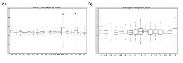
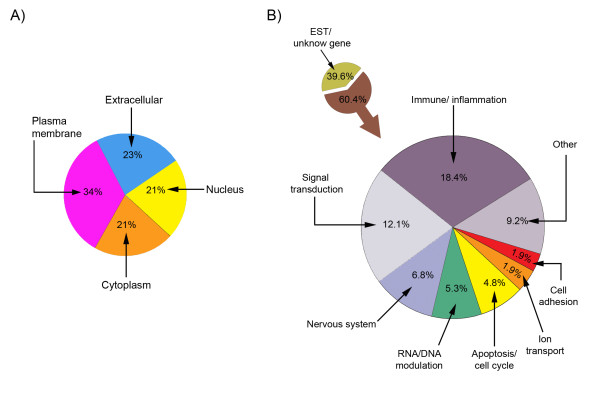
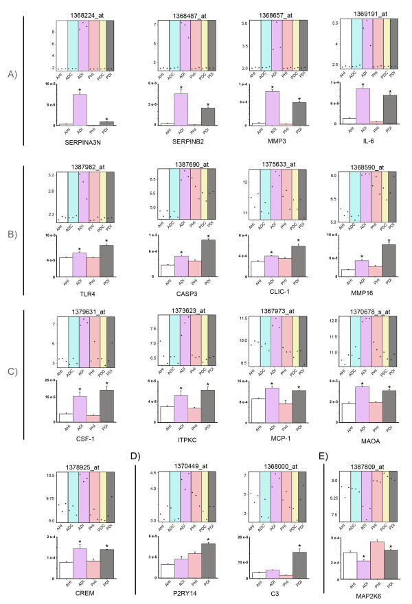
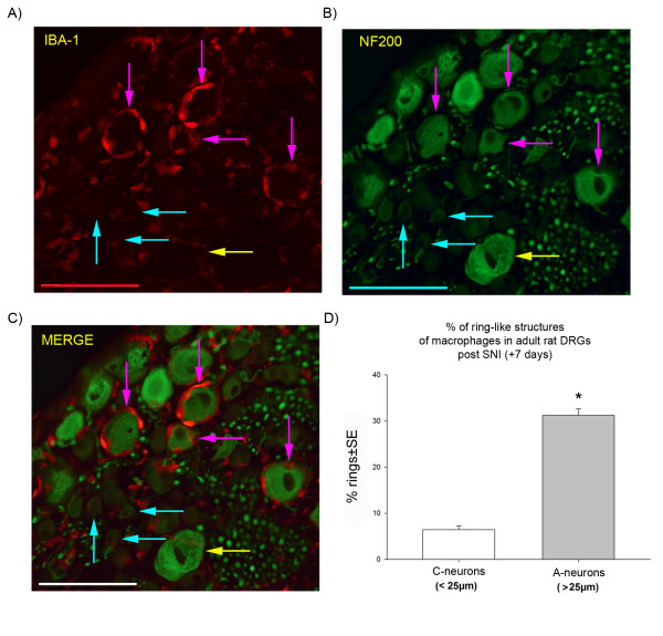
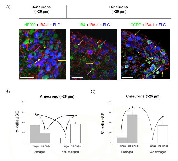
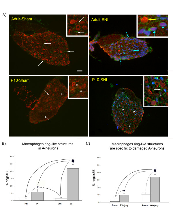
Results: We have performed a microarray analysis of the rat L4/L5 dorsal root ganglia (DRG), 7 days post spared nerve injury, a model of neuropathic pain. Genes that are regulated in adult rats displaying neuropathic behaviour were compared to those regulated in young rats (10 days old) that did not show the same neuropathic behaviour. The results show a set of genes, differentially regulated in the adult DRG, that are principally involved in immune system modulation. A functional consequence of this different immune response to injury is that resident macrophages cluster around the large A sensory neuron bodies in the adult DRG seven days post injury, whereas the macrophages in young DRG remain scattered evenly throughout the ganglion, as in controls.

Conclusions: The results show, for the first time, a major difference in the neuroimmune response to nerve injury in the dorsal root ganglion of young and adult rats. Differential analysis reveals a new set of immune related genes in the ganglia, that are differentially regulated in adult neuropathic pain, and that are consistent with the selective activation of macrophages around adult, but not young large A sensory neurons post injury. These differences may contribute to the reduced incidence of neuropathic pain in infants.


Figures

Figure 1
Figure 1
Quality assessment of the microarrays. The figure shows the relative expression signal of the RMA-normalised microarrays before (a) and after (b) removal of the outliers (*). A: adult; P: 10 day old rat; I: spared nerve injury group H: sham group and C: contralateral group (all 7 days after surgery).
Figure 2
Figure 2
Cellular location (A) and functional groups (B) of the mRNA Affy_IDs from adult dorsal root ganglia 7 days post spared nerve injury. Unknown refers to those Affymetrix entries that are either unknown genes or expressed sequences tag (ETS) that correspond to "transcribed loci" and "similar-to-genes" mRNAs.
Figure 3
Figure 3
Microarray and quantitative Real-time PCR (qPCR) validation of genes differentially regulated in the dorsal root ganglia 7 days after SNI surgery. The figure shows microarray plots of the selected Affy_IDs and counterpart graphs of qPCR validation of the corresponding gene. For clarity, genes have been grouped into A) 'preferentially expressed in adult SNI', B) 'preferentially expressed in young SNI', C) 'genes equally expressed in both adult and young SNI groups', D) 'genes specific to young SNI' and E) 'downregulated gene group'. In the microarray plots, the y-axis indicates the normalised log2 value of expression and each point represents the level of a particular Affy_ID in a single array. In the qPCR validation graphs, the y-axis indicates the mean fluorescence intensity (± standard error). White: Adult sham rats (AHI); Blue: Adult SNI rats, contralateral (ADC); Purple: Adult SNI rats, ipsilateral (ADI); Pink: Young sham rats (PHI); Yellow: young SNI rats, contralateral (PDC); Grey: Young SNI rats, ipsilateral (PDI). For each gene, an ANOVA was performed and p < 0.001. * represent the differences with all experimental groups p < 0.05 in a 'post-hoc' Tukey test or SNK-test, power>80%.
Figure 4
Figure 4
Reactive macrophages in L4/L5 adult rat dorsal root ganglia 7 days after spared nerve injury. The immunohistochemistry shows A) macrophages (IBA-1 marker, in red) forming clusters around the large (>25 μm) A-neurons (pink arrows), but not around small C-neurons (blue arrows); B) the same sections stained with NF200 (in green) to show A-neurons, the yellow arrow indicates a large A-neuron without macrophages; C) the merged image of IBA-1 and NF200 staining clearly shows the 'ring-like' structure of macrophages surrounding the A-neurons (pink arrows), while the small C-neurons (< 25 μm) has no macrophage ring (blue arrows); D) a graph showing the percentage of macrophage 'ring-like' formations in DRG neurons 7 days post SNI according to large and small sensory neurons. *: Statistical significance between A- and C-neurons is indicated (t-test, p < 0.001; n = 660 cells, pooled from 3 animals; power> 80%); SE: standard error, scale bar: 100 μm.
Figure 5
Figure 5
Analysis of the macrophage invasion of adult dorsal root ganglia 7 days after spared nerve injury. Fluorogold (FLG, blue) was used as a retrograde tracer injected at the site of nerve injury, to label damaged neurons. A) Macrophages (IBA-1 positive, in red) surround large A-neurons (> 25 μm and NF200 +ve, in green) and are distributed as a 'ring-like' structures. The macrophages have large cell bodies and processes directed toward the damaged neurons (yellow arrows) and undamaged neurons (red arrow). The left panel shows that these 'ring-like' structures surround the many of Fluorogold labelled A-neurons (yellow arrows). Some non-Fluorogold labelled (undamaged) A-neurons are also surrounded by macrophages (red arrow) but others are not (pink arrow). The small diameter (< 25 μm) C-neurons (middle panel: non peptidergic, IB4 +ve, or right panel: peptidergic, CGRP +ve; both in green) do not generally display ring-like displays of macrophages (white arrows) although there are some exceptions (blue arrow). Scale bar: 100 μm. B) Graph showing the relative proportion of damaged (Fluorogold +ve) or undamaged A-neurons (> 25 μm) with macrophage rings (n = 559 A-neurons from 3 animals). C) Graph showing the relative proportion of damaged (Fluorogold +ve) or undamaged in C-neurons (< 25 μm) (n = 284 C-neurons, from 3 animals). Statistical significance is indicated by * (ANOVA p < 0.001, SNK p < 0.05, power>80%). Grey boxes indicate damaged neurons; white boxes indicate non-damaged neurons. Lines indicate the statistical differences between the several experimental groups. SE: standard error.
Figure 6
Figure 6
The effect of postnatal age on the distribution of macrophages in the dorsal root ganglia after spared nerve injury (SNI). A) Shows large A-neurons (in red, NF200 +ve cells) and macrophages (in green, IBA-1 +ve) in adult or young rats in sham and injured groups (7 days after nerve injury). Both young and adult sham groups have inactivated macrophages (white arrows). In contrast, in adult animals post injury, the macrophages have large cell bodies (blue arrows) which form a 'ring-like' structure that surround the large, injured A-neurons. The majority of non-injured neurons do not have these rings (yellow arrow). In young SNI animals (P10, 10 days old at the time of nerve injury) the macrophage distribution is similar to the sham groups (white arrows) with a small increase in 'ring-like' structures which is only significant when compared to the sham groups. B) Counts of 'ring-like' structures in young and adult DRG 7 days post SNI surgery. PI: ipsilateral SNI surgery at 10 days old; PH: sham at 10 days old, AI: ipsilateral to SNI surgery in adult; AH: sham in adult. *: indicates statistically significance when compared to the sham groups; #: indicates statistical significance when compared to all experimental groups (ANOVA p < 0.001; SNK p < 0.05; n = 2219 A-neurons from 3 animals; power>80%). C) Counts of 'ring-like' structures in damaged or non-damaged neurons in adult and young SNI operated animals. A preferential attraction of macrophages towards damaged large A-neurons is evident in the adult SNI group. P-non: non damaged A-neurons in young rats; P-injury: damaged A-neurons in young rats; A-non: non damaged A-neurons in adult rats; A-injury: damaged A-neurons in adult rats. Fluorogold (in blue) was used as a retrograde tracer injected at the site of the injury, to label damaged neurons. #: indicates statistical significance in all experimental groups (ANOVA p < 0.001; SNK p < 0.05; n = 559 A-neurons from 3 animals; power>80%) *: indicates statistical significance (t-test p < 0.005) when an independent t-test was performed on the young groups only (dashed lines). SE: standard error. Scale bar: 100 μm.

References

    1. Baron R. Mechanisms of disease: neuropathic pain--a clinical perspective. Nat Clin Pract Neurol. 2006;2:95–106. doi: 10.1038/ncpneuro0113. - DOI - PubMed
    1. Howard RF. Current status of pain management in children. JAMA. 2003;290:2464–2469. doi: 10.1001/jama.290.18.2464. - DOI - PubMed
    1. Anand P, Birch R. Restoration of sensory function and lack of long-term chronic pain syndromes after brachial plexus injury in human neonates. Brain. 2002;125:113–122. doi: 10.1093/brain/awf017. - DOI - PubMed
    1. Howard RF, Walker SM, Mota PM, Fitzgerald M. The ontogeny of neuropathic pain: postnatal onset of mechanical allodynia in rat spared nerve injury (SNI) and chronic constriction injury (CCI) models. Pain. 2005;115:382–389. doi: 10.1016/j.pain.2005.03.016. - DOI - PubMed
    1. Ririe DG, Eisenach JC. Age-dependent responses to nerve injury-induced mechanical allodynia. Anesthesiology. 2006;104:344–350. doi: 10.1097/00000542-200602000-00021. - DOI - PubMed

Publication types