. 2009 Dec 10:9:65.
doi: 10.1186/1471-213X-9-65.

Identification of known and novel pancreas genes expressed downstream of Nkx2.2 during development

Affiliations

Identification of known and novel pancreas genes expressed downstream of Nkx2.2 during development

Keith R Anderson et al. BMC Dev Biol. .

Abstract

Background: The homeodomain containing transcription factor Nkx2.2 is essential for the differentiation of pancreatic endocrine cells. Deletion of Nkx2.2 in mice leads to misspecification of islet cell types; insulin-expressing beta cells and glucagon-expressing alpha cells are replaced by ghrelin-expressing cells. Additional studies have suggested that Nkx2.2 functions both as a transcriptional repressor and activator to regulate islet cell formation and function. To identify genes that are potentially regulated by Nkx2.2 during the major wave of endocrine and exocrine cell differentiation, we assessed gene expression changes that occur in the absence of Nkx2.2 at the onset of the secondary transition in the developing pancreas.

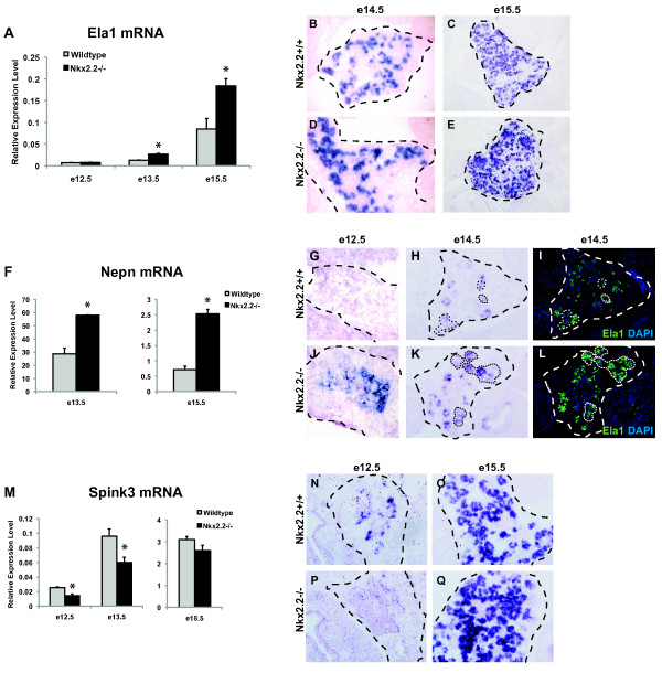
Results: Microarray analysis identified 80 genes that were differentially expressed in e12.5 and/or e13.5 Nkx2.2-/- embryos. Some of these genes encode transcription factors that have been previously identified in the pancreas, clarifying the position of Nkx2.2 within the islet transcriptional regulatory pathway. We also identified signaling factors and transmembrane proteins that function downstream of Nkx2.2, including several that have not previously been described in the pancreas. Interestingly, a number of known exocrine genes are also misexpressed in the Nkx2.2-/- pancreas.

Conclusions: Expression profiling of Nkx2.2-/- mice during embryogenesis has allowed us to identify known and novel pancreatic genes that function downstream of Nkx2.2 to regulate pancreas development. Several of the newly identified signaling factors and transmembrane proteins may function to influence islet cell fate decisions. These studies have also revealed a novel function for Nkx2.2 in maintaining appropriate exocrine gene expression. Most importantly, Nkx2.2 appears to function within a complex regulatory loop with Ngn3 at a key endocrine differentiation step.


Figures

Figure 1
Figure 1
Categorization and confirmation of Nkx2.2-/- gene expression changes. (A, B) Gene changes categorized by gene ontology (GO) terms determined in Tables 1, 2, and 3. Quantification of downregulated (A) and upregulated (B) genes is represented as percentages when compared to total affected genes. (C) qRTPCR verification of genes of interest at e13.5. Genes are listed in alphabetical order, except Tm4sf4, which was graphed separately due to a larger y-axis data range. Error bars represent SEM and asterisks denotes statistical significance < 0.05. Wild type = grey bars, Nkx2.2-/- = black bars.
Figure 2
Figure 2
Loss of Nkx2.2 affects expression of critical early endocrine progenitor transcription factors; Ngn3, NeuroD1, Myt1, and MafB. qRTPCR of (A) Ngn3 levels were assessed at e12.5, e13.5, and e15.5; (D) NeuroD1, (E) Myt1, and (J) MafB transcript levels were determined at e13.5, e15.5, and e18.5 and all genes compared wild type (grey bars) and Nkx2.2-/- pancreata (black bars). In situ hybridization comparing wild type and Nkx2.2-/- pancreas at e15.5 of Ngn3 (B and C), at e14.5 and e17.5 of Myt1 (F, G, H, and I), at e15.5 and e17.5 of MafB (L, N, P, and R). Immunofluorescence staining of MafB (green) was compared between wild type (K) and Nkx2.2 null (O) pancreata at e12.5. Immunofluorescence staining of Ela1 (green) on adjacent sections at e15.5 shows restriction to the endocrine compartment (M and Q). DAPI (blue) represents nuclei. Error bars represent SEM and asterisks denote statistical significance < 0.05. Magnification 20× for all except N and R, which are 40×. Large dashed lines outline the pancreas at e12.5, e14.5 and e15.5. Small dashed lines outline MafB expression.
Figure 3
Figure 3
Loss of Nkx2.2 affects expression of known endocrine transmembrane proteins, Tmem27, Adra2a, and a novel tetraspanin, Tm4sf4. qRTPCR of (A) Tmem27, (L) Adra2a, and (O) Tm4sf4 mRNA transcript levels were determined at e13.5, e15.5, and e18.5 and all genes compared wild type (grey bars) and Nkx2.2-/- pancreata (black bars). In situ hybridization comparing wild type and Nkx2.2-/- pancreas at e12.5, e14.5, and e17.5 of Tmem27 (B, C, D, G, H, and I), e14.5 of Adra2a (M and N), and e12.5, e14.5, and e17.5 of Tm4sf4 (P, Q, R, U, V, and W). Error bars represent SEM and asterisks denote statistical significance < 0.05. To assess co-expression, immunofluoresence staining of Tmem27 (E and J;green) and Ghrelin (F and K;copper) was performed on adjacent sections. Similarly, adjacent sections to the Tm4sf4 in situ hybridizations were stained for ghrelin (T and Y;green) and overlayed (T and Y: inset 2× zoom). DAPI (blue) indicates nuclei. Magnification 20× for e12.5, e14.5, R, S, T, W, X, Y, while 40× for E, F, J, and K. Large black dashed lines outline the pancreas at e12.5 and e14.5. Small solid white lines outline Tmem27 and ghrelin co-expression areas. Dashed white lines outline Tmem27+ cells that do not co-express ghrelin.
Figure 4
Figure 4
Loss of Nkx2.2 affects expression of known exocrine factors, Ela1, Spink3, and a novel secreted proteoglycan, Nepn. qRTPCR of (A) Ela1 mRNA was assessed at e12.5, e13.5, and e15.5, (F) Nepn mRNA was assessed at e13.5 and e15.5, and (M) Spink3 mRNA transcript levels were determined at e12.5 and e13.5, while all gene expression analyses compared wild type (grey bars) and Nkx2.2-/- pancreata (black bars). In situ hybridization comparing wild type and Nkx2.2-/- pancreas at e14.5 and e15.5 for Ela1 (B, C, D, and E), e12.5 and e14.5 for Nepn (G, H, J, and K), e12.5 and e15.5 for Spink3 (N, O, P, and Q). To show co-expression of Nepn in the exocrine compartment and confirm increased protein levels, Ela1 immunofluorescence was performed on adjacent sections (I and L;green). DAPI (C and E; blue) indicates nuclei. Error bars represent SEM and asterisks denote statistical significance < 0.05. Magnification 20×. Large dashed lines outline the pancreas at e12.5, e14.5, and e15.5. Small dashed lines outline areas of Nepn and Ela1 co-expression.
Figure 5
Figure 5
A proposed model for an Nkx2.2 role during endocrine cell specification at the secondary transition. The solid arrows indicate direct activation that is based on published regulatory data, while dashed arrows represent untested direct regulatory interactions proposed here as well as by other groups and in some cases known indirect regulation.

Similar articles

Cited by

References

    1. Gu G, Brown JR, Melton DA. Direct lineage tracing reveals the ontogeny of pancreatic cell fates during mouse embryogenesis. Mech Dev. 2003;120:35–43. doi: 10.1016/S0925-4773(02)00330-1. - DOI - PubMed
    1. Kawaguchi Y, Cooper B, Gannon M, Ray M, MacDonald RJ, Wright CV. The role of the transcriptional regulator Ptf1a in converting intestinal to pancreatic progenitors. Nat Genet. 2002;32:128–34. doi: 10.1038/ng959. - DOI - PubMed
    1. Krapp A, Knofler M, Ledermann B, Burki K, Berney C, Zoerkler N, Hagenbuchle O, Wellauer PK. The bHLH protein PTF1-p48 is essential for the formation of the exocrine and the correct spatial organization of the endocrine pancreas. Genes Dev. 1998;12:3752–63. doi: 10.1101/gad.12.23.3752. - DOI - PMC - PubMed
    1. Guz Y, Montminy MR, Stein R, Leonard J, Gamer LW, Wright CV, Teitelman G. Expression of murine STF-1, a putative insulin gene transcription factor, in beta cells of pancreas, duodenal epithelium and pancreatic exocrine and endocrine progenitors during ontogeny. Development. 1995;121:11–8. - PubMed
    1. Jorgensen MC, Ahnfelt-Ronne J, Hald J, Madsen OD, Serup P, Hecksher-Sorensen J. An illustrated review of early pancreas development in the mouse. Endocr Rev. 2007;28:685–705. doi: 10.1210/er.2007-0016. - DOI - PubMed

Publication types

LinkOut - more resources