. 2009 Dec 11:9:19.
doi: 10.1186/1471-2482-9-19.

Laparoscopic retrograde (fundus first) cholecystectomy

Affiliations

Laparoscopic retrograde (fundus first) cholecystectomy

Michael D Kelly. BMC Surg. .

Abstract

Background: Retrograde ("fundus first") dissection is frequently used in open cholecystectomy and although feasible in laparoscopic cholecystectomy (LC) it has not been widely practiced. LC is most simply carried out using antegrade dissection with a grasper to provide cephalad fundic traction. A series is presented to investigate the place of retrograde dissection in the hands of an experienced laparoscopic surgeon using modern instrumentation.

Methods: A prospective record of all LCs carried out by an experienced laparoscopic surgeon following his appointment in Bristol in 2004 was examined. Retrograde dissection was resorted to when difficulties were encountered with exposure and/or dissection of Calot's triangle.

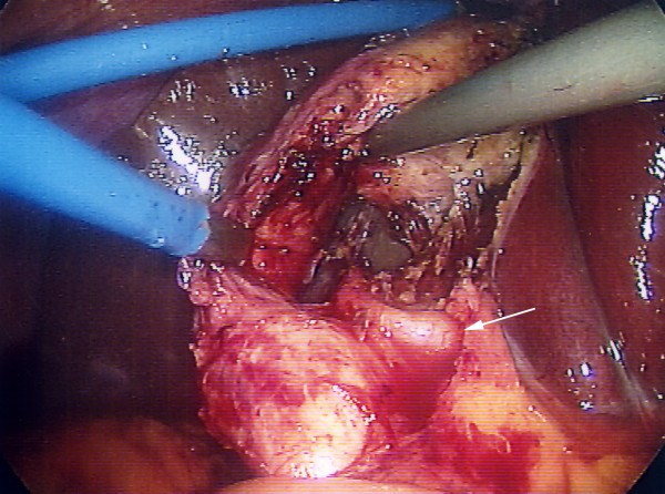
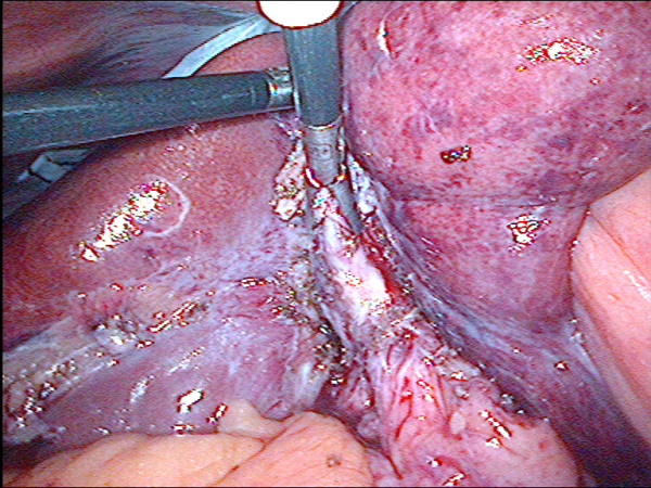
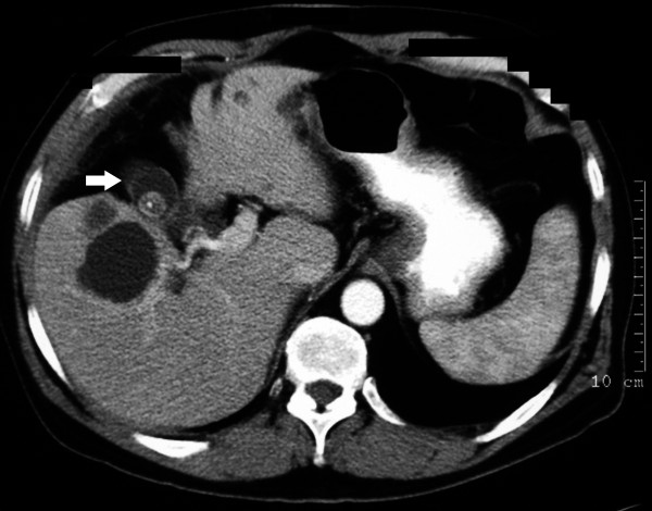
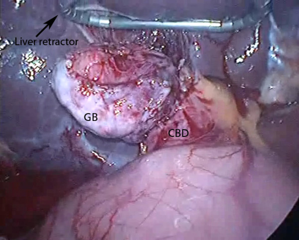
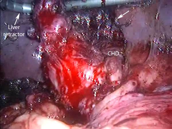
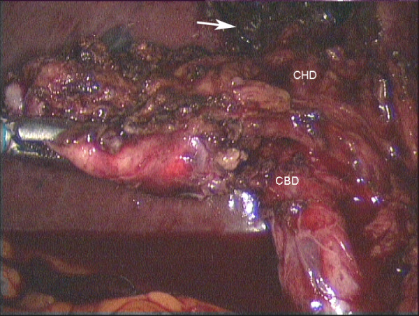
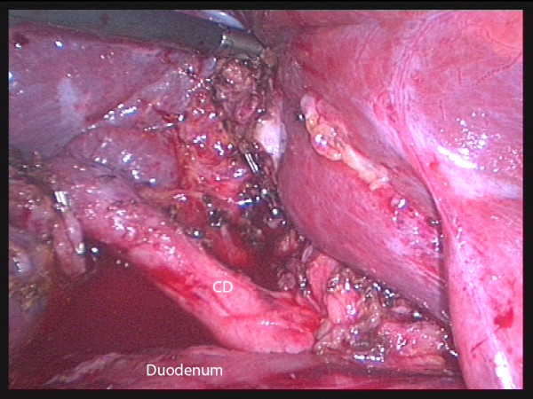
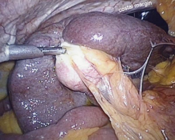
Results: 1041 LCs were carried out including 148 (14%) emergency operations and 131 (13%) associated bile duct explorations. There were no bile duct injuries although conversion to open operation was required in six patients (0.6%). Retrograde LC was attempted successfully in 11 patients (1.1%). The age ranged from 28 to 80 years (mean 61) and there were 7 males. Indications were; fibrous, contracted gallbladder 7, Mirizzi syndrome 2 and severe kyphosis 2. Operative photographs are included to show the type of case where it was needed and the technique used. Postoperative stay was 1/2 to 5 days (mean 2.2) with no delayed sequelae on followup. Histopathology showed; chronic cholecystitis 7, xanthogranulomatous cholecystitis 3 and acute necrotising cholecystitis 1.

Conclusions: In this series, retrograde laparoscopic dissection was necessary in 1.1% of LCs and a liver retractor was needed in 9 of the 11 cases. This technique does have a place and should be in the armamentarium of the laparoscopic surgeon.


Figures

Figure 1
Figure 1
Laparoscopic view of standard dissection technique in a Mirizzi type I arrangement. Fundic traction gave good exposure of Calot's triangle and there was no need for retrograde (fundus first) dissection. Accurate transcystic cholangiography would, however, have been very difficult. Arrow points to the right hepatic artery crossing the common hepatic duct.
Figure 2
Figure 2
Laparoscopic view showing failure of standard technique. There is poor exposure and inability to safely dissect Calot's triangle with standard cephalad fundic traction (case 1).
Figure 3
Figure 3
Preoperative CT scan. The scan shows a severely fibrotic, contracted gallbladder (arrow) and multiple simple liver cysts. Retrograde dissection was required in this patient (case 3) (Somatom Volume Zoom - 4 slice, Siemens AG, Erlangen, Germany).
Figure 4
Figure 4
Laparoscopic view of liver retraction. The gallbladder (GB) has been mobilized prior to bile duct exploration via the large defect in the common bile duct (CBD) at its junction with the GB (case 5) (angled triangular Diamond-Flex liver retractor, Surgical Innovations Group, England http://www.sigroupplc.com. Elemental Healthcare, England, UK http://www.elementalhealthcare.co.uk).
Figure 5
Figure 5
Laparoscopic view showing liver retraction and retrograde dissection in acute Mirizzi syndrome. Mobilisation of the inflamed and shrunken gallbladder was made possible by liver retraction (case 7). The arrow points to absorbable haemostatic gauze (surgicel, Ethicon, Somerville NJ, USA) in the gallbladder bed of the liver (CHD = common hepatic duct).
Figure 6
Figure 6
Laparoscopic view of a mobilized, contracted gallbladder with a grasper retracting it laterally. The anatomy is obvious now that the fundic traction has been relaxed and the GB freed from the liver, however the initial dissection was carried inadvertently to the medial side of the common bile duct (CBD) while there was strong cephalad fundic traction (case 9). The arrow points to surgicel in the gallbladder bed of the liver. (CHD = common hepatic duct).
Figure 7
Figure 7
Laparoscopic view of satisfactory retraction of the liver using a grasper. There was a shrunken gallbladder around a cast of stones with aberrant cystic duct (CD) anatomy (case 10).
Figure 8
Figure 8
Laparoscopic view showing the combination of kyphosis and a large, rigid right lobe of liver. Antegrade dissection using fundic traction was not possible and "fundus first" dissection was needed (case 11).

Similar articles

Cited by

References

    1. McIntyre RC Jr, Bensard DD, Stiegman GV, Pearlman NW, Durham J. Exposure for laparoscopic cholecystectomy dissection alters biliary ductal anatomy. Surg Endosc. 1996;10:41–3. doi: 10.1007/s004649910010. - DOI - PubMed
    1. Hunter JG. Avoidance of bile duct injury during laparoscopic cholecystectomy. Am J Surg. 1991;162:71–6. doi: 10.1016/0002-9610(91)90207-T. - DOI - PubMed
    1. Strasberg SM. Avoidance of biliary injury during laparoscopic cholecystectomy. J Hepatobiliary Pancreat Surg. 2002;9:543–547. doi: 10.1007/s005340200071. - DOI - PubMed
    1. Hugh TB, Kelly MD, Mekisic A. Rouviere's sulcus: A useful landmark in laparoscopic cholecystectomy. Br J Surg. 1997;84(9):1253–1254. doi: 10.1002/bjs.1800840916. - DOI - PubMed
    1. Hugh TB. New strategies to prevent laparoscopic bile duct injury-surgeons can learn from pilots. Surgery. 2002;132:826–35. doi: 10.1067/msy.2002.127681. - DOI - PubMed

LinkOut - more resources