. 2009 Dec 14:9:66.
doi: 10.1186/1471-213X-9-66.

Mechanism of primitive duct formation in the pancreas and submandibular glands: a role for SDF-1

Affiliations

Mechanism of primitive duct formation in the pancreas and submandibular glands: a role for SDF-1

Anne-Christine Hick et al. BMC Dev Biol. .

Abstract

Background: The exocrine pancreas is composed of a branched network of ducts connected to acini. They are lined by a monolayered epithelium that derives from the endoderm and is surrounded by mesoderm-derived mesenchyme. The morphogenic mechanisms by which the ductal network is established as well as the signaling pathways involved in this process are poorly understood.

Results: By morphological analyzis of wild-type and mutant mouse embryos and using cultured embryonic explants we investigated how epithelial morphogenesis takes place and is regulated by chemokine signaling. Pancreas ontogenesis displayed a sequence of two opposite epithelial transitions. During the first transition, the monolayered and polarized endodermal cells give rise to tissue buds composed of a mass of non polarized epithelial cells. During the second transition the buds reorganize into branched and polarized epithelial monolayers that further differentiate into tubulo-acinar glands. We found that the second epithelial transition is controlled by the chemokine Stromal cell-Derived Factor (SDF)-1. The latter is expressed by the mesenchyme, whereas its receptor CXCR4 is expressed by the epithelium. Reorganization of cultured pancreatic buds into monolayered epithelia was blocked in the presence of AMD3100, a SDF-1 antagonist. Analyzis of sdf1 and cxcr4 knockout embryos at the stage of the second epithelial transition revealed transient defective morphogenesis of the ventral and dorsal pancreas. Reorganization of a globular mass of epithelial cells in polarized monolayers is also observed during submandibular glands development. We found that SDF-1 and CXCR4 are expressed in this organ and that AMD3100 treatment of submandibular gland explants blocks its branching morphogenesis.

Conclusion: In conclusion, our data show that the primitive pancreatic ductal network, which is lined by a monolayered and polarized epithelium, forms by remodeling of a globular mass of non polarized epithelial cells. Our data also suggest that SDF-1 controls the branching morphogenesis of several exocrine tissues.


Figures
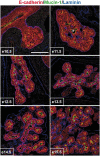
Figure 1
Figure 1
Remodeling of the pancreatic epithelial cell mass into polarized monolayers. Pancreatic sections from e10.5 to e15.5 embryos were examined by immunofluorescence using antibodies directed against E-cadherin, mucin-1 and laminin. Pancreatic development starts by the formation of a mass of non-polarized epithelial cells (e10.5-e11.5). This mass is then remodeled (e12.5-e13.5) and finally every epithelial cell become polarized, with its basal pole contacting laminin and its apical pole facing a lumen (e14.5-e15.5). * indicates the central duct, in connection with the duodenum. Scale bar, 100 μm.
Figure 2
Figure 2
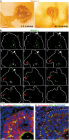
The pancreatic buds consist of an epithelial cell mass with peripheral lumina. (A) Whole mount immunoanalyzis of dorsal and ventral pancreatic buds at e11.5 with an anti-mucin-1 antibody. The staining delineates the apical pole of cells lining the lumen of the primitive gut tube, the pancreatic central duct (*) and its extensions. It also shows blind lumina at the periphery of the pancreatic bud. (B) Gallery of pictures from a 3D confocal acquisition on a whole-mount immunofluorescence analyzis of e11.5 dorsal pancreas with anti Mucin-1 (green) antibody. Blind peripheral lumina can be observed from b to g (white arrowheads) and from f to k (red arrowheads). (C, D) Sections of e11.5 dorsal pancreatic bud stained with antibodies against E-cadherin and mucin-1 (C) and pericentrin and GM-130 (D). Nuclei are labeled with TOPRO. Epithelial cells at the periphery of the pancreatic bud (E-cadherin, dashed line) show polarized staining of mucin-1 (arrows), pericentrin and GM-130. * indicates the central duct. Scale bars, 50 μm (A); 20 μm (B); 10 μm (C, D).
Figure 3
Figure 3
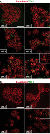
Primitive duct morphogenesis occurs by transition from epithelial mass to monolayers in the pancreas and in the SMG. Pancreatic sections from e10.5 to e15.5 (A) and SMG sections from e13.5 to e16.5 (B) embryos were examined by immunofluorescence using antibodies directed against ZO-1 and E-cadherin. Both organs show that duct development occurs by remodeling of a mass of epithelial cells into polarized monolayers. Before remodeling, E-cadherin is expressed all around the cells, whereas it is restricted to the baso-lateral membranes in the monolayered epithelium. In the latter, ZO-1 (tight junction) separates the E-cadherin-negative apical pole from the E-cadherin-positive lateral domains (insets). * indicates the central duct, in connection with the duodenum (A). Scale bars, 50 μm.
Figure 4
Figure 4
SDF-1 and its receptor CXCR4 are expressed in developing mouse pancreas and SMG. Pancreatic (A-D) and SMG (E-H) sections at e12.5 (A, B, E and F) and e14.5 (C, D, G and H) were processed for in situ hybridizations using antisense probes for SDF-1 and for CXCR4. SDF-1 is expressed in the mesenchyme and its receptor in epithelial cells (pancreatic epithelium is delineated by a dotted line). At e14.5, SDF-1 staining is also found in pancreatic epithelial cells. Capillaries are positive for both the ligand and receptor (arrows). Scale bars, 100 μm (A-D); 200 μm (E-H).
Figure 5
Figure 5
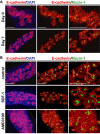
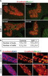
SDF-1 signaling controls pancreatic branching morphogenesis. Immunofluorescence analyzis of pancreatic tissue stained for E-cadherin, mucin-1 and DNA. (A) The dorsal pancreatic bud was dissected from e12.5 embryos and either fixed and processed for immunostainings (= e12.5 = day 0) or cultured on filter for 7 days (= day 7) prior to immunostaining analyzis. Dissected mouse e12.5 pancreas before the culture (Day 0) appears as a mass of epithelial cells (red; E-cadherin-positive) surrounded by mesenchyme (E-cadherin-negative). Some cells accumulate mucin-1 at their apical pole, but no lumina are formed. After 7 days in culture (Day 7), the epithelial mass has been remodeled and cells are polarized and form monolayers delineating lumina. (B) e12.5 pancreatic explants dissected from wild-type mouse and cultured for 7 days. Control and SDF-1 (300 ng/ml) treated explants form polarized monolayers delineating lumina and imbricated with mesenchyme. In contrast, treatment of the pancreatic explants with 20 μM AMD3100, a specific pharmacological inhibitor of CXCR4, inhibits epithelial morphogenesis, as seen by the maintenance of the epithelial cells in clusters. Scale bars, 50 μm.
Figure 6
Figure 6
SDF-1 signaling controls SMG branching morphogenesis. (A) Representative whole-mount immunostaining of E-cadherin in explants cultured for three days with the control medium, SDF-1, AMD3100 or CCX733. Control and SDF-1-treated cultures show that the epithelium has invaded the whole culture and forms a complex arborescence. In contrast, treatment of the SMG explants with 20 μM AMD3100 inhibits epithelial branching morphogenesis, as seen by the failure to colonize the explant mesenchyme and the reduced number of buds. Similarly, CCX733 reduces the branching as well as the invasion of the mesenchyme. (B) The numbers of terminal buds in each cultured SMG explants were counted at day 0, 1, 2 and 3, and the budding index was calculated by dividing the number of buds at day 1, 2 and 3 by the number of buds at day 0. t-test: *, P < 0.001. Scale bar, 500 μm.
Figure 7
Figure 7
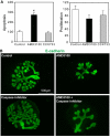
SDF-1 signaling promotes epithelial cell survival in the SMG. (A) Quantification of the number of epithelial cells positive for the cleaved caspase 3 (left panel), or phosphohistone H3 (right panel) in SMG explants cultured for 24 hours. All the epithelial cells positive for the marker were counted on every sixth sections of the explant and total number was normalised to its size. Values for control explants were set as 100. t-test: *, P < 0.05. (B) Representative whole-mount immunostaining of E-cadherin in explants cultured for two days in the presence or absence of a general caspase inhibitor. Blocking caspase activity does not affect branching activity in control or AMD3100-treated explants. Scale bars, 500 μm.
Figure 8
Figure 8
SDF-1 signaling controls pancreatic branching morphogenesis in vivo. (A) Representative immunostainings of two e12.5 control and sdf1 knockout pancreata with E-cadherin and mucin-1 antibodies. Reduced branching activity is observed in sdf1-/- ventral and dorsal pancreas (B) Quantification of the degree of branching in four e12.5 control and four sdf1 knockout pancreata. The organs were sectioned and the number of buds and clefts were counted on every sixth sections and the average numbers per ventral pancreata sections are given. (P < 0.0005) (C) Pancreatic explants were dissected at e12.5 from wild-type or cxcr4-/- mouse embryos and cultured for 7 days. CXCR4-deficient explants do not remodel into polarized epithelial monolayers and exhibit the same morphogenetic defects as wild-type explants treated with AMD3100. Scale bar, 50 μm.
Figure 9
Figure 9
Development and banching morphogenesis of the pancreas. Pancreatic specification of polarized endodermal cells leads to the formation of a multilayered and then globular mass of epithelial cells with a central duct lumen (pancreatic bud). This lumen, connected to the gut tube lumen, forms extensions within the pancreatic bud, and is surrounded by several layers of cells, linked by adherens junctions (E-cadherin), but lacking tight junctions (ZO-1). Around e11.5, before remodeling of the mass, some cells at the periphery acquire apico-basal polarity and delineate blind lumina (secondary lumina). Initial branching morphogenesis events result from the coalescence of secondary and central lumina, allowing the progression towards epithelial monolayers.

Similar articles

Cited by

References

    1. Hogan BL. Building organs from buds, branches and tubes. Differentiation. 2006;74:323–325. doi: 10.1111/j.1432-0436.2006.00107.x. - DOI - PubMed
    1. Andrew DJ, Ewald AJ. Morphogenesis of epithelial tubes: Insights into tube formation, elongation, and elaboration. Dev Biol. 2009. in press . - PMC - PubMed
    1. Lu P, Werb Z. Patterning mechanisms of branched organs. Science. 2008;322:1506–1509. doi: 10.1126/science.1162783. - DOI - PMC - PubMed
    1. Bryant DM, Mostov KE. From cells to organs: building polarized tissue. Nat Rev Mol Cell Biol. 2008;9:887–901. doi: 10.1038/nrm2523. - DOI - PMC - PubMed
    1. Slack JM. Developmental biology of the pancreas. Development. 1995;121:1569–1580. - PubMed

Publication types

LinkOut - more resources