. 2009 Dec 15:9:67.
doi: 10.1186/1471-213X-9-67.

Developmental expression of BK channels in chick cochlear hair cells

Affiliations

Developmental expression of BK channels in chick cochlear hair cells

Yi Li et al. BMC Dev Biol. .

Abstract

Background: Cochlear hair cells are high-frequency sensory receptors. At the onset of hearing, hair cells acquire fast, calcium-activated potassium (BK) currents, turning immature spiking cells into functional receptors. In non-mammalian vertebrates, the number and kinetics of BK channels are varied systematically along the frequency-axis of the cochlea giving rise to an intrinsic electrical tuning mechanism. The processes that control the appearance and heterogeneity of hair cell BK currents remain unclear.

Results: Quantitative PCR results showed a non-monotonic increase in BK alpha subunit expression throughout embryonic development of the chick auditory organ (i.e. basilar papilla). Expression peaked near embryonic day (E) 19 with six times the transcript level of E11 sensory epithelia. The steady increase in gene expression from E11 to E19 could not explain the sudden acquisition of currents at E18-19, implicating post-transcriptional mechanisms. Protein expression also preceded function but progressed in a sequence from diffuse cytoplasmic staining at early ages to punctate membrane-bound clusters at E18. Electrophysiology data confirmed a continued refinement of BK trafficking from E18 to E20, indicating a translocation of BK clusters from supranuclear to subnuclear domains over this critical developmental age.

Conclusions: Gene products encoding BK alpha subunits are detected up to 8 days before the acquisition of anti-BK clusters and functional BK currents. Therefore, post-transcriptional mechanisms seem to play a key role in the delayed emergence of calcium-sensitive currents. We suggest that regulation of translation and trafficking of functional alpha subunits, near voltage-gated calcium channels, leads to functional BK currents at the onset of hearing.


Figures

Figure 1
Figure 1
The expression of BK transcripts during development of the chick auditory epithelium. (A) Cycle-by-cycle amplification data are shown for S16 using serial dilutions of total RNA from chick basilar papilla. Threshold (dotted line) was manually drawn at a point during the log-linear phase of amplification. Cycle threshold was determined by identifying the threshold crossing-point for each amplification curve. (B) Total input RNA was serially diluted starting from a concentrated sample of chick basilar papilla. Diluted RNA was reverse transcribed and analyzed by qPCR. Efficiency data were fit by linear least-squares regression to Ct = M*log2(dilution)+B. The slope (M) for each curve was between -1.0 and -1.1, indicating equal efficiency and 2-fold amplification per cycle. (C) The expression of BK transcripts, assessed by the non-variant-specific probe αX, gradually increased during late-stage maturation but decreased again in the first week after hatching. The time course for acquiring calcium-sensitive BK currents is illustrated (black dots) based on data from Fuchs and Sokolwski (1990). (D) The proportion of STREX splice variants among the total population of BK transcripts steadily increased during development, whereas the proportion of β1 decreased at the onset of hearing.
Figure 2
Figure 2
The expression of BK subunits during development of the chick basilar papilla. Cross-sections of the chick cochlear were stained with anti-BK (Chemicon) to qualitatively correlate protein levels with gene expression levels. Panels are organized by tonotopic position (base-to-apex, left-to-right) and age (E19-to-E12, top-to-bottom). Each cross-section is positioned so that the inferior to superior radial axis lies from left-to-right. Cochlear nerve projections were immunopositive as were the base of tall hair cells lying along the superior-most portions of most sections. Antibody specificity was confirmed using Western blot on membrane fractions from chick brain (inset in panel A). The dominant band appeared near 140 kDa, matching the predicted weight of the long C-terminus BK α variant. Additional bands near this molecular weight may be other splice forms or proteolytic by-products. No other spurious bands were observed in the blot. Scale bar in (B) is applicable to all panels. TM, tectorial membrane. HC, hair cell layer. CG, cochlear ganglion projections.
Figure 3
Figure 3
BK channel clusters appear at E18 during hair cell maturation. (A) Western blot analysis on membrane-bound protein from chick brain reveals a 120 kDa band, near the predicted weight of the minimal BK α variant (127 kDa; no inclusion of alternative exons). A fainter, lower-weight band is also apparent. No other spurious bands were observed in the blot. Both bands were absent when the primary antibody was preadsorbed with the immunogen. (B) Isolated hair cells from E10 to posthatch basilar papilla are shown labeled with antibodies to BK channels (Alomone). The apical surface and hair bundle are outlined to show orientation of the cell (dotted lines). Punctate clusters appear at E18. Bright label at E10 to E12 is inside the cell at the level of the nucleolus. (C) Several preparations were triple labeled for anti-BK (Alomone; red), hair bundles (phalloidin; green), and a nuclear stain (Hoechst; blue). Two examples for each age are shown (left two columns) along with no primary controls (far right column). Anti-BK clusters appear supranuclear at E18 and subnuclear from E20 onward. Scale bars = 10 μm.
Figure 4
Figure 4
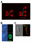
BK channel clusters are associated with the hair cell membrane. (A) Isolated hair cells were imaged using confocal microscopy to determine whether anti-BK clusters were associated with the membrane or found intracellularly. Two exemplar hair cells are shown, both oriented with hair bundles toward the top of the image. Between 13 and 17 optical sections were taken at increments of 0.5 μm. Projections of full Z-stack are shown alongside orthogonal cuts through the reconstructed stack. Cuts were made at the level of each anti-BK puncta and positioned to the right of each hair cell example. In all cases, puncta were present along the outer edge of the cut-view, indicating overlap with the hair cell plasma membrane. (B) Hair cells and auditory neurons were acutely isolated onto the same slide in some preparations and stained for glutamate receptor 2 (GluR2) in order to detect residual afferent terminals on the dissociated hair cells. Afferent auditory neurons were identified according to cell size and the presence of bipolar axonal projections. Under epifluorescence and using constant exposure conditions, anti-GluR2 label was found on the cell soma of auditory neurons but was absent from hair cells. (C) Anti-BK puncta were distant from synaptic ribbons in isolated hair cells. Isolated hair cells were co-labeled with anti-BK (red) and anti-ctbp2/RIBEYE (green), a marker for presynaptic ribbons. An exemplar tall hair cell is shown using epifluorescence imaging. Debris that might be associated with afferent terminals is absent from the matching differential interference contrast image (left). The image shown is representative of over 5 tall hair cells imaged in this co-labeling experiment. Scale bars in all panels = 10 μm.
Figure 5
Figure 5
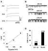
The density of functional BK channels at the basolateral pole increases with age. (A) Whole-cell recordings were made from embryonic and posthatch hair cells to confirm the onset of BK currents around E18/19. Example traces from three ages are shown. In these examples, currents were elicited by voltage steps from -80 mV to -20 or 0 mV. A fast-activating outward current was present at E18 under whole-cell recording conditions, but no such current could be recorded from earlier ages. (B) Single-channel current traces are shown for a cell-attached patch from a posthatch hair cell held at three different pipette potentials. Bath and pipette salines consisted of standard ECF and high potassium electrode solutions, respectively. Channel openings are downward. In this configuration, more negative pipette voltages correspond to increasing depolarization. The channel was voltage-sensitive, as evidenced by an increased open probability for more depolarized voltages. (C) Single-channel current amplitudes were estimated from all-points histograms using traces in (B). Linear regression to these data (dotted line) indicates a unitary conductance of 150 pS. Reversal was estimated to be about -60 mV, near the presumed resting potential of the cell. Therefore, all inclusion criteria were satisfied, establishing the identity of this channel as BK. (D) The percent of cell-attached patches (C.A.P.) exhibiting channel fluctuations attributed to BK are plotted for cells from embryonic and posthatch basilar papilla. The total number of attempts is shown in parentheses. The breakdown for the E17-E19 data set was: E17 (6%), E18 (7%), E19 (17%).

Similar articles

Cited by

References

    1. Kros CJ, Ruppersberg JP, Rusch A. Expression of a potassium current in inner hair cells during development of hearing in mice. Nature. 1998;394:281–4. doi: 10.1038/28401. - DOI - PubMed
    1. Marcotti W, Johnson SL, Holley MC, Kros CJ. Developmental changes in the expression of potassium currents of embryonic, neonatal and mature mouse inner hair cells. J Physiol. 2003;548:383–400. doi: 10.1113/jphysiol.2002.034801. - DOI - PMC - PubMed
    1. Eatock RA, Rusch A. Developmental changes in the physiology of hair cells. Semin Cell Dev Biol. 1997;8:265–275. doi: 10.1006/scdb.1997.0142. - DOI - PubMed
    1. Kros CJ, Rusch A, Richardson GP. Mechano-electrical transducer currents in hair cells of the cultured neonatal mouse cochlea. Proc Biol Sci. 1992;249:185–93. doi: 10.1098/rspb.1992.0102. - DOI - PubMed
    1. Si F, Brodie H, Gillespie PG, Vazquez AE, Yamoah EN. Developmental assembly of transduction apparatus in chick basilar papilla. J Neurosci. 2003;23:10815–26. - PMC - PubMed

Publication types

MeSH terms

Substances

LinkOut - more resources