

ATP stimulates chemokine production via a store-operated calcium entry pathway in C6 glioma cells
- PMID: 20003523
- PMCID: PMC2807438
- DOI: 10.1186/1471-2407-9-442
ATP stimulates chemokine production via a store-operated calcium entry pathway in C6 glioma cells
Abstract
Background: Glioma present as one of the most challenging cancers to treat, however, understanding of tumor cell biology is not well understood. Extracellular adenosine triphosphate (ATP) could serve as a critical signaling molecule regulating tumor development. This study has examined pharmacological modulation of calcium (Ca2+) entry through store-operated channels (SOC) on cellular expression and production of immune-cell mobilizing chemokines in ATP-stimulated C6 glioma cells.
Methods: Calcium spectrofluorometry was carried out to measure mobilization of intracellular Ca2+ [Ca2+]i following ATP stimulation of rat C6 glioma cells. Pretreatment with two inhibitors of SOC, SKF96365 or gadolinium, was used to examine for effects on [Ca2+]i. RT-PCR was performed to determine effects of purinergic stimulation on C6 cell expression of metabotropic P2Y receptors (P2YR) and the chemokines, monocyte chemoattractant protein-1 (MCP-1) and interleukin-8 (IL-8). ELISA was carried out to measure production of MCP-1 and IL-8 with ATP stimulation of glioma cells.
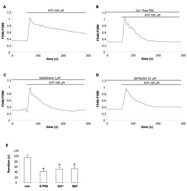
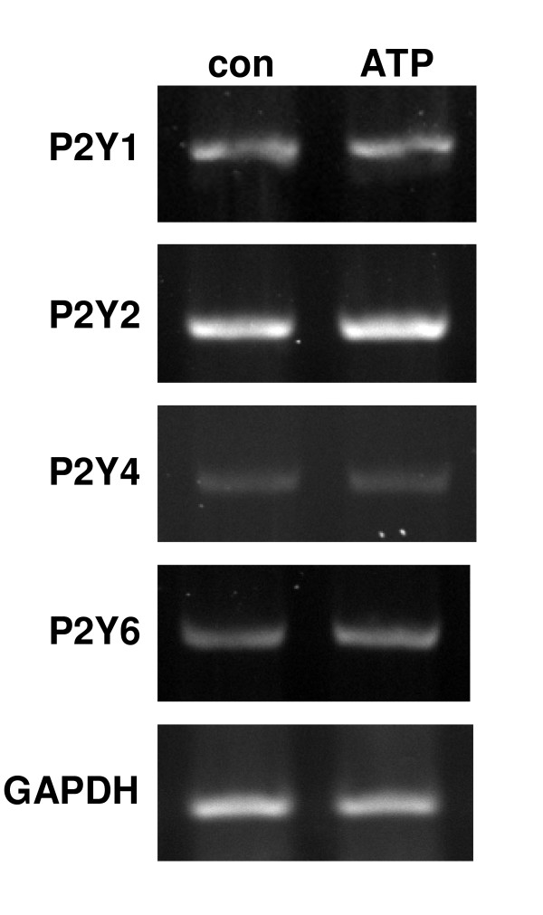
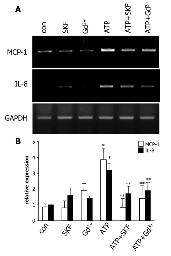
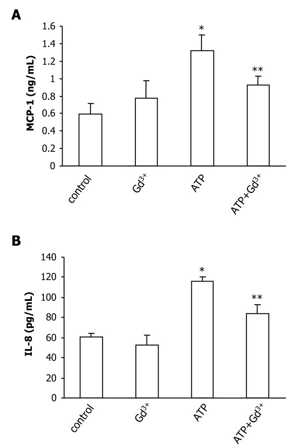
Results: Application of ATP (at 100 microM) to C6 glioma induced an increase in [Ca2+]i with the response exhibiting two components of decay. In the presence of the SOC inhibitors, SKF96365 or gadolinium, or with Ca2+-free solution, ATP responses lacked a slow phase suggesting the secondary component was due to SOC-mediated influx of Ca2+. RT-PCR confirmed expression of purinergic P2Y-subtype receptors in C6 cells which would serve as a precursor to activation of SOC. In addition, ATP-stimulated C6 cells showed enhanced expression of the chemokines, MCP-1 and IL-8, with SKF96365 or gadolinium effective in reducing chemokine expression. Gadolinium treatment of ATP-stimulated C6 cells was also found to inhibit the production of MCP-1 and IL-8.
Conclusion: These results suggest ATP-induced Ca2+ entry, mediated by activation of SOC in C6 glioma, as a mechanism leading to increased cellular expression and release of chemokines. Elevated levels of MCP-1 and IL-8 are predicted to enhance the mobility of tumor cells and promote recruitment of microglia into developing tumors thereby supporting tumor growth.
Figures
Similar articles
-
Expression and function of the P2X(7) receptor in rat C6 glioma cells.Cancer Lett. 2008 Feb 18;260(1-2):79-87. doi: 10.1016/j.canlet.2007.10.025. Epub 2007 Nov 26. Cancer Lett. 2008. PMID: 18039556
-
Purinergic mediated changes in Ca2+ mobilization and functional responses in microglia: effects of low levels of ATP.J Neurosci Res. 2005 Aug 1;81(3):349-56. doi: 10.1002/jnr.20475. J Neurosci Res. 2005. PMID: 15948175 Review.
-
Calcium dependence of purinergic subtype P2Y₁ receptor modulation of C6 glioma cell migration.Neurosci Lett. 2011 Jun 22;497(2):80-4. doi: 10.1016/j.neulet.2011.04.034. Epub 2011 Apr 22. Neurosci Lett. 2011. PMID: 21540076
-
Purinergic responses of calcium-dependent signaling pathways in cultured adult human astrocytes.BMC Neurosci. 2014 Jan 22;15:18. doi: 10.1186/1471-2202-15-18. BMC Neurosci. 2014. PMID: 24447580 Free PMC article.
-
Role of the store-operated Ca2+ channel in ATP-induced Ca2+ signalling in mesenchymal stem cells and regulation of cell functions.Front Biosci (Landmark Ed). 2021 Dec 30;26(12):1737-1745. doi: 10.52586/5065. Front Biosci (Landmark Ed). 2021. PMID: 34994186 Review.
Cited by
-
The Role of Ion Channels in Microglial Activation and Proliferation - A Complex Interplay between Ligand-Gated Ion Channels, K(+) Channels, and Intracellular Ca(2.).Front Immunol. 2015 Oct 22;6:497. doi: 10.3389/fimmu.2015.00497. eCollection 2015. Front Immunol. 2015. PMID: 26557116 Free PMC article. Review.
-
Purinergic signalling and cancer.Purinergic Signal. 2013 Dec;9(4):491-540. doi: 10.1007/s11302-013-9372-5. Purinergic Signal. 2013. PMID: 23797685 Free PMC article. Review.
-
Extracellular ATP promotes breast cancer chemoresistance via HIF-1α signaling.Cell Death Dis. 2022 Mar 2;13(3):199. doi: 10.1038/s41419-022-04647-6. Cell Death Dis. 2022. PMID: 35236823 Free PMC article.
-
The roles of microglia/macrophages in tumor progression of brain cancer and metastatic disease.Front Biosci (Landmark Ed). 2017 Jun 1;22(10):1805-1829. doi: 10.2741/4573. Front Biosci (Landmark Ed). 2017. PMID: 28410147 Free PMC article. Review.
-
Ectonucleotidases in tumor cells and tumor-associated immune cells: an overview.J Biomed Biotechnol. 2012;2012:959848. doi: 10.1155/2012/959848. Epub 2012 Oct 14. J Biomed Biotechnol. 2012. PMID: 23118517 Free PMC article. Review.
References
Publication types
MeSH terms
Substances
Grants and funding
LinkOut - more resources
Full Text Sources
Research Materials
Miscellaneous
