. 2009 Dec 15:9:101.
doi: 10.1186/1472-6750-9-101.

Unrestricted somatic stem cells from human umbilical cord blood grow in serum-free medium as spheres

Affiliations

Unrestricted somatic stem cells from human umbilical cord blood grow in serum-free medium as spheres

Faten Zaibak et al. BMC Biotechnol. .

Abstract

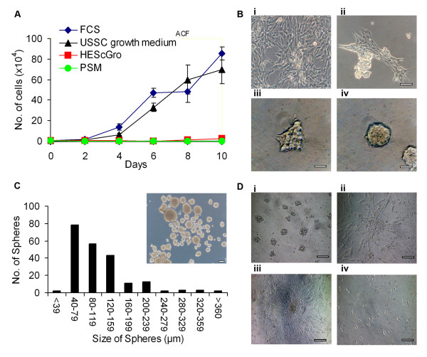
Background: Human umbilical cord blood-derived unrestricted somatic stem cells (USSCs), which are capable of multilineage differentiation, are currently under investigation for a number of therapeutic applications. A major obstacle to their clinical use is the fact that in vitro expansion is still dependent upon fetal calf serum, which could be a source of pathogens. In this study, we investigate the capacity of three different stem cell culture media to support USSCs in serum-free conditions; HEScGRO, PSM and USSC growth medium ACF. Our findings demonstrate that USSCs do not grow in HEScGRO or PSM, but we were able to isolate, proliferate and maintain multipotency of three USSC lines in USSC growth medium ACF.

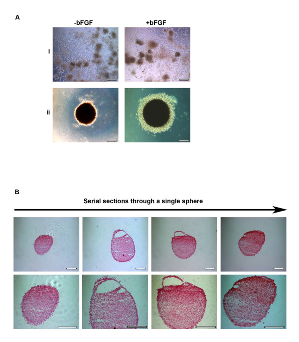
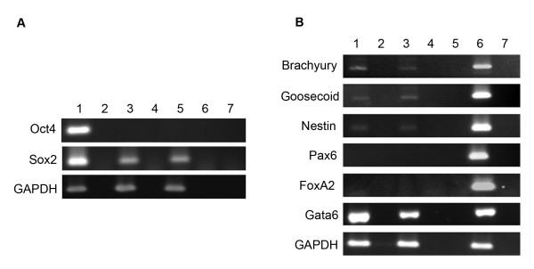
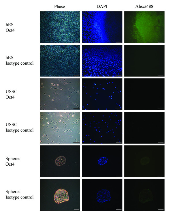
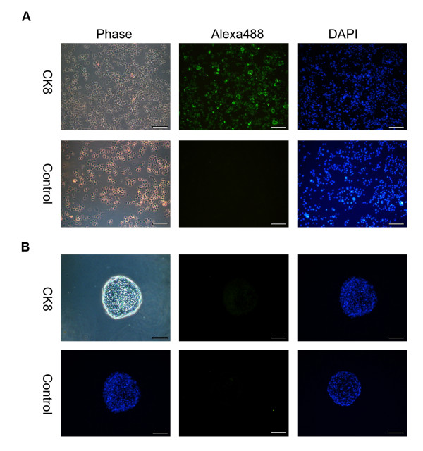
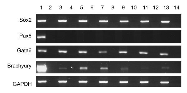
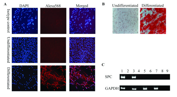
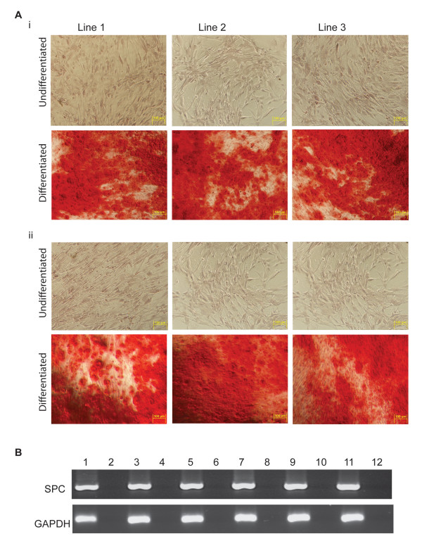
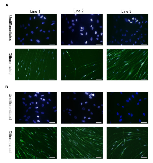
Results: For the first one to three passages, cells grown in USSC growth medium ACF proliferate and maintain their morphology, but with continued passaging the cells form spherical cell aggregates. Upon dissociation of spheres, cells continue to grow in suspension and form new spheres. Dissociated cells can also revert to monolayer growth when cultured on extracellular matrix support (fibronectin or gelatin), or in medium containing fetal calf serum. Analysis of markers associated with pluripotency (Oct4 and Sox2) and differentiation (FoxA2, Brachyury, Goosecoid, Nestin, Pax6, Gata6 and Cytokeratin 8) confirms that cells in the spheres maintain their gene expression profile. The cells in the spheres also retain the ability to differentiate in vitro to form cells representative of the three germline layers after five passages.

Conclusions: These data suggest that USSC growth medium ACF maintains USSCs in an undifferentiated state and supports growth in suspension. This is the first demonstration that USSCs can grow in a serum- and animal component-free medium and that USSCs can form spheres.


Figures

Figure 1
Figure 1
Derivation and maintenance of spheres. A, Growth kinetics of USSCs cultured in stem cell proliferation medium, HEScGRO and PSM and USSC growth mediumACF. Cells were seeded at low density and total cell number was determined every two days. B, Time course of the formation of spheres. i, USSCs in stem cell proliferation medium; ii-iv, USSCs in USSC growth mediumACF; ii, after three passages USSCs begin to cluster; iii, within 24 hours of formation, clusters detach from the surface; iv, with continued culturing, clusters increase in size and have defined boundaries. (Magnification, ×200; scale bar is 100 μm)C, Size of spheres on day seven. (Magnification, ×100; scale bar is 100 μm)D, Growth of spheres after mechanical dissociation and re-plating on extracellular matrix-coated and non-ECM-coated surfaces; i, clusters of cells continue to grow in USSC growth mediumACF; ii, dissociated spheres can grow as an adherent monolayer on fibronectin and collagen-coated surfaces in USSC growth mediumACF; iii, dissociated spheres can grown as an adherent monolayer on gelatin-coated surfaces in USSC growth mediumACF; iv, in the absence of mechanical dissociation, spheres cultured in stem cell proliferation media grow as an adherent monolayer. (Magnification, ×200; scale bar is 100 μm).
Figure 2
Figure 2
Characterisation of spheres. A, Morphology of spheres seven days after seeding at high density (1 × 10⁴ cells/30 μl) as hanging drops in the presence or absence of bFGF in: i, stem cell proliferation medium without fetal calf serum; or ii, USSC growth mediumACF. (Magnification, ×200; scale bar is 100 μm) B, Serial sections of spheres stained with haematoxylin and eosin, generated by culturing in USSC growth mediumACF for seven days at both ×200 and ×400 magnification; scale bar is 100 μm.
Figure 3
Figure 3
Gene expression profile seven days after sphere formation. A, RT-PCR for pluripotency-associated genes. RT-PCR shows an absence of Oct4 gene expression in both USSCs and seven day spheres, yet demonstrates expression of the another pluripotency-associated marker Sox2. Lane 1, hES cells; Lane 2, RT-negative control of the hES cells; Lane 3, USSCs; Lane 4, RT-negative control of the USSCs; Lane 5, day seven spheres; Lane 6, RT-negative control of the day seven spheres; and Lane 7, water control. B, RT-PCR of genes representative of the three germline layers show that day seven spheres maintain the same expression profile as adherent USSCs. Lane 1, USSCs; Lane 2, RT-negative of the USSCs; Lane 3, day seven spheres; Lane 4, RT-negative for the day seven spheres; Lane 5, water control; Lane 6, positive control, spontaneously differentiated hES cells; Lane 7, RT-negative of the spontaneously differentiated hES cells. The house keeping gene GAPDH was used as a positive control.
Figure 4
Figure 4
Oct4 staining on USSC-derived spheres. Representative microscopy images of seven day spheres fixed and stained with an Oct4 antibody (Alexa488; green) and counter stained with 4, 6-diamidino-2-phenylindole (DAPI; blue). As a positive control, the nuclear localisation of the Oct4 antibody was confirmed in hES cells. USSCs were also probed to confirm the Oct4 status of the starting cell population. The respective negative isotype control images are also shown. USSCs and seven day spheres do not express Oct4 at the protein level. (Magnification, ×200; scale bar is 100 μm).
Figure 5
Figure 5
Cytokeratin 8 staining on USSC-derived spheres. A, As a positive control for CK8 expression, primary human bronchial epithelial (16HBE14o-) cells were stained. B, Representative microscopy images of seven day spheres fixed, sectioned and stained with a Cytokeratin 8 (CK8) antibody (Alexa488; green) and counter stained with 4, 6-diamidino-2-phenylindole (DAPI; blue). The respective negative isotype control images are also shown. (Magnification ×200; scale bar is 100 μm.)
Figure 6
Figure 6
Gene expression profile of three different stem cell lines after five passages in USSC growth mediumACF. RT-PCR for pluripotency (Sox2), ectodermal (Pax6), mesodermal (Gata6) and endodermal (Brachyury), associated genes after five passages in USSC growth mediumACF. The house keeping gene GAPDH was used as a positive control. Lane 1, hES cells; Lane 2, RT-negative control for hES cells. Lanes 3-8 are USSCs cultured in fetal calf serum: Lane 3, USSC Line 1; Lane 4, RT-negative control for USSC Line 1; Lane 5, USSC Line 2; Lane 6, RT-negative control for USSC Line 2; Lane 7, USSC Line 3; and Lane 8, RT-negative control for USSC Line 3. Lanes 9-14 are USSCs passaged five times in USSC growth mediumACF: Lane 9, USSC Line 1; Lane 10, RT-negative control for USSC Line 1; Lane 11, USSC Line 2; Lane 12, RT-negative control for USSC Line 2; Lane 13, USSC Line 3; and Lane 14, RT-negative control for USSC Line 3.
Figure 7
Figure 7
Differentiation of spheres into neuronal, bone and epithelial-like cells. Seven days after sphere formation, cells were dissociated and plated on coated plates, as described in the materials and methods. The next day, cells were cultured in differentiation medium. A, Neuronal differentiation was confirmed by staining with a neuronal-specific β-tubulin III antibody (Alexa568; red) and counter stained with 4, 6-diamidino-2-phenylindole (DAPI; blue). B, Bone differentiation cultures were stained with Alizarin Red S to test for mineral deposition. C, Epithelial differentiation was confirmed by RT-PCR of SPC. Lane 1, human bronchial epithelial cell line, 16HBE14o-; Lane 2, RT-negative control of the human bronchial epithelial cell line; Lane 3, differentiated spheres; Lane 4, RT-negative control of the differentiated spheres; Lane 5, undifferentiated spheres; Lane 6, RT-negative control of the undifferentiated spheres; Lane 7, USSC Line 1; Lane 8, RT-negative control of the USSC Line 1; and Lane 9, water control.
Figure 8
Figure 8
Differentiation capacity of three different stem cell lines after five passages in USSC growth mediumACF. After five passages in USSC growth mediumACF, cells were dissociated and plated on coated plates, as described in Materials and Methods. The next day, differentiation medium was added. A, Bone differentiation cultures were stained with Alizarin Red S to test for mineral deposition. (i) USSC cultured in fetal calf serum; and (ii) cells cultured in USSC growth mediumACF for five passages. B, Epithelial differentiation was confirmed by RT-PCR of SPC of cells differentiated in Small Airway Growth Medium. Lane 1-6 USSCs passaged in fetal calf serum: Lane 1, USSC Line 1; Lane 2, RT-negative control for USSC Line 1; Lane 3, USSC Line 2; Lane 4, RT-negative control for USSC Line 2; Lane 5, USSC Line 3; and Lane 6, RT-negative control for USSC Line 3. Lanes 7-12 USSCs passaged five times in USSC growth mediumACF: Lane 7, USSC Line 1; Lane 8, RT-negative control for USSC Line 1; Lane 9, USSC Line 2; Lane 10, RT-negative control for USSC Line 2; Lane 11, USSC Line 3; and Lane 12, RT-negative control for USSC Line 3.
Figure 9
Figure 9
Neuronal differentiation capacity of three different stem cell lines after five passages in USSC growth mediumACF. Neuronal differentiation was confirmed by staining with a neuronal-specific β-tubulin III antibody (Alexa488). A, USSCs cultured in fetal calf serum; and B, USSCs cultured in USSC growth mediumACF for five passages. DAPI (blue) and Alexa488 (green) merged images shown. (Magnification ×200; scale bar is 100 μm).

Similar articles

Cited by

References

    1. Kögler G, Sensken S, Airey JA, Trapp T, Muschen M, Feldhahn N, Liedtke S, Sorg RV, Fischer J, Rosenbaum C. A new human somatic stem cell from placental cord blood with intrinsic pluripotent differentiation potential. J Exp Med. 2004;200(2):123–135. doi: 10.1084/jem.20040440. - DOI - PMC - PubMed
    1. Fallahi-Sichani M, Soleimani M, Najafi SM, Kiani J, Arefian E, Atashi A. In vitro differentiation of cord blood unrestricted somatic stem cells expressing dopamine-associated genes into neuron-like cells. Cell Biol Int. 2007;31(3):299–303. doi: 10.1016/j.cellbi.2006.11.011. - DOI - PubMed
    1. Jager M, Degistirici O, Knipper A, Fischer J, Sager M, Krauspe R. Bone healing and migration of cord blood-derived stem cells into a critical size femoral defect after xenotransplantation. J Bone Miner Res. 2007;22(8):1224–1233. doi: 10.1359/jbmr.070414. - DOI - PubMed
    1. Sensken S, Waclawczyk S, Knaupp AS, Trapp T, Enczmann J, Wernet P, Kogler G. In vitro differentiation of human cord blood-derived unrestricted somatic stem cells towards an endodermal pathway. Cytotherapy. 2007;9(4):362–378. doi: 10.1080/14653240701320254. - DOI - PubMed
    1. Kim BO, Tian H, Prasongsukarn K, Wu J, Angoulvant D, Wnendt S, Muhs A, Spitkovsky D, Li RK. Cell transplantation improves ventricular function after a myocardial infarction: a preclinical study of human unrestricted somatic stem cells in a porcine model. Circulation. 2005;112(9 Suppl):I96–104. - PubMed

Publication types

Substances