. 2009 Dec 16:9:261.
doi: 10.1186/1471-2180-9-261.

The hemolytic and cytolytic activities of Serratia marcescens phospholipase A (PhlA) depend on lysophospholipid production by PhlA

Affiliations

The hemolytic and cytolytic activities of Serratia marcescens phospholipase A (PhlA) depend on lysophospholipid production by PhlA

Ken Shimuta et al. BMC Microbiol. .

Abstract

Background: Serratia marcescens is a gram-negative bacterium and often causes nosocomial infections. There have been few studies of the virulence factors of this bacterium. The only S. marcescens hemolytic and cytotoxic factor reported, thus far, is the hemolysin ShlA.

Results: An S. marcescens shlAB deletion mutant was constructed and shown to have no contact hemolytic activity. However, the deletion mutant retained hemolytic activity on human blood agar plates, indicating the presence of another S. marcescens hemolytic factor. Functional cloning of S. marcescens identified a phospholipase A (PhlA) with hemolytic activity on human blood agar plates. A phlAB deletion mutant lost hemolytic activity on human blood agar plates. Purified recombinant PhlA hydrolyzed several types of phospholipids and exhibited phospholipase A1 (PLA1), but not phospholipase A2 (PLA2), activity. The cytotoxic and hemolytic activities of PhlA both required phospholipids as substrates.

Conclusion: We have shown that the S. marcescens phlA gene produces hemolysis on human blood agar plates. PhlA induces destabilization of target cell membranes in the presence of phospholipids. Our results indicated that the lysophospholipids produced by PhlA affected cell membranes resulting in hemolysis and cell death.


Figures
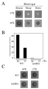
Figure 1
Figure 1
Hemolytic activity of S. marcescens. (A) Hemolytic activity of S. marcescens strain niid 298 on several blood agars. Cells (1 × 10⁶) were cultured overnight, and then inoculated on various blood agars and incubated at 30°C or 37°C for 16 h. Clear zones indicated hemolysis. (B) Contact hemolysis assay for human RBC. Cells harvested in log phase were mixed with washed human RBC and incubated at 30°C or 37°C for 1 h with shaking. Released hemoglobin was measured spectrophotometrically as absorbance at 405 nm. Results are shown as percent lysis compared to complete lysis of RBC in distilled water. (C) Hemolytic activity of the shlBA deletion mutant on human blood agar. Experiments were performed as in (A).
Figure 2
Figure 2
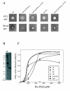
Phospholipase and hemolytic activities of S. marcescens PhlA. (A) Overnight cultures of wild-type strain S. marcescens niid 298, E. coli DH5αcells carrying pGEMeasy, E. coli DH5αcarrying pGEMeasy-phlAB, S. marcescens niid 298 phlAB deletion mutant, and S. marcescens niid 298 phlAB deletion mutant carrying pGEMeasy-phlAB (1 × 10⁶ cells) were inoculated on blood agar plates and PCY agar plates and incubated at 37°C for 16 and 24 h, respectively. (B) Purified His-PhlA (1 μg) was separated by 12.5% SDS-PAGE, and then was stained with Coomassie blue. Protein standards were in lane M, with relative molecular masses (kDa) at the left. (C) Various phospholipids were mixed with His-PhlA and incubated at 37°C for 1 h. Free fatty acids (FFA) released from phospholipids were detected using a NEFA-C kit. The amount of FFA was determined from an oleic acid calibration curve. Values are averages ± SE of three independent experiments.
Figure 3
Figure 3
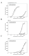
PLA1 and PLA2 activities of PhlA. PhlA activity was evaluated in a fluorescence enhancement assay using the following PLA fluorescence substrates: (A) bis-BODIPYFLC11-PC, (B) PED-A1, and (C) PED6. Fluorescence intensity was measured at 485 nm excitation and 530 nm emission using a fluorescence microplate reader (Appliskan; Thermo Electron Corporation). Open circles show His-PhlA; filled circles show PLA2 from bovine pancreas as a control. Values are averages ± SE from three independent experiments.
Figure 4
Figure 4
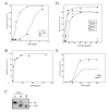
Phospholipid requirements of PhlA hemolytic and cytotoxic activities. (A) Human RBC were mixed with various concentrations of His-PhlA in the absence (open circles) or presence of lecithin (filled circles) or phosphatidylcholine (filled squares) and incubated at 37°C for 1 h. (B) Human RBC were mixed with various concentrations of lysophosphatidylcholine (LPC) and incubated at 37°C for 1 h. (C) Products of the reaction of PC with (+) or without (-) His-PhlA were analyzed by thin-layer chromatography. (D) Human (circles), sheep (triangles), and horse (squares) RBC were mixed with 8.3 μM PhlA (filled symbols) or no PhlA (open symbols) and incubated at 37°C for 1 h in the presence of various concentrations of lecithin with 2 mM CaCl2. (E) HeLa and 5637 cells were exposed to various concentrations of His-PhlA for 1 h in the presence of lecithin. His-PhlA cytotoxicity was evaluated with a CytoTox 96 Non-Radioactive Cytotoxicity Assay kit (Promega). Open and filled circles show HeLa and 5637 cells, respectively. Values are averages ± SE from three independent experiments. (A), (B), and (D) Results are expressed as percent lysis compared with lysis of RBC in distilled water, as in the contact hemolysis assay (Fig. 1).

Similar articles

Cited by

References

    1. Yu VL. Serratia marcescens: historical perspective and clinical review. The New England Journal of Medicine. 1979;300:887–893. - PubMed
    1. Hejazi A, Falkiner FR. Serratia marcescens. J Med Microbiol. 1997;46:903–912. doi: 10.1099/00222615-46-11-903. - DOI - PubMed
    1. Hertle R. The family of Serratia type pore forming toxins. Curr Protein Pept Sci. 2005;4:313–325. doi: 10.2174/1389203054546370. - DOI - PubMed
    1. Palmer M. The family of thiol-activated, cholesterol-binding cytolysins. Toxicon. 2001;39:1681–1689. doi: 10.1016/S0041-0101(01)00155-6. - DOI - PubMed
    1. Shinoda S, Matsuoka H, Tsuchie T, Miyoshi S, Yamamoto S, Taniguchi H, Mizuguchi Y. Purification and characterization of a lecithin-dependent haemolysin from Escherichia coli transformed by a Vibrio parahaemolyticus gene. J Gen Microbiol. 1991;137:2705–2711. - PubMed

Publication types

MeSH terms

LinkOut - more resources