

Cell type-specific and light-dependent expression of Rab1 and Rab6 GTPases in mammalian retinas
- PMID: 20003598
- PMCID: PMC2858915
- DOI: 10.1017/S0952523809990277
Cell type-specific and light-dependent expression of Rab1 and Rab6 GTPases in mammalian retinas
Abstract
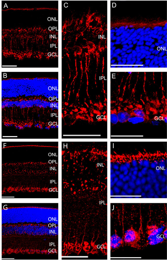
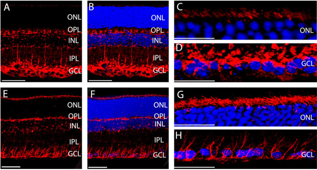
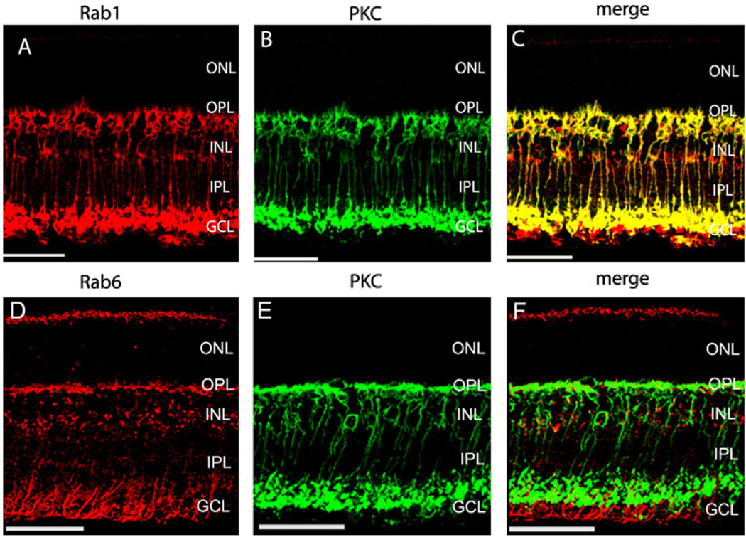
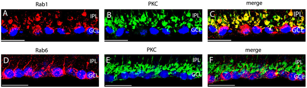
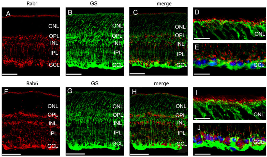
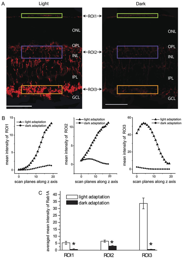
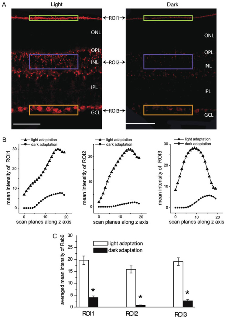
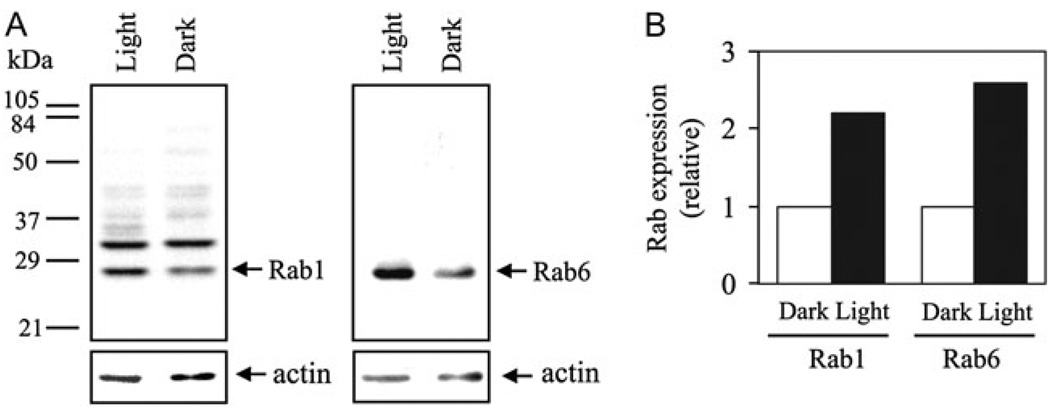
The Ras-like Rab1 and Rab6 GTPases modulate protein traffic along the early secretory pathway and are involved in the regulation of maturation of rhodopsin in the outer retina. However, Rab GTPases have not been studied in the inner retinas. Here, we analyzed the anatomatic distribution and expression of Rab1 and Rab6 in the mouse and rat retinas by immunohistochemistry and immunoblotting. We found that Rab1 was specifically expressed in the rod bipolar cells, while Rab6 was expressed in a different cell type(s) from rod bipolar cells in the inner retina. We also demonstrated that expression of Rab1 and Rab6 was increased with light. These data provided the first evidence implicating that Rab1 and Rab6 may be involved in the regulation of the retinal adaptation.
Figures
Similar articles
-
Giantin interacts with both the small GTPase Rab6 and Rab1.Exp Cell Res. 2007 Jul 1;313(11):2318-25. doi: 10.1016/j.yexcr.2007.03.031. Epub 2007 Apr 1. Exp Cell Res. 2007. PMID: 17475246
-
Identification and characterization of interacting partners of Rab GTPases by yeast two-hybrid analyses.Methods Mol Biol. 2008;440:111-25. doi: 10.1007/978-1-59745-178-9_9. Methods Mol Biol. 2008. PMID: 18369941
-
The fungal RABOME: RAB GTPases acting in the endocytic and exocytic pathways of Aspergillus nidulans (with excursions to other filamentous fungi).Mol Microbiol. 2021 Jul;116(1):53-70. doi: 10.1111/mmi.14716. Epub 2021 Apr 3. Mol Microbiol. 2021. PMID: 33724562 Review.
-
Regulation of anterograde transport of adrenergic and angiotensin II receptors by Rab2 and Rab6 GTPases.Cell Signal. 2007 Nov;19(11):2388-99. doi: 10.1016/j.cellsig.2007.07.017. Epub 2007 Aug 1. Cell Signal. 2007. PMID: 17716866 Free PMC article.
-
Regulation of G-protein-coupled receptor activity by rab GTPases.Recept Channels. 2002;8(2):87-97. Recept Channels. 2002. PMID: 12448790 Review.
Cited by
-
Rab6A as a Pan-Astrocytic Marker in Mouse and Human Brain, and Comparison with Other Glial Markers (GFAP, GS, Aldh1L1, SOX9).Cells. 2021 Jan 5;10(1):72. doi: 10.3390/cells10010072. Cells. 2021. PMID: 33466322 Free PMC article.
-
Rab1 in cell signaling, cancer and other diseases.Oncogene. 2016 Nov 3;35(44):5699-5704. doi: 10.1038/onc.2016.81. Epub 2016 Apr 4. Oncogene. 2016. PMID: 27041585 Free PMC article. Review.
-
GCAP1, Rab6, and HSP27: Novel Autoantibody Targets in Cancer-Associated Retinopathy and Autoimmune Retinopathy.Transl Vis Sci Technol. 2016 May 2;5(3):1. doi: 10.1167/tvst.5.3.1. eCollection 2016 May. Transl Vis Sci Technol. 2016. PMID: 27152249 Free PMC article.
References
-
- Antony C, Cibert C, Geraud G, Santa Maria A, Maro B, Mayau V, Goud B. The small GTP-binding protein rab6p is distributed from medial Golgi to the trans-Golgi network as determined by a confocal microscopic approach. Journal of Cell Science. 1992;103(Pt 3):785–796. - PubMed
-
- Behrens UD, Kasten P, Wagner HJ. Adaptation-dependent plasticity of rod bipolar cell axon terminal morphology in the rat retina. Cell and Tissue Research. 1998;294:243–251. - PubMed
-
- Bui BV, Hu RG, Acosta ML, Donaldson P, Vingrys AJ, Kalloniatis M. Glutamate metabolic pathways and retinal function. Journal of Neurochemistry. 2009;111:589–599. - PubMed
-
- Caminos E, Velasco A, Jarrin M, Lillo C, Jimeno D, Aijon J, Lara JM. A comparative study of protein kinase C-like immunoreactive cells in the retina. Brain, Behavior and Evolution. 2000;56:330–339. - PubMed
Publication types
MeSH terms
Substances
Grants and funding
LinkOut - more resources
Full Text Sources
Other Literature Sources
Research Materials
