. 2009 Dec;8(6):305-18.

Translational modulation of proteins expressed from bicistronic vectors

Affiliations

Translational modulation of proteins expressed from bicistronic vectors

Prasun J Mishra et al. Mol Imaging. 2009 Dec.

Abstract

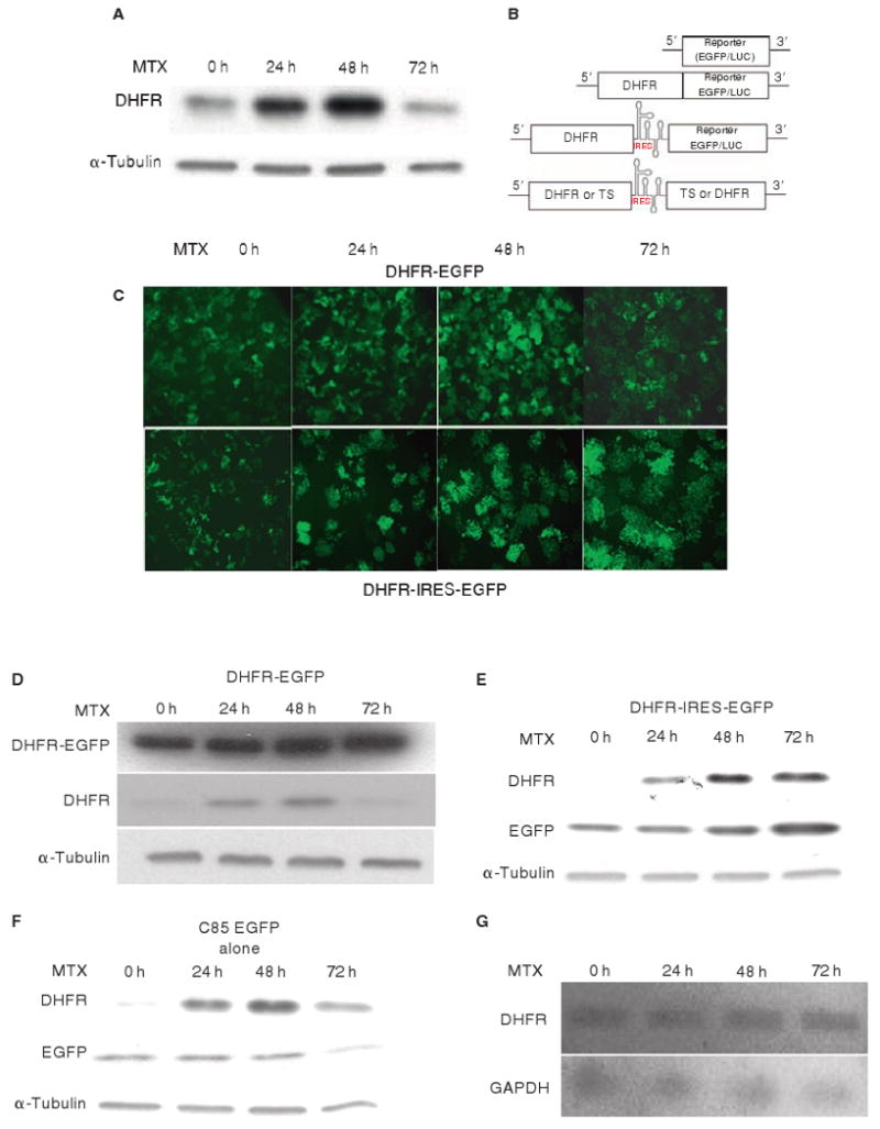
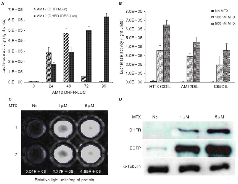
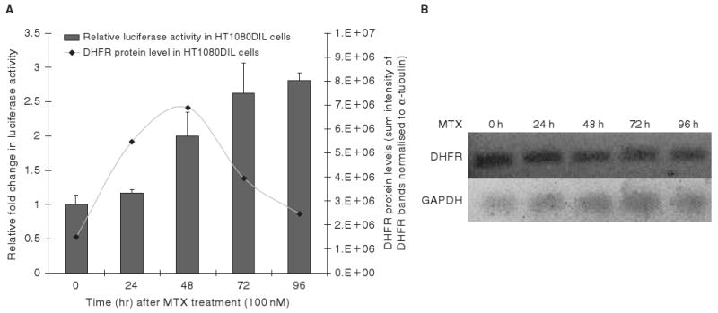
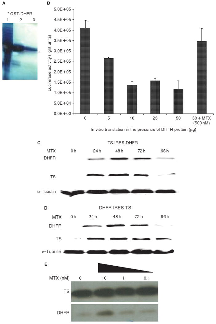
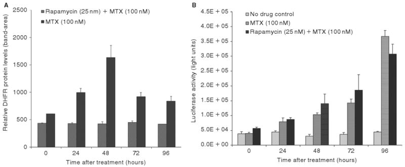
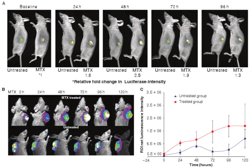
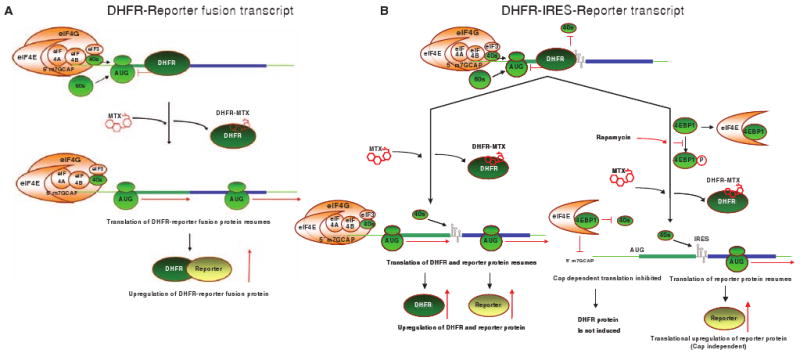
Bicistronic vectors are useful tools for exogenous expression of two gene products from a single promoter element; however, reduced expression of protein from the second cistron compared with the first cistron is a common limitation to this approach. To overcome this limitation, we explored use of dihydrofolate reductase (DHFR) complementary DNA encoded in bicistronic vectors to induce a second protein of interest by methotrexate (MTX) treatment. Previous studies have demonstrated that levels of DHFR protein and DHFR fusion protein can be induced translationally following MTX treatment of cells. We demonstrated that in response to MTX treatment, DHFR partner protein in a bicistronic construct is induced for longer periods of time when compared with endogenous DHFR and DHFR fusion protein, in vitro and in vivo. Using rapamycin pretreatment followed by MTX treatment, we also devised a strategy to modulate levels of two proteins expressed from a bicistronic construct in a cap-independent manner. To our knowledge, this is the first report demonstrating that levels of proteins in DHFR-based bicistronic constructs can be induced and modulated using MTX and rapamycin treatment.


Figures

Figure 1
Figure 1
EGFP reporter protein downstream of IRES can be upregulated on MTX treatment. A, Levels of endogenous DHFR can be translationally upregulated on MTX treatment; DHFR levels peak at 48 hours and drop at 72 hours. B, Schematic representation of various vectors used in the study. C, C85 colon cancer cells expressing EGFP linked to DHFR as a fusion protein (DHFR-EGFP) and in a bicistronic construct (DHFR-IRES-EGFP) were treated with MTX (100 nM), and EGFP expression was monitored using a fluorescent microscope. Notably, in a bicistronic construct, GFP remains upregulated for a longer period of time in response to MTX. D and E, Similar to the DHFR-EGFP fusion protein (D), EGFP protein levels were upregulated in C85 cells expressing the bicistronic construct DHFR-IRES-EGFP as determined by Western blotting. F, In a control experiment, EGFP levels in C85 cells expressing vector-containing EGFP alone, in the absence of DHFR c-DNA, on MTX treatment (100 nM), are not induced. G, DHFR and GAPDH (control) mRNA levels were unchanged on MTX treatment, indicating that MTX-mediated induction of the DHFR in the bicistronic construct DHFR-IRES-EGFP is at the translational level as determined by RT-PCR.
Figure 2
Figure 2
Levels of luciferase reporter proteins expressed downstream of IRES were upregulated on MTX treatment in three different cell lines. A, Other than EGFP, luciferase in a DHFR-IRES-Luc (DIL) vector was used as a second reporter construct to confirm that levels of second protein in a bicistronic construct can be upregulated. AM12 cells expressing DIL or expressing a fusion protein (DHFR-EGFP) were treated with MTX (100 nM), and cells were assayed for the luciferase activity. In bicistronic construct, luciferase remains upregulated for a longer period of time, at least 96 hours, in response to MTX compared with fusion protein. B, Reproducibility of the phenomenon was tested in three different cell lines, HT1080, C85, and AM12 cells, expressing DIL vector. The cells were treated with MTX (100 and 500 nM). The luciferase levels were upregulated significantly on MTX treatment in all three cell lines containing the bicistronic construct. C and D, In a control experiment, AM12 cells expressing DHFR-TK-GFP-Luc (DTGL) quadruple fusion protein expressing the luciferase (C) and DHFR and GFP protein levels were induced (D).
Figure 3
Figure 3
On MTX treatment, the levels of second protein downstream of IRES element remain translationally upregulated for a longer period of time, at least up to 96 hours in vitro compared with the first protein upstream of IRES element. A, The HT1080 fibrosarcoma cells were transfected with DIL and treated (untreated as control) with MTX (100 nM), and cell pellets at various time points were collected at 0, 24, 48, 72, and 96 hours. Luciferase activity was assayed using bioluminescence. The relative fold change in MTX-treated cells compared with untreated cells was determined. Following MTX treatment, DHFR and α-tubulin protein levels were determined using Western blotting. Bands were quantitated using Kodak Image Station 2000MM, and sum intensities of DHFR bands were normalized to α-tubulin. DHFR protein upstream of IRES element peaks at 48 hours and then drops at 72 to 96 hours. Luciferase downstream of the IRES element is inducible and stays up for at least 96 hours. B, DHFR mRNA levels were unchanged on MTX treatment, indicating that MTX-mediated induction of the DHFR in the bicistronic construct is at the translational level as determined by RT-PCR. GAPDH mRNA levels were used as the control.
Figure 4
Figure 4
DHFR regulates expression of its partner protein in a bicistronic construct, and MTX-mediated induction of partner protein is DHFR position independent. A, GST-DHFR (wt DHFR) protein was purified (see Materials and Methods); various steps of purification are shown: (1) cell lysate after induction with isopropyl β-D-1-thiogalactopyranoside, (2) elute after column purification, and (3) purified GST-DHFR protein after dialysis. B, DHFR-IRES-luc mRNA was used as a template in an in vitro translation reaction using a rabbit reticulocyte lysate system. Addition of GST-DHFR (wt DHFR) protein (5–50 μg) to the reaction mixture inhibited the expression of luciferase gene, which is located in the second citron, indicating that DHFR can regulate the expression of its partner protein in a bicistronic construct by interfering with IRES function. C and D, Two bicistronic constructs with altered positions of two genes, DHFR and thymidylate synthase (TS) (DHFR-IRES-TS and TS-IRES-DHFR), were designed and expressed in C85 cells. On MTX treatment (100 nM), the cells were lysed at various time points, and DHFR and TS protein levels were determined using Western blotting. On MTX treatment, TS protein levels were induced in both TS-IRES-DHFR- (B) and DHFR-IRES-TS- (C) expressing C85 cells. Notably, increased expression of second protein was sustained over a longer period of time when placed downstream of IRES, whereas the levels of upstream protein were induced at 48 hours and dropped at 72 hours after MTX treatment. E, In a control experiment, the C85 cells alone (untransfected) were treated with varying concentrations of MTX (10–0.1 nM). As expected, endogenous DHFR protein was induced on MTX treatment; however, endogenous TS protein levels were uninduced on MTX treatment.
Figure 5
Figure 5
Antifolate-mediated translational upregulation of the first protein is 5′ cap dependent, whereas the second protein is 5′ cap independent in a bicistronic construct. A and B, The HT1080 cells expressing DIL (containing wt DHFR cDNA) were pretreated with rapamycin (25 nM) to inhibit cap-dependent translation followed by MTX (100 nM) treatment. Cell pellets were collected at various time points (0–96 hours), and DHFR protein levels were determined using Western blotting. A, Rapamycin pretreatment inhibited MTX-mediated induction of the DHFR protein, located upstream to the IRES element, compared with MTX alone where DHFR levels were induced. B, Luciferase levels were still induced in the cells after rapamycin pretreatment and MTX treatment and were up for at least 96 hours, indicating that the induction was mediated by cap-independent translation. In untreated control cells, there was no significant induction of luciferase activity.
Figure 6
Figure 6
Protein downstream of IRES remains induced at least up to 120 hours in vivo. A–C, Results of studies using DHFR-TK-GFP-Luc (DTGL) fusion protein expressing C85 cells are shown in A; B and C show results using HT1080 DIL tumors grown in nude mice. The animals were treated with a single dose of MTX (5 mg/kg) (untreated animals were used as controls). Luciferase levels were monitored 0, 24, 48, 72, 96, and 120 hours after MTX injection using bioluminescence imaging. One representative mouse from the treated and the untreated group (DHFR-Luc and DHFR-IRES-Luc) is shown. A, Similar to the in vitro observations, in DHFR-Luc-expressing tumors, on MTX treatment, the luciferase levels peaked at 48 hours and dropped back to normal, whereas in the untreated group, there was no significant change in luciferase intensity. The relative fold difference in MTX-treated tumors compared with the untreated tumors was determined. B, An additional time point of 120 hours, beyond 96 hours, was included in the MTX-treated and untreated animals expressing wt DHFR-IRES-Luc. Luciferase levels were induced on MTX treatment and remained up for at least 120 hours (upper panel) compared with the levels for the untreated group (bottom panel). C, Luminescent intensity of the region of interest (ROI) was quantitated using the Kodak Image Station 2000MM. Luciferase gene was induced in all of the animals treated by MTX compared with the untreated animals. This additional time point (120 hours) in this in vivo study further strengthens the feasibility to control a “second” gene expression for a long period of time.
Figure 7
Figure 7
Model demonstrating the possible mechanism of induction and modulation of proteins in DHFR-based bicistronic vectors using MTX and rapamycin. A, We have previously shown that DHFR binds to its mRNA and autoregulates its expression. , B, In this report, we suggest that DHFR protein may bind and autoregulates its partner proteins in a bicistronic mRNA (DHFR-IRES-reporter). MTX is a tight binding inhibitor of DHFR. MTX-bound DHFR protein cannot bind to its mRNA and cannot autoregulate its and its partner protein's expression, leading to upregulation of DHFR and reporter proteins from a bicistronic mRNA. Rapamycin treatment inhibits cap-dependent translation (by inhibiting 4E-BP1 phosphorylation, leading to increased association between 4E-BP1 and the cap-binding protein eIF-4E). – Antifolate-mediated upregulation of DHFR protein upstream to IRES is inhibited by rapamycin pretreatment, indicating that the upregulation of DHFR protein upstream IRES is 5′ cap dependent, whereas translational upregulation of reporter protein downstream to IRES was not inhibited by rapamycin pretreatment, indicating that MTX-mediated induction of downstream reporter protein is 5′ cap independent in a bicistronic mRNA. Given that cap-dependent translation is more stringently regulated in the cell than IRES-mediated translation, the levels of upstream protein induce early but drop back to normal as soon as the drug is metabolized in the cell, whereas levels of downstream protein stay induced for longer periods.

Similar articles

Cited by

References

    1. Pyronnet S, Pradayrol L, Sonenberg N. A cell cycle-dependent internal ribosome entry site. Mol Cell. 2000;5:607–16. - PubMed
    1. Bochkov YA, Palmenberg AC. Translational efficiency of EMCV IRES in bicistronic vectors is dependent upon IRES sequence and gene location. Biotechniques. 2006;41:283–4. 286, 288. - PubMed
    1. Holcik M, Korneluk RG. Functional characterization of the X-linked inhibitor of apoptosis (XIAP) internal ribosome entry site element: role of La autoantigen in XIAP translation. Mol Cell Biol. 2000;20:4648–57. - PMC - PubMed
    1. Mitchell SA, Brown EC, Coldwell MJ, et al. Protein factor requirements of the Apaf-1 internal ribosome entry segment: roles of polypyrimidine tract binding protein and upstream of N-ras. Mol Cell Biol. 2001;21:3364–74. - PMC - PubMed
    1. Mitchell SA, Spriggs KA, Coldwell MJ, et al. The Apaf-1 internal ribosome entry segment attains the correct structural conformation for function via interactions with PTB and unr. Mol Cell. 2003;11:757–71. - PubMed

Publication types

MeSH terms

LinkOut - more resources