. 2009 Dec 9;17(12):1573-1581.
doi: 10.1016/j.str.2009.10.012.

Crystallographic insight into collagen recognition by discoidin domain receptor 2

Affiliations

Crystallographic insight into collagen recognition by discoidin domain receptor 2

Federico Carafoli et al. Structure. .

Abstract

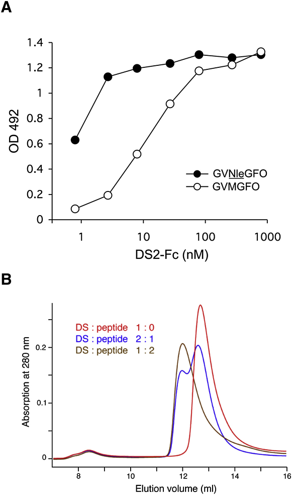
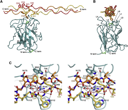
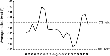
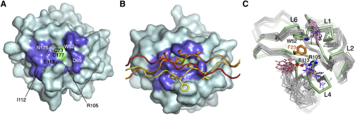
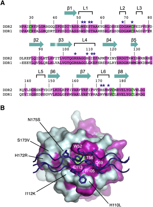
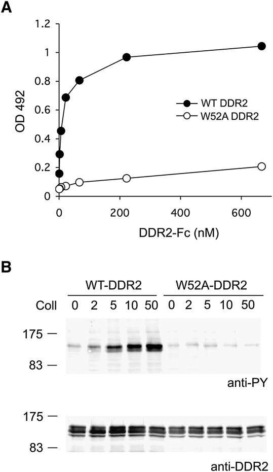
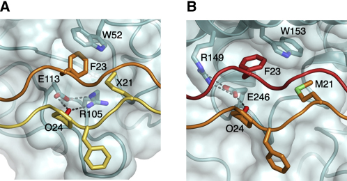
The discoidin domain receptors, DDR1 and DDR2, are widely expressed receptor tyrosine kinases that are activated by triple-helical collagen. They control important aspects of cell behavior and are dysregulated in several human diseases. The major DDR2-binding site in collagens I-III is a GVMGFO motif (O is hydroxyproline) that also binds the matricellular protein SPARC. We have determined the crystal structure of the discoidin domain of human DDR2 bound to a triple-helical collagen peptide. The GVMGFO motifs of two collagen chains are recognized by an amphiphilic pocket delimited by a functionally critical tryptophan residue and a buried salt bridge. Collagen binding results in structural changes of DDR2 surface loops that may be linked to the process of receptor activation. A comparison of the GVMGFO-binding sites of DDR2 and SPARC reveals a striking case of convergent evolution in collagen recognition.


Figures

Figure 1
Figure 1
Collagen Peptide Binding by the DDR2 DS Domain (A) Solid-phase binding assay with recombinant DS2-Fc protein (Leitinger, 2003) added to 96-well plates coated with triple-helical collagen peptides at 10 μg/ml: GPC-(GPP)5-GPRGQOGVXGFO-(GPP)5-GPC-NH2, where X is either methionine or norleucine. Shown is a representative of three independent experiments, each performed in duplicate. (B) Analytical size exclusion chromatograms of the free DDR2 DS domain and its complex with the triple-helical collagen peptide Ac-GPOGPOGPOGPR-GQOGVNleGFOGPOGPOG-NH2. The DS domain and peptide were mixed in the indicated molar ratios. A globular molecular mass standard of 29 kDa, carbonic anhydrase, elutes at 12.3 ml from this column.
Figure 2
Figure 2
Crystal Structure of the DDR2 DS Domain-Collagen Complex (A) Cartoon representation of the DS domain (cyan) and the collagen peptide (yellow, leading chain; orange, middle chain; red, trailing chain). The β strands of the DS domain are numbered sequentially. Disulfide bonds are in green. The side chains of the collagen GVMGFO motif are shown as sticks. Selected residues are labeled. X denotes norleucine. (B) Orthogonal view of the complex, related to (A) by a 90° rotation about a vertical axis. The collagen peptide is viewed from N to C terminus. Loops at the top of the DS domain are labeled as follows: L1-3, β1-β2; L4, β3-β4; L5, β5-β6; and L6, β7-β8. (C) Stereo view of the DDR2-collagen interface. Selected DDR2 and collagen residues are shown as sticks, in the same colors as in (A). The trailing collagen chain is shown as a semitransparent coil. Water molecules are shown as red spheres. Dashed lines indicate hydrogen bonds.
Figure 3
Figure 3
Helix Parameters of the Collagen Peptide Residues [i–1 (leading), i (middle), i+1 (trailing)] were fitted to residues [i (leading), i+1 (middle), i+2 (trailing)], and the associated rotation was taken as the helical twist at position i. The sequence of the collagen peptide is indicated at the bottom. X denotes norleucine. The twists of ideal left-handed 7/2 and 10/3 helices are −103° and −108°, respectively (Okuyama et al., 2006).
Figure 4
Figure 4
Structural Changes in the DDR2 DS Domain upon Collagen Binding (A) Surface representation of the free DDR2 DS domain in solution (Ichikawa et al., 2007). The collagen-binding residues identified by this study are in green (disulfide) and blue (all other residues). Selected residues are labeled. (B) Surface representation of the DS domain in complex with the collagen peptide (yellow, leading chain; orange, middle chain; red, trailing chain). The side chains of the GVMGFO motif are shown as sticks. The view direction and coloring of the DS domain surface are the same as in (A). (C) Superposition of the NMR ensemble of the free DDR2 DS domain (Ichikawa et al., 2007) (gray Cα traces; Trp52, Arg105, and Glu113 side chains in pink) and the crystal structure of the DDR2 DS-collagen (green, DS domain; orange, Phe23 of the collagen middle chain). The structures were superimposed using 43 Cα atoms of the DS domain β-barrel (rmsd 0.82 Å). Model 5 of the NMR ensemble is most similar overall to the crystal structure (rmsd 1.8 Å) and was taken as the reference. Selected residues and loops are labeled.
Figure 5
Figure 5
Sequence Conservation of the Collagen-Binding Site (A) Sequence alignment of the DS domains of human DDR1 and DDR2. The sequence numbering and secondary structure elements of the DDR2 DS domain are indicated above the alignment. Conserved residues and cysteines are highlighted in magenta and green, respectively. Residues that lose ≥5 Å2 of their solvent-accessible surface upon collagen binding are indicated by purple stars. (B) Mapping of conserved residues onto the molecular surface of the DDR2 DS domain. Residues that are identical in DDR1 and DDR2 are in magenta. The Cys73-Cys177 disulfide bridge is in green. Selected conserved residues and nonconserved substitutions in DDR1 are indicated. The collagen peptide is in purple, and the side chains of the GVMGFO motifs of the leading and middle chains are shown as sticks.
Figure 6
Figure 6
Essential Role of Trp52 in DDR2 Function (A) Solid-phase binding assay with recombinant wild-type or W52A DDR2-Fc proteins added for 3 hr at room temperature to 96-well plates coated with either collagen I or BSA. Shown is a representative of three independent experiments, each performed in duplicate. (B) Full-length wild-type or W52A DDR2 was transiently expressed in HEK293 cells. After stimulation for 90 min with collagen I (Coll), aliquots of cell lysates were analyzed by SDS-PAGE and Western blotting. The blots were probed with anti-phosphotyrosine (anti-PY) monoclonal antibody 4G10 (upper blot) or polyclonal anti-DDR2 antibodies (lower panel). The positions of molecular markers (in kilodaltons) are indicated. Collagen I was used at different concentrations as indicated (in μg/ml). The experiment was performed three times with very similar results.
Figure 7
Figure 7
Comparison of Collagen Recognition by DDR2 and SPARC DDR2 (A) (this work) and SPARC (B) (Hohenester et al., 2008) are in cyan and shown as cartoons with semitransparent surfaces. The leading, middle, and trailing chains of the collagen peptides are in yellow, orange, and red, respectively. Selected residues are shown as sticks. X denotes norleucine. Dashed lines indicate hydrogen bonds.
Figure 8
Figure 8
Possible mechanisms of DDR activation DDR1 and DDR2 are dimeric in the absence of collagen (Noordeen et al., 2006). The mechanism of autoinhibition in the inactive dimer is unknown, but is likely to involve the second domain of the ectodomain and/or the large cytosolic juxtamembrane domain. DDR activation may result from the simultaneous binding of both DS domains in the dimer to a single collagen triple helix (“composite binding site”), or the DS domains may bind collagen independently (“independent binding sites”). In any case, collagen binding is proposed to release the autoinhibition, resulting in activation of the cytoplasmic tyrosine kinase domains.

References

    1. Abdulhussein R., McFadden C., Fuentes-Prior P., Vogel W.F. Exploring the collagen-binding site of the DDR1 tyrosine kinase receptor. J. Biol. Chem. 2004;279:31462–31470. - PubMed
    1. Abdulhussein R., Koo D.H., Vogel W.F. Identification of disulfide-linked dimers of the receptor tyrosine kinase DDR1. J. Biol. Chem. 2008;283:12026–12033. - PMC - PubMed
    1. Appleton B.A., Wu P., Maloney J., Yin J., Liang W.C., Stawicki S., Mortara K., Bowman K.K., Elliott J.M., Desmarais W. Structural studies of neuropilin/antibody complexes provide insights into semaphorin and VEGF binding. EMBO J. 2007;26:4902–4912. - PMC - PubMed
    1. Bargal R., Cormier-Daire V., Ben-Neriah Z., Le Merrer M., Sosna J., Melki J., Zangen D.H., Smithson S.F., Borochowitz Z., Belostotsky R., Raas-Rothschild A. Mutations in DDR2 gene cause SMED with short limbs and abnormal calcifications. Am. J. Hum. Genet. 2009;84:80–84. - PMC - PubMed
    1. Brandl M., Weiss M.S., Jabs A., Suhnel J., Hilgenfeld R. C-H…π-interactions in proteins. J. Mol. Biol. 2001;307:357–377. - PubMed

Publication types

MeSH terms