. 2009 Dec 9;17(12):1591-1604.
doi: 10.1016/j.str.2009.09.015.

Comprehensive molecular structure of the eukaryotic ribosome

Affiliations

Comprehensive molecular structure of the eukaryotic ribosome

Derek J Taylor et al. Structure. .

Abstract

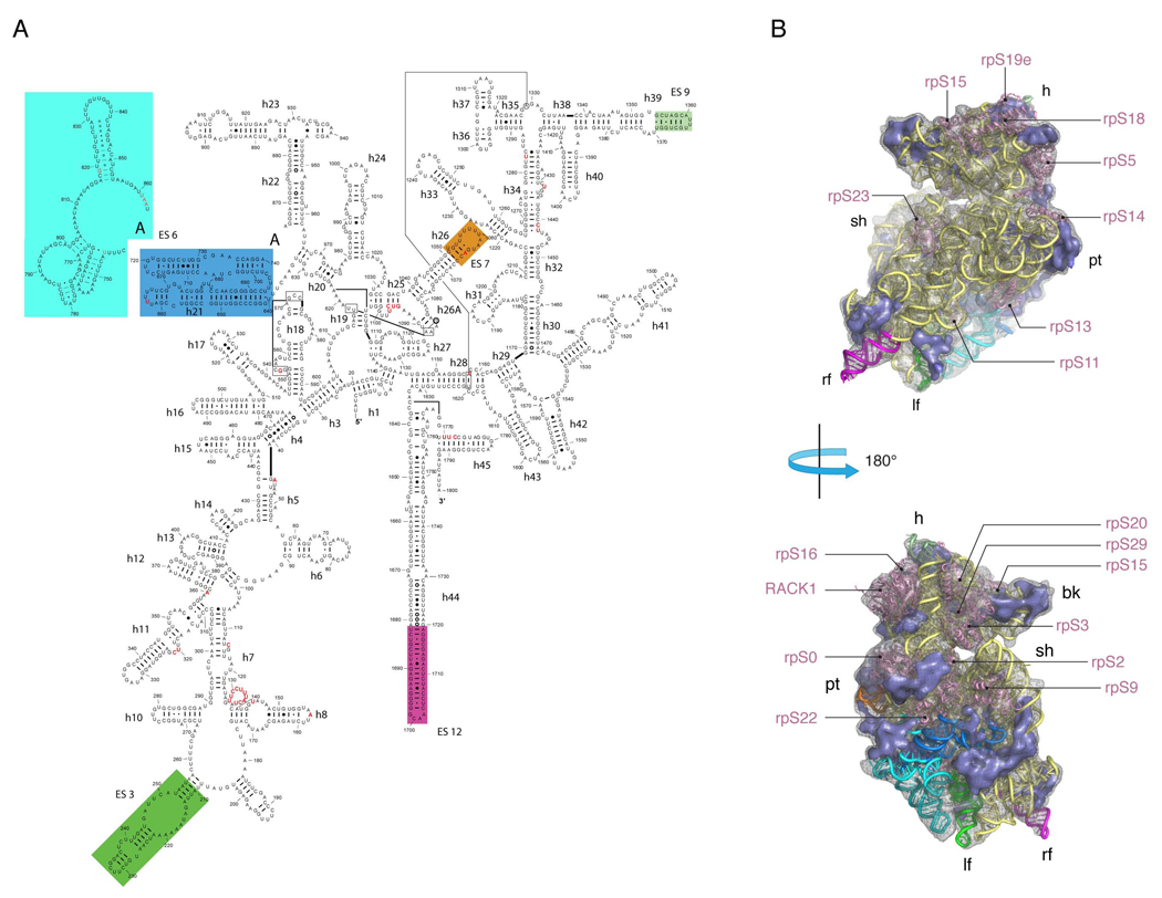
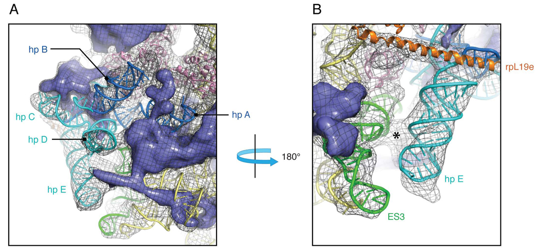
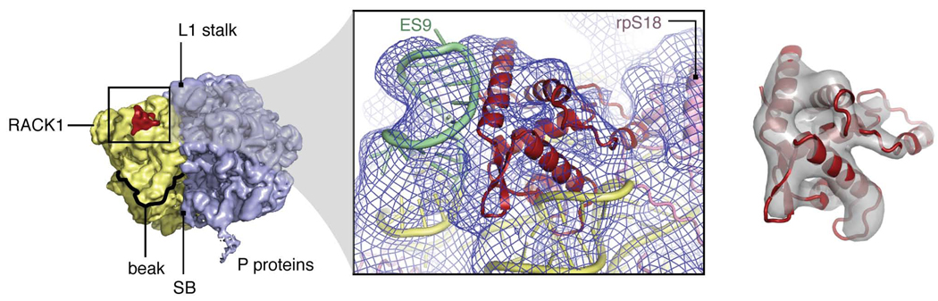
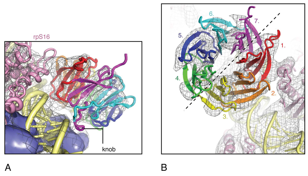
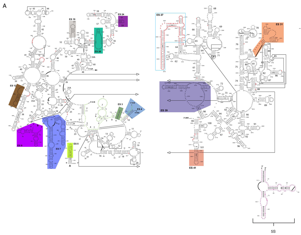
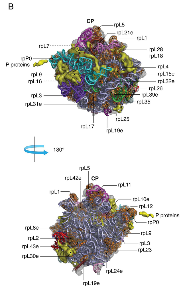
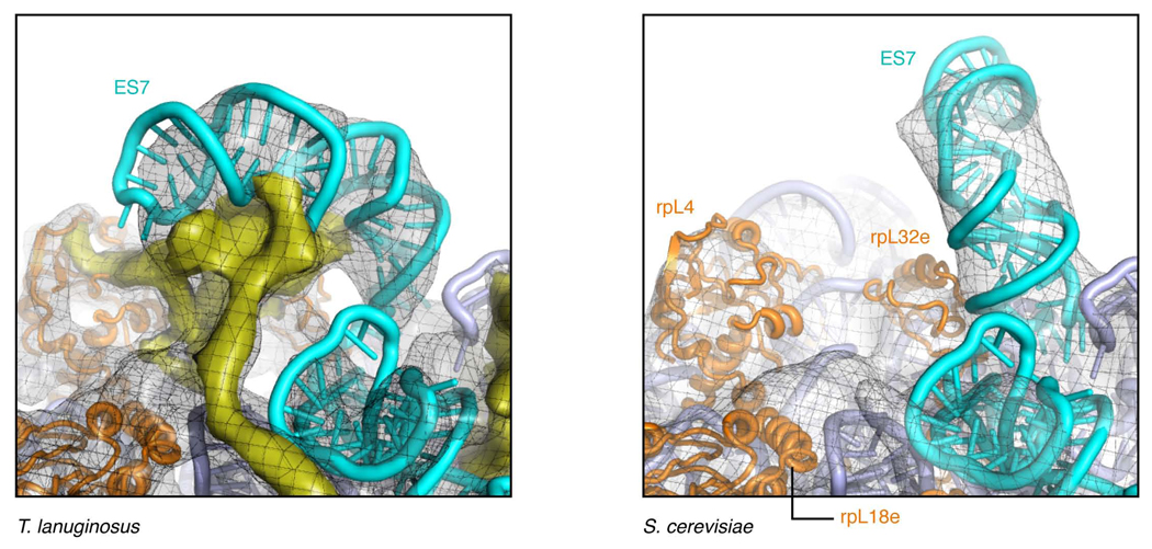
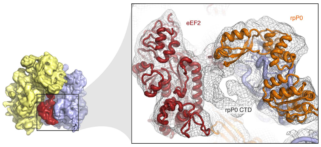
Despite the emergence of a large number of X-ray crystallographic models of the bacterial 70S ribosome over the past decade, an accurate atomic model of the eukaryotic 80S ribosome is still not available. Eukaryotic ribosomes possess more ribosomal proteins and ribosomal RNA than do bacterial ribosomes, which are implicated in extraribosomal functions in the eukaryotic cells. By combining cryo-EM with RNA and protein homology modeling, we obtained an atomic model of the yeast 80S ribosome complete with all ribosomal RNA expansion segments and all ribosomal proteins for which a structural homolog can be identified. Mutation or deletion of 80S ribosomal proteins can abrogate maturation of the ribosome, leading to several human diseases. We have localized one such protein unique to eukaryotes, rpS19e, whose mutations are associated with Diamond-Blackfan anemia in humans. Additionally, we characterize crucial interactions between the dynamic stalk base of the ribosome with eukaryotic elongation factor 2.


Figures

Figure 1
Figure 1
Structure of the eukaryotic 40S subunit. (a) Secondary structure prediction and rRNA helix numbering based primarily on that of the Comparative RNA Web Site of 18S rRNA of S. cerevisiae (http://www.rna.ccbb.utexas.edu). The secondary structure of ES6, however, is modeled based on that reported by Wuyts et al. (Wuyts et al., 2002). Expansion segments are color-coded. (b) The atomic model of the 40S subunit with ribosomal RNA represented as tubes, with expansion segments color-coded to match their secondary structure position presented in (a). Ribosomal proteins, with known homologs and placement, are shown as pink cartoons. Density accounting for proteins that are unique to eukaryotes with no structural homolog available or that have not been localized within the context of the ribosome is represented in slate. The boundary of the cryo-EM density of the 40S subunit is shown as a grey mesh.
Figure 2
Figure 2
The quaternary structure of ES6. (a) Solvent view of the 40S subunit. ES6 is represented by two 5’ hairpins (A and B; blue tubes) followed by three hairpins on the 3’ end (C, D, E; cyan tubes). All five hairpins interact intimately with unknown proteins (slate density) on the solvent side of the 40S subunit. (b) Interaction of ES3 (green) with hairpin E of ES6 (cyan tube) as viewed from the intersubunit space of the ribosome. Strong density in the EM map (marked with an “*”) supports phylogenic data indicating that base pairing interaction exists between the two ESs. Prominent density seen for the C-terminal helix of rpL19e, reaching over from the 60S subunit, allowed the modeling of this region in our structure. This helix makes extensive interactions with ES6, particularly at the base of hairpin E.
Figure 3
Figure 3
Localization of rpS19e in the 40S ribosomal subunit. rpS19e (red) localizes to the top of the head of the small subunit. Homology model of rpS19e crystal structure displayed within transparent cryo-EM density. Coloring is the same as in figure 1.
Figure 4
Figure 4
Interactions of RACK1 with the 40S subunit. (a) Side-view of RACK1 with its interaction to the 40S subunit. An unknown protein resides near RACK1, but the density between the two moieties is rather weak. Primary interactions between RACK1 and the 40S subunit are coordinated through 18S rRNA and rpS16 (rpS9p). (b) Top-view of RACK1 with the seven propellers color-coded. EM density indicates that primary interactions with the 40S subunit are coordinated via blade 1 of RACK1. Density of the RACK1 structure further indicates the molecule to have two distinct hemicycles with one half formed by tight interactions between blades 1–3 and a second half from blades 4–7.
Figure 5
Figure 5
Structure of the eukaryotic 60S subunit. (a) Secondary structure prediction and rRNA helix numbering according to that of the Comparative RNA Web Site of rRNA of S. cerevisiae (http://www.rna.ccbb.utexas.edu). Expansion segments are color-coded. (b) The atomic model of the 60S subunit with ribosomal rna represented as tubes, with expansion segments color-coded to match their secondary structure position presented in (a). 5S and 5.8S rRNA are shown as magenta and green tubes, respectively. Ribosomal proteins, with known homologs and placement, are shown as orange cartoons. Density accounting for proteins that are unique to eukaryotes with no structural homolog available or that have not been localized within the context of the ribosome is represented in yellow. The boundary of the cryo-EM map of the 60S subunit is shown as a grey mesh.
Figure 5
Figure 5
Structure of the eukaryotic 60S subunit. (a) Secondary structure prediction and rRNA helix numbering according to that of the Comparative RNA Web Site of rRNA of S. cerevisiae (http://www.rna.ccbb.utexas.edu). Expansion segments are color-coded. (b) The atomic model of the 60S subunit with ribosomal rna represented as tubes, with expansion segments color-coded to match their secondary structure position presented in (a). 5S and 5.8S rRNA are shown as magenta and green tubes, respectively. Ribosomal proteins, with known homologs and placement, are shown as orange cartoons. Density accounting for proteins that are unique to eukaryotes with no structural homolog available or that have not been localized within the context of the ribosome is represented in yellow. The boundary of the cryo-EM map of the 60S subunit is shown as a grey mesh.
Figure 6
Figure 6
Protein density anchors ES7 to the body of the 60S. ES7 of T. lanuginosus (left) is tethered to the body of the 60S subunit by an unidentified protein mass (yellow), presenting a “closed” conformation of ES7. This protein was absent in a previously reported structure of S. cerevisiae (right) (Spahn et al., 2001), leading to an “open” conformation of ES7.
Figure 7
Figure 7
Interaction of rpP0 CTD with eEF2. The NTD of rpP0 was modeled into the density of our cryo-EM map, based on the orthologous x-ray structure of L10 from T. maritime (Diaconu et al., 2005). Density for the CTD of rpP0 is clearly visible and indicative of α-helical secondary structure. The density further indicates a clear interaction between the rpP0 CTD and a long α-helix residing domain I of eEF2.

Similar articles

Cited by

References

    1. Alkemar G, Nygard O. Probing the secondary structure of expansion segment ES6 in 18S ribosomal RNA. Biochemistry. 2006;45:8067–8078. - PubMed
    1. Altschul SF, Madden TL, Schaffer AA, Zhang J, Zhang Z, Miller W, Lipman DJ. Gapped BLAST and PSI-BLAST: a new generation of protein database search programs. Nucleic Acids Res. 1997;25:3389–3402. - PMC - PubMed
    1. Berk V, Zhang W, Pai RD, Cate JH. tructural basis for mRNA and tRNA positioning on the ribosome. Proc Natl Acad Sci U S A. 2006;103:15830–15834. - PMC - PubMed
    1. Blaha G, Stanley RE, Steitz TA. Formation of the first peptide bond: the structure of EF-P bound to the 70S ribosome. Science. 2009;325:966–970. - PMC - PubMed
    1. Buchhaupt M, Meyer B, Kotter P, Entian KD. Genetic evidence for 18S rRNA binding and an Rps19p assembly function of yeast nucleolar protein Nep1p. Mol Genet Genomics. 2006;276:273–284. - PubMed

Publication types

LinkOut - more resources