. 2009 Dec 9;17(12):1636-1648.
doi: 10.1016/j.str.2009.10.008.

Identification of structural mechanisms of HIV-1 protease specificity using computational peptide docking: implications for drug resistance

Affiliations

Identification of structural mechanisms of HIV-1 protease specificity using computational peptide docking: implications for drug resistance

Sidhartha Chaudhury et al. Structure. .

Abstract

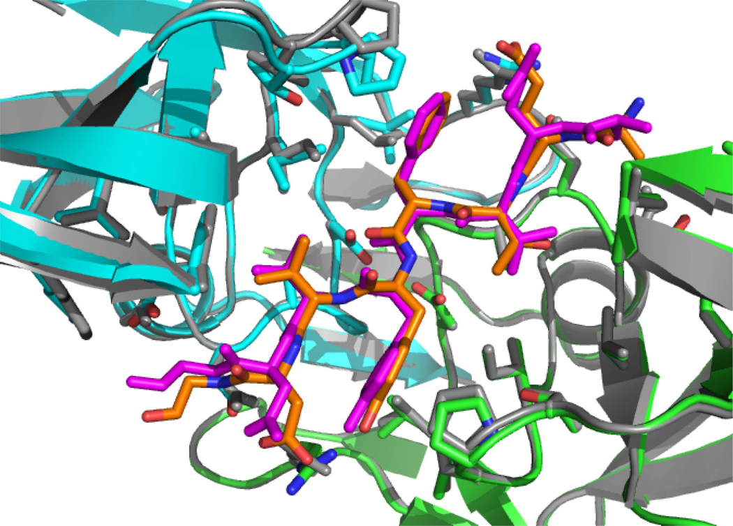
Drug-resistant mutations (DRMs) in HIV-1 protease are a major challenge to antiretroviral therapy. Protease-substrate interactions that are determined to be critical for native selectivity could serve as robust targets for drug design that are immune to DRMs. In order to identify the structural mechanisms of selectivity, we developed a peptide-docking algorithm to predict the atomic structure of protease-substrate complexes and applied it to a large and diverse set of cleavable and noncleavable peptides. Cleavable peptides showed significantly lower energies of interaction than noncleavable peptides with six protease active-site residues playing the most significant role in discrimination. Surprisingly, all six residues correspond to sequence positions associated with drug resistance mutations, demonstrating that the very residues that are responsible for native substrate specificity in HIV-1 protease are altered during its evolution to drug resistance, suggesting that drug resistance and substrate selectivity may share common mechanisms.


Figures

Figure 1
Figure 1. Peptide-docking algorithm
Flow chart of the Monte-Carlo minimization algorithm for peptide docking.
Figure 2
Figure 2. Protease-peptide complex structure prediction
The structure predictions for the peptides (A) RT-RH, (B) MA-CA, and (C) RH-IN. The crystal structure of each complex is shown in gray with the peptide in magenta. The predicted structure is colored with the HIV-1 protease dimer in green and cyan and the peptide substrate in orange. Active site and substrate residues are shown as sticks. P4 is the peptide residue on the bottom-left, P4’ is the peptide residue at the top-right.
Figure 3
Figure 3. Energy distributions of cleavable and non-cleavable peptides
Histograms of energies summed over different subsets of residues in the peptide-protease complex for cleavable (blue) and non-cleavable (red) peptides. (A) Energies from all active-site and peptide residues. (B) Energies from peptide residues alone. (C) Energies from active-site DRM-associated residues (protease residues 23, 30, 47, 48, 50, 76, 82, 84) (D) Energies from all active-site residues not associated with DRMs.
Figure 4
Figure 4. Specificity-determining residues in the protease-substrate complex
Residues that were significant at p < 0.001 are colored red, those that were significant at p < 0.01 are colored orange, and those that were significant at p < 0.05 are colored yellow. Protease residues are shown in sticks, peptide residues shown as spheres.
Figure 5
Figure 5. Structural and energetic mechanisms of specificity
Left, histograms for the individual residue energies for specificity determinant residues (A) I47', (B) I84', (C) V82, and (D) D30' for cleavable (blue) and non-cleavable (red) peptides. Right, structures of representative cleavable and non-cleavable peptides for each specificity-determining residue. The specificity-determining residue is colored with respect to its residue energy (spectrum from blue to red for low energy to high energy). Important interacting residues as well as the specificity-determining residues are shown as sticks for the peptide (dark gray) and the protease (light gray). The cleavable peptides selected for illustration are the ten endogenous substrates, the non-cleavable peptides are the ten with the highest residue energy for each respective specificity-determining residue.
Figure 6
Figure 6. Evaluation of the substrate envelope hypothesis
(A) Peptide residues P2-P2' are shown for cleavable (left) and non-cleavable (right) peptides. Peptide side-chain atoms are shown as sticks (magenta), atoms that protrude from the substrate envelope are shown as yellow spheres. Protease specificity-determining residues are shown as sticks (green and cyan). (B) Plot of number of heavy atoms that protrude from the substrate envelope (Natoms) vs. the energy of the protease-peptide complex, for cleavable (red) and non-cleavable (blue) peptides.
Figure 7
Figure 7. Substrate and binding envelope in the protease active-site
(A) The substrate envelope (red surface) and binding (blue mesh) envelope volumes are illustrated for substrate residues P3-P3'. Drug resistant mutation residues 30, 47, 48, 50, 82, and 84 are shown as sticks. (B) The inhibitor volume consisting of the combined volumes of inhibitors Nelfinavir, Saquinavir, Indinavir, Ritonavir, Amprenavir, Lopinavir, and Atazanavir, is shown in orange.

Similar articles

Cited by

References

    1. Altman MD, Ali A, Reddy GS, Nalam MN, Anjum SG, Cao H, Chellappan S, Kairys V, Fernandes MX, Gilson MK, et al. HIV-1 protease inhibitors from inverse design in the substrate envelope exhibit subnanomolar binding to drug-resistant variants. J Am Chem Soc. 2008a;130:6099–6113. - PMC - PubMed
    1. Altman MD, Nalivaika EA, Prabu-Jeyabalan M, Schiffer CA, Tidor B. Computational design and experimental study of tighter binding peptides to an inactivated mutant of HIV-1 protease. Proteins. 2008b;70:678–694. - PMC - PubMed
    1. Bagossi P, Sperka T, Feher A, Kadas J, Zahuczky G, Miklossy G, Boross P, Tozser J. Amino acid preferences for a critical substrate binding subsite of retroviral proteases in type 1 cleavage sites. Journal of virology. 2005;79:4213–4218. - PMC - PubMed
    1. Beck ZQ, Lin YC, Elder JH. Molecular basis for the relative substrate specificity of human immunodeficiency virus type 1 and feline immunodeficiency virus proteases. Journal of virology. 2001;75:9458–9469. - PMC - PubMed
    1. Beck ZQ, Morris GM, Elder JH. Defining HIV-1 protease substrate selectivity. Current drug targets. 2002;2:37–50. - PubMed

Publication types

LinkOut - more resources