Review
. 2010 Feb 15;494(2):107-20.
doi: 10.1016/j.abb.2009.12.005. Epub 2009 Dec 11.

Butyrylcholinesterase for protection from organophosphorus poisons: catalytic complexities and hysteretic behavior

Affiliations
Review

Butyrylcholinesterase for protection from organophosphorus poisons: catalytic complexities and hysteretic behavior

Patrick Masson et al. Arch Biochem Biophys. .

Abstract

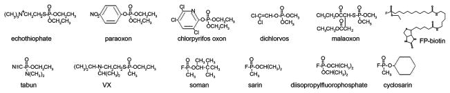
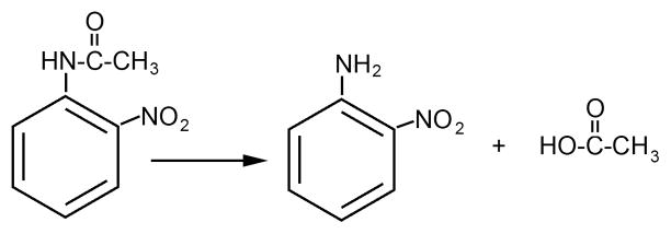
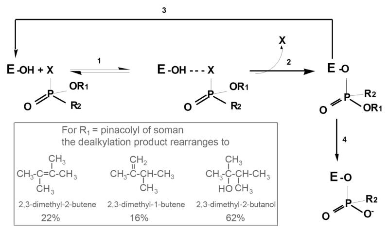
Butyrylcholinesterase is a promiscuous enzyme that displays complex kinetic behavior. It is toxicologically important because it detoxifies organophosphorus poisons (OP) by making a covalent bond with the OP. The OP and the butyrylcholinesterase are both inactivated in the process. Inactivation of butyrylcholinesterase has no adverse effects. However, inactivation of acetylcholinesterase in nerve synapses can be lethal. OP-inhibited butyrylcholinesterase and acetylcholinesterase can be reactivated with oximes provided the OP has not aged. Strategies for preventing the toxicity of OP include (a) treatment with an OP scavenger, (b) reaction of non-aged enzyme with oximes, (c) reactivation of aged enzyme, (d) slowing down aging with peripheral site ligands, and (e) design of mutants that rapidly hydrolyze OP. Option (a) has progressed through phase I clinical trials with human butyrylcholinesterase. Option (b) is in routine clinical use. The others are at the basic research level. Butyrylcholinesterase displays complex kinetic behavior including activation by positively charged esters, ability to hydrolyze amides, and a lag time (hysteresis) preceding hydrolysis of benzoylcholine and N-methylindoxyl acetate. Mass spectrometry has identified new OP binding motifs on tyrosine and lysine in proteins that have no active site serine. It is proposed, but not yet proven, that low dose exposure involves OP modification of proteins that have no active site serine.


Figures

Figure 1
Figure 1
Structures of organophosphorus agents. Paraoxon, chlorpyrifos oxon, dichlorvos, and malaoxon are the active metabolites of pesticides. Echothiophate has been used in eyedrops to treat glaucoma. Tabun, VX, soman, sarin, and cyclosarin are nerve agents developed for use as chemical warfare agents. FP-biotin and diisopropylfluorophosphate are research reagents.
Figure 2
Figure 2
Model of the human butyrylcholinesterase tetramer. Four subunits assemble through the tetramerization domain at the C-terminus. The tetramerization domain has four parallel alpha helices wrapped around a single antiparallel polyproline helix. The polyproline peptide derives from lamellipodin. The tetramer is a dimer of dimers. Dimers have an interchain disulfide bond at Cys571. A) Viewed from the top, with the tetramerization domain and the polyproline peptide in the center. Two of the active sites are exposed to solvent, while two face the central cavity of the butyrylcholinesterase tetramer. B) Viewed from the side. Reproduced from [185].
Figure 3
Figure 3
Hydrolysis of o-nitroacetanilide by acetylcholinesterase and butyrylcholinesterase.
Figure 4
Figure 4
Reaction of butyrylcholinesterase and acetylcholinesterase with OP. 1) formation of reversible complex; 2) phosphylation of the active site serine, with departure of the leaving group X and inversion of the stereochemistry of the OP; 3) reactivation of phosphylated enzyme by oximes or by 2 M potassium fluoride; 4) aging to produce a negatively charged OP stabilized by interaction with the positively charged histidine of the catalytic triad. Inset. When the OP is soman, R1 is pinacolyl alcohol. Dealkylation does not yield pinacolyl alcohol, but instead yields 3 rearranged products [75].
Figure 5
Figure 5
Oxime structures
Figure 6
Figure 6
Beta-elimination. OP-labeled serine loses the OP and a molecule of water in the beta-elimination reaction. The active site serine is converted to dehydroalanine.
Figure 7
Figure 7
MS spectrum of tryptic digest of soman-labeled human butyrylcholinesterase. Peptides were separated by HPLC and 1 ml fractions collected. A 0.5 microliter aliquot of each fraction was analyzed in the MALDI-TOF mass spectrometer. Panel A shows that fraction 35 contains the aged soman-labeled peptide at 3006.5 m/z and a small amount of unlabeled peptide at 2928.5 m/z. Panel B shows that fraction 36 contains the dehydroalanine form of the peptide at 2910.5 m/z, unlabeled peptide at 2928.5 m/z, and a small amount of aged-soman labeled peptide at 3006.5 m/z. The active site serine of human butyrylcholinesterase is Ser198 (accession # gi:116353).
Figure 8
Figure 8
Salt bridge between the negatively charged aged soman bound to Ser198 and the positively charged catalytic His438 of human butyrylcholinesterase. The crystal structure is in pdb code 1p0q [81].
Figure 9
Figure 9
Soman-inhibited butyrylcholinesterase tetramer is resistant to unfolding. A mixture of native unlabeled butyrylcholinesterase, nonaged soman-butyrylcholinesterase, and aged soman-butyrylcholinesterase, 0.5 mg butyrylcholinesterase protein total, was layered on a polyacrylamide gel containing a 0–8 M transverse gradient of urea. After electrophoresis the gel was stained with Coomassie blue. Three well-separated curves of butyrylcholinesterase protein are seen. Curve 1) not labeled butyrylcholinesterase tetramer swells into an unfolded structure as the urea concentration increases from 0 to 8 M. The unfolded butyrylcholinesterase migrates more slowly through the gel. The urea concentration at the mid-point for unfolding is 3.5 M. 2) Nonaged soman-phosphylated butyrylcholinesterase begins to unfold at a higher urea concentration. 3) Aged soman-butyrylcholinesterase retains its folded structure up to 4 M urea and has a midpoint for unfolding at 6 M urea. N indicates the migration of normal folded tetrameric butyrylcholinesterase in the absence of urea. U indicates migration of unfolded butyrylcholinesterase in 8 M urea. The direction of migration is from minus (−) to plus (+).
Scheme 1
Scheme 1
Scheme 2
Scheme 2

Similar articles

Cited by

References

    1. Saxena A, Sun W, Luo C, Myers TM, Koplovitz I, Lenz DE, Doctor BP. J Mol Neurosci. 2006;30:145–148. - PubMed
    1. Manoharan I, Boopathy R, Darvesh S, Lockridge O. Clin Chim Acta. 2007;378:128–135. - PubMed
    1. Darvesh S, Hopkins DA. J Comp Neurol. 2003;463:25–43. - PubMed
    1. Wetherell JR, French MC. Biochem Pharmacol. 1986;35:939–945. - PubMed
    1. Duysen EG, Li B, Xie W, Schopfer LM, Anderson RS, Broomfield CA, Lockridge O. J Pharmacol Exp Ther. 2001;299:528–535. - PubMed

Publication types

MeSH terms

Substances