Distinct central representations for sensory fibers innervating either the conjunctiva or cornea of the rat
- PMID: 20004193
- PMCID: PMC2824013
- DOI: 10.1016/j.exer.2009.11.018
Distinct central representations for sensory fibers innervating either the conjunctiva or cornea of the rat
Abstract
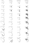
The laminar sheet of epithelium (e.g., skin and mucous membrane) enclosing our bodies is represented in the dorsal horns of the medulla and spinal cord. The eyeball however indents this laminar sheet and is shrouded by different layers: the cornea/sclera, the conjunctiva, and hairy skin. This involution of the orb confounds defining the central representation of the cornea and its surrounding mucosa and skin. We used herein the transganglionic transport of a cocktail of HRP conjugated to cholera toxin and wheat germ agglutinin to determine the central representation of these epithelia in the dorsal horns of the rat. The HRP cocktail was injected either into the stroma of the cornea, the mucosa of the conjunctiva, or the supraorbital and infraorbital nerves. Injections of the cornea produced dense label in the interstitial islands in the ventral medullary dorsal horn (MDH), probably lamina I, and in neuropil in the ventromedial tip of the MDH, probably lamina II. There sometimes was variable, diffuse label in the C1 dorsal horn after corneal injections but more rostral parts of the trigeminal sensory complex were never labeled. Injections of the conjunctiva densely labeled laminae I-III in the C1 dorsal horn, while laminae IV-V were diffusely labeled. Sparser reaction product also was seen in lamina I in positions similar to the cornea projection. Label was seen ventrally in subnuclei interpolaris and oralis, as well as the principal trigeminal nucleus. Projections of the infraorbital nerve included all laminae in the trigeminocervical complex as well as large portions of the rostral subnuclei in the spinal trigeminal nucleus. The projections of the supraorbital nerve were similar, but were restricted to ventral parts of the trigeminal sensory complex. In other cases the cornea was injected either after cutting the supraorbital and infraorbital nerves or the conjunctiva was injected after enucleating the eyeball. Any reaction product from corneal injections was reduced dramatically in the C1 dorsal horn after transection of the infraorbital and supraorbital nerves. Injecting the conjunctiva after enucleating the eyeball densely labeled the C1 projection to the dorsal horn, a small patch in lamina I in the MDH, as well as the rostral trigeminal complex. We propose that the cornea has but a single representation in the trigeminocervical complex in its ventral part near the caudal end of the medulla. We also propose the palpebral conjunctiva mucosa is represented in the C1 dorsal horn, and speculate that the bulbar conjunctiva overlaps with that of the cornea in lamina I. We discuss these projections in relation to the circuitry for the supraorbital-evoked and corneal-evoked blink reflexes. The relationship of the cornea and conjunctiva is intimate, and investigators must be very careful when attempting to stimulate them in isolation.
Figures


References
-
- Acosta MC, Tan ME, Belmonte C, Gallar J. Sensations evoked by selective mechanical, chemical, and thermal stimulation of the conjunctiva and cornea. Invest Ophthalmol Vis Sci. 2001b;42:2063–2067. - PubMed
-
- Aramideh M, De Visser BWO. Brainstem reflexes: Electrodiagnostic techniques, physiology, normative data, and clinical applications. Muscle Nerve. 2002;26:14–30. - PubMed
-
- Belmonte C, Garcia-Hirshfeld J, Gallar J. Neurobiology of ocular pain. Prog Retinal Eye Res. 1997;16:117–156.
-
- Belmonte CGJ. Corneal nociceptors. In: Belmonte C, Cervero F, editors. Neurobiology of Nociceptors. Oxford University Press; New York: 1996. pp. 146–183.
Publication types
MeSH terms
Substances
Grants and funding
LinkOut - more resources
Full Text Sources

