

A deficiency in the B cell response of C57BL/6 mice correlates with loss of macrophage-mediated killing of Leishmania amazonensis
- PMID: 20004204
- PMCID: PMC2814795
- DOI: 10.1016/j.ijpara.2009.11.010
A deficiency in the B cell response of C57BL/6 mice correlates with loss of macrophage-mediated killing of Leishmania amazonensis
Abstract
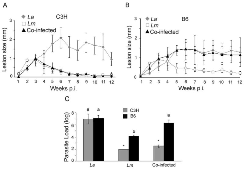
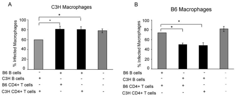
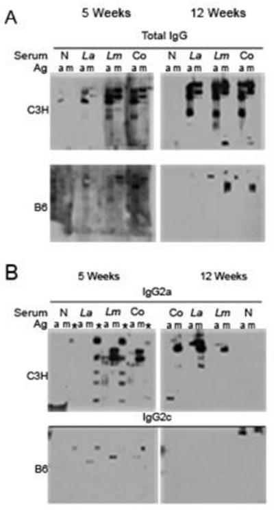
Infection of C3HeB/FeJ and C57BL/6 mice with Leishmania major stimulates a healing cell-mediated immune response, while Leishmania amazonensis infection leads to chronic disease. Here we show C3HeB/FeJ mice co-infected with both species of Leishmania heal, while co-infected C57BL/6 mice do not. Using an in vitro killing assay we determined B cells from infected C57BL/6 mice are ineffective in promoting parasite killing compared with B cells from infected C3HeB/FeJ mice. Furthermore, infected C57BL/6 mice produce less antigen-specific antibodies compared with infected C3HeB/FeJ mice. These findings suggest B cells play a required role in the cell-mediated immune response against L. amazonensis.
2009 Australian Society for Parasitology Inc. Published by Elsevier Ltd. All rights reserved.
Figures
Similar articles
-
An in vitro model of antibody-enhanced killing of the intracellular parasite Leishmania amazonensis.PLoS One. 2014 Sep 5;9(9):e106426. doi: 10.1371/journal.pone.0106426. eCollection 2014. PLoS One. 2014. PMID: 25191842 Free PMC article.
-
Promotion of a functional B cell germinal center response after Leishmania species co-infection is associated with lesion resolution.Am J Pathol. 2012 May;180(5):2009-17. doi: 10.1016/j.ajpath.2012.01.012. Epub 2012 Mar 17. Am J Pathol. 2012. PMID: 22429963 Free PMC article.
-
Protection of C3HeB/FeJ mice against Leishmania amazonensis challenge after previous Leishmania major infection.Am J Trop Med Hyg. 2004 Oct;71(4):407-11. Am J Trop Med Hyg. 2004. PMID: 15516635
-
The Paradox of a Phagosomal Lifestyle: How Innate Host Cell-Leishmania amazonensis Interactions Lead to a Progressive Chronic Disease.Front Immunol. 2021 Sep 7;12:728848. doi: 10.3389/fimmu.2021.728848. eCollection 2021. Front Immunol. 2021. PMID: 34557194 Free PMC article. Review.
-
A detrimental role for IgG and FcgammaR in Leishmania mexicana infection.Immunol Res. 2008;42(1-3):197-209. doi: 10.1007/s12026-008-8074-5. Immunol Res. 2008. PMID: 19002608 Review.
Cited by
-
CD4 T cell activation by B cells in human Leishmania (Viannia) infection.BMC Infect Dis. 2014 Feb 25;14:108. doi: 10.1186/1471-2334-14-108. BMC Infect Dis. 2014. PMID: 24568275 Free PMC article.
-
Characterization of the B cell response to Leishmania infection after anti-CD20 B cell depletion.Int J Clin Exp Pathol. 2015 Jun 1;8(6):6192-202. eCollection 2015. Int J Clin Exp Pathol. 2015. PMID: 26261496 Free PMC article.
-
An in vitro model of antibody-enhanced killing of the intracellular parasite Leishmania amazonensis.PLoS One. 2014 Sep 5;9(9):e106426. doi: 10.1371/journal.pone.0106426. eCollection 2014. PLoS One. 2014. PMID: 25191842 Free PMC article.
-
Effect of Local Administration of Meglumine Antimoniate and Polyhexamethylene Biguanide Alone or in Combination with a Toll-like Receptor 4 Agonist for the Treatment of Papular Dermatitis due to Leishmania infantum in Dogs.Pathogens. 2023 Jun 10;12(6):821. doi: 10.3390/pathogens12060821. Pathogens. 2023. PMID: 37375511 Free PMC article.
-
Promotion of a functional B cell germinal center response after Leishmania species co-infection is associated with lesion resolution.Am J Pathol. 2012 May;180(5):2009-17. doi: 10.1016/j.ajpath.2012.01.012. Epub 2012 Mar 17. Am J Pathol. 2012. PMID: 22429963 Free PMC article.
References
-
- Gonzalez-Lombana CZ, Santiago HC, Macedo JP, Seixas VA, Russo RC, Tafuri WL, Afonso LC, Vieira LQ. Early infection with Leishmania major restrains pathogenic response to Leishmania amazonensis and parasite growth. Acta Trop. 2008;106:27–38. - PubMed
-
- Hjelm F, Carlsson F, Getahun A, Heyman B. Antibody-Mediated Regulation of the Immune Response. Scand J Immunol. 2006;64:177–184. - PubMed
-
- Hulett MD, Hogarth PM. Molecular basis of Fc receptor function. Adv Immunol. 1994;57:1–127. - PubMed
-
- Ji J, Sun J, Qi H, Soong L. Analysis of T helper cell responses during infection with Leishmania amazonensis. Am J Trop Med Hyg. 2002;66:338–345. - PubMed
Publication types
MeSH terms
Grants and funding
LinkOut - more resources
Full Text Sources
