. 2010 Feb 26;396(3):510-27.
doi: 10.1016/j.jmb.2009.12.003. Epub 2009 Dec 11.

Light activation of rhodopsin: insights from molecular dynamics simulations guided by solid-state NMR distance restraints

Affiliations

Light activation of rhodopsin: insights from molecular dynamics simulations guided by solid-state NMR distance restraints

Viktor Hornak et al. J Mol Biol. .

Abstract

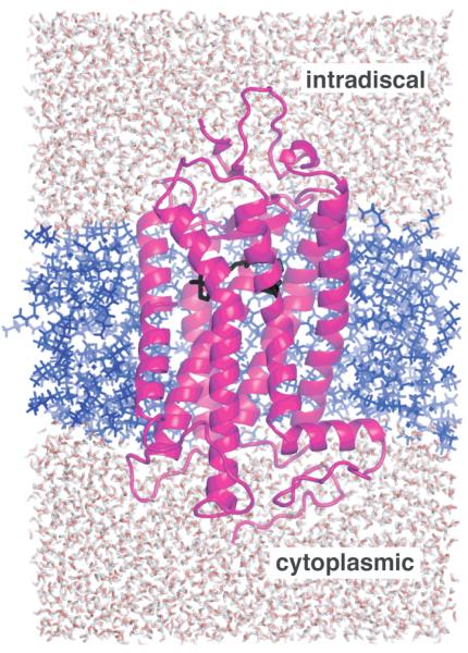
Structural restraints provided by solid-state NMR measurements of the metarhodopsin II intermediate are combined with molecular dynamics simulations to help visualize structural changes in the light activation of rhodopsin. Since the timescale for the formation of the metarhodopsin II intermediate (>1 ms) is beyond that readily accessible by molecular dynamics, we use NMR distance restraints derived from 13C dipolar recoupling measurements to guide the simulations. The simulations yield a working model for how photoisomerization of the 11-cis retinylidene chromophore bound within the interior of rhodopsin is coupled to transmembrane helix motion and receptor activation. The mechanism of activation that emerges is that multiple switches on the extracellular (or intradiscal) side of rhodopsin trigger structural changes that converge to disrupt the ionic lock between helices H3 and H6 on the intracellular side of the receptor.


Figures

Fig. 1
Fig. 1
Crystal structure of rhodopsin. The photoreactive 11-cis retinal chromophore (red) is buried within the bundle of seven TM helices on the extracellular (or intradiscal) side of the receptor. Transmembrane helices H1-H4 (gray) form a rigid framework that is stabilized by tight packing mediated by group conserved amino acids and hydrogen bonding interactions. Guided MD simulations are used to characterize the motion of TM helices H5 (green), H6 (blue) and H7 (purple) upon isomerization of the retinal and deprotonation of the retinal – Lys296 Schiff base linkage.
Fig. 2
Fig. 2
Trajectory of the C20 methyl group and Schiff base proton upon retinal isomerization. (A) The packing of residues around the retinal chromophore in the crystal structure of rhodopsin is visualized by calculating a cavity surface for the binding site. The shape of this cavity likely determines the motion of the C19 and C20 methyl groups upon isomerization. The C19 methyl group is tightly packed against Ile189 and Tyr191, whereas a void exists above the retinal C20 methyl group in the direction of the EL2 loop. The view in this panel is from the β-ionone ring end of the retinal. (B) Structure of the retinal binding site in rhodopsin highlighting the position of the retinal in rhodopsin (red) and in a model from MD simulations immediately after isomerization (orange). (C) and (D) present views of the retinal binding site in rhodopsin viewed from the Schiff base end of the retinal highlighting the position, orientation and interactions of the retinal SB nitrogen. (C) In rhodopsin, the SB proton is oriented toward the extracellular side of the receptor and interacts with Glu113 (H3) and Ser186 (EL2). (D) In the guided MD simulations of Meta II, the electron pair on the SB nitrogen has rotated to a more hydrophobic environment and is pointing toward the cytoplasmic side of the protein.
Fig. 3
Fig. 3
Two-dimensional residue map of close (< 6 Å) interactions near the retinal-binding site determined by 2D DARR NMR distance measurements in rhodopsin and Meta II. There are three main types interactions shown: 1) retinal-protein interactions observed in both rhodopsin and Meta II (blue dotted lines), 2) interactions that are observed in rhodopsin and are lost upon conversion to Meta II (orange dotted lines) and 3) interactions that are observed only in Meta II (broken magenta lines). See also Table 1.
Fig. 4
Fig. 4
Displacement of EL2 upon rhodopsin activation. (A) The β4 strand of EL2 forms a lid of the retinal binding site and is connected to transmembrane helix H5. The rhodopsin crystal structure (gray) is superimposed on the Meta II model (orange) obtained from the guided MD simulations to illustrate the displacement of EL2 needed to satisfy the NMR restraints. (B) and (C) present space filling models of the retinal binding site highlighting the packing of the C20 and C19 methyl groups, respectively. (B) The retinal C20 methyl group is in contact with Trp265 and Tyr268 on H6. Clockwise rotation of the C20 methyl group upon isomerization changes its orientation toward EL2. (C) The C19 methyl group packs against Thr118 on H3, Tyr268 on H6 and Ile189 and Tyr191 on EL2. Counterclockwise rotation of the C19 methyl group would result in a steric clash with Tyr191 and Tyr268, and may be responsible for a shift in the hydrogen bonding interactions involving EL2.
Fig. 5
Fig. 5
Hydrogen bonding of His211 in rhodopsin (A) and Meta II (B). Meta II formation is associated with a change in the hydrogen bonding network centered on His211. In rhodopsin, the backbone carbonyl of His211 forms a hydrogen bond with the side chain of Glu122, while the imidazole side chain of His211 interacts with Trp126. A shift in the position of the retinal β-ionone ring upon activation disrupts the interaction between the backbone carbonyl of His211 and Glu122, and a new hydrogen bond forms with the imidazole side chain δ-nitrogen.
Fig. 6
Fig. 6
Hydrogen bonding changes involving EL2. (A) Crystal structure of rhodopsin highlighting the hydrogen bonding network centered on Glu181. Glu181 is hydrogen bonded (dotted blue lines) to Tyr192 and Tyr191 on EL2 and Tyr268 on H6. Tyr191 is also hydrogen bonded to Tyr268 on H6. Black dotted lines highlight the close interactions between Tyr268 and the retinal C19/C20 methyl groups, and between Tyr191 and the retinal C19 methyl group. (B) The guided MD simulations indicate that retinal isomerization and displacement of EL2 upon activation leads to a rearrangement of the hydrogen bonding network involving Glu181 on EL2. The Glu181 side chain remains hydrogen bonded to Tyr191, Tyr192 and Tyr268 in Meta II. However, Tyr191 is now hydrogen bonded to both Tyr192 and Tyr268. The Meta II model also shows that the retinal C19 methyl group is no longer packed against Tyr191 or Tyr268, although Tyr268 is still in close contact with the retinal C20 methyl group.
Fig. 7
Fig. 7
Coupling of retinal isomerization and displacement of EL2 to motion of H7. (A) The extracellular end of H7 contacts EL2 from Pro285 to Ala292. The H5-EL2 segment is presented in blue and EL3-H7 is presented in grey. In rhodopsin, Met288 on H7 is packed against Glu181 on EL2 and Pro285 on EL3 is packed against Gly182 on EL2. The displacement of EL2 upon retinal isomerization and deprotonation of the retinal SB linkage may allow EL3 and H7 to shift into their active conformations. (B) A view of the water-mediated hydrogen bonding network involving H7. The network extends from Trp265 (red; on H6) to Phe313 (green; on H8) through the conserved NPxxY (Asn302…Tyr306) on H7. Blue filled circles represent structural water. Positions of the all-trans retinal and Trp265 from guided MD simulations are shown in orange. In rhodopsin, Trp265 is packed between the side chain of Ala295 on H7 and the retinal β-ionone ring. The Meta II model shows a movement of the Trp265 side chain toward the extracellular side of the receptor away from the water cluster surrounding Asn302 (H7). (C) and (D) present a view of the protein pocket surrounding Asn302 on H7 in rhodopsin. One side of the pocket containing Asn302 is hydrophobic and composed of Leu76, Leu79, Ala124, Leu131 and Leu128. The other side of the pocket is hydrophilic containing Asn55 and Asp83. (C) In the Meta II model from guided MD simulations, Asn302 (orange) shifts toward and hydrogen bonds with Asp83. (D) An overlap of the rhodopsin (grey) and opsin (orange) structures shows a large movement of the side chain of Tyr306 on H7 from its position in rhodopsin to its new position facing H6 in opsin, which was previously occupied by the Met257 side chain on H6. The side chain of Tyr306 is now packed against the surrounding leucine residues on H3 and H2. Asn302 in opsin does not move significantly relative to its position in rhodopsin. (E) and (F) presents the interaction of amino acids around the conserved ERY motif on H3 in rhodopsin and opsin respectively.
Fig. 8
Fig. 8
Working model of rhodopsin activation. Retinal isomerization leads to the strain in retinal binding site; all-trans retinal is not accommodated in the binding site in the dark. There are several studies probing the sequence of structural changes following isomerization. In detergent, Hofmann and coworkers, have shown that SB deprotonation precedes the motion of H6 and proton uptake by Glu134. However, changes in the IR bands of Glu122 already accompany the formation of Meta I and the retinal SB in Meta I is accessible to hydroxylamine. Together these observations suggest that retinal isomerization first leads to displacement of EL2 and (at least limited) motion of H5 prior to deprotonation. Deprotonation of the SB then allows the retinal to shift into an active position that either guides or drives rotation of Trp265 and H6. This sequence of events is consistent with kinetic FTIR measurements indicating that there may be 15-20% deprotonation of the retinal Schiff base before changes in the IR bands between 1755 and 1740 cm−1 associated with Asp83 and Glu122. Rotation of Trp265 side chain towards EL2 disrupts the network of hydrogen bonding interactions involving the NPxxY motif on H7 and responsible for maintaining the inactive state of the receptor. The outward rotation of H6 is accompanied by the rotation of Tyr223 and Tyr306 into the H3-H6 interface. These residues face Met257 and hydrogen bond with Arg135, disrupting the ionic lock.
Fig. 9
Fig. 9
Computational model used in the MD simulations. Rhodopsin (magenta) is embedded in an octane bilayer (blue) and immersed in a box of water (red/white) which is ~32 Å on each side. The retinal chromophore (black) is close to extracellular side of the receptor (top).

Similar articles

Cited by

References

    1. Menon ST, Han M, Sakmar TP. Rhodopsin: Structural basis of molecular physiology. Physiol. Rev. 2001;81:1659–1688. - PubMed
    1. Schwartz TW, Frimurer TM, Holst B, Rosenkilde MM, Elling CE. Molecular mechanism of 7TM receptor activation - A global toggle switch model. Annu. Rev. Pharmacol. Toxicol. 2006;46:481–519. - PubMed
    1. Kobilka B, Schertler GFX. New G-protein-coupled receptor crystal structures: insights and limitations. Trends Pharmacol. Sci. 2008;29:79–83. - PubMed
    1. Palczewski K, Kumasaka T, Hori T, Behnke CA, Motoshima H, Fox BA, et al. Crystal structure of rhodopsin: A G protein-coupled receptor. Science. 2000;289:739–745. - PubMed
    1. Okada T, Sugihara M, Bondar AN, Elstner M, Entel P, Buss V. The retinal conformation and its environment in rhodopsin in light of a new 2.2 Å crystal structure. J. Mol. Biol. 2004;342:571–583. - PubMed

Publication types