. 2010 Feb 10;28(6):1454-62.
doi: 10.1016/j.vaccine.2009.11.073. Epub 2009 Dec 8.

Immunization with the attenuated plasmidless Chlamydia trachomatis L2(25667R) strain provides partial protection in a murine model of female genitourinary tract infection

Affiliations

Immunization with the attenuated plasmidless Chlamydia trachomatis L2(25667R) strain provides partial protection in a murine model of female genitourinary tract infection

Norma Olivares-Zavaleta et al. Vaccine. .

Abstract

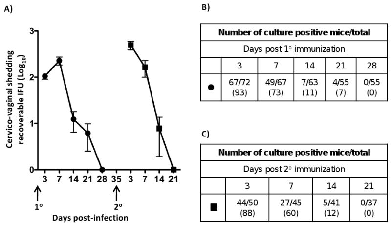
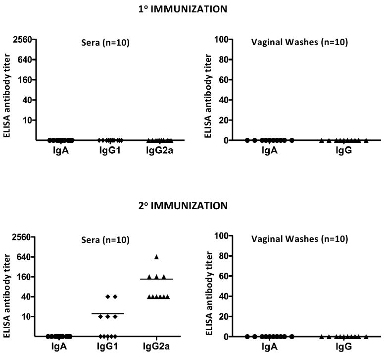
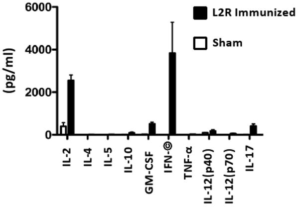
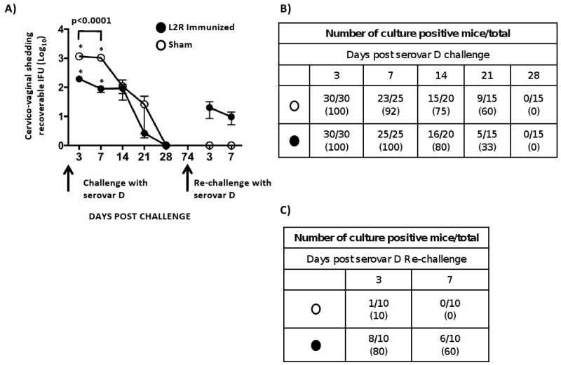
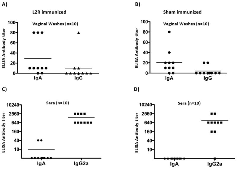
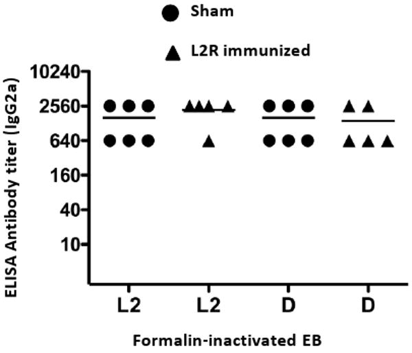
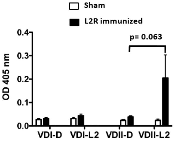
Here we report on the safety, immunogenicity, and vaccine efficacy of the naturally occurring plasmid-free attenuated Chlamydia trachomatis L2-25667R (L2R) strain in a murine infection model. Intravaginal immunization induced both chlamydial specific serum antibody and systemic CD4(+) Th1 biased immune responses but failed to induce local IgA antibodies. Immunization induced no pathological changes in the urogenital tract. Protective immunity was evaluated by vaginal challenge with a natural occurring non-attenuated plasmid positive C. trachomatis urogenital strain (serovar D). Vaccinated mice were not protected from colonization/infection but exhibited a reduction in infectious burden at early time periods (1-2 weeks) post-challenge. Partial protective immunity did not protect against inflammatory disease. Thus, intravaginal vaccination with the live-attenuated L2R stain is safe, induces a systemic antibody and CD4(+) Th1 biased immune response, but its protective efficacy is limited to reducing chlamydial burden at early time periods post-infection.


Figures

Figure 1
Figure 1. Immunization of C3H/HeJ mice with C. trachomatis L2R
A) A group of 72 progesterone treated female C3H/HeJ mice were infected intravaginally (1° immunization) with 4×10⁷ IFU/mouse (10 ID50). Control mice were sham infected with SPG only. Chlamydial cervico-vaginal shedding was monitored by swabbing the vaginal vault and performing cultures on monolayers of HeLa 299 cells at 3, 7, 14, 21 and 28 days post infection (dpi). Infectious loads in cervico-vaginal swabs were determined following immunostaining of methanol fixed cells and calculating inclusion forming units (IFU). Fifty mice received a second infection (2° immunization) at 35 dpi, 7 days after clearance of the 10 infection. The filled circles and squares represent the mean number of recoverable IFUs ± SEM in 10 and 20 infected mice, respectively. The number of culture-positive mice per total mice at each culture time point during 1° L2R infection (B), or 2° (C). The numbers in parenthesis represent the percent of infectivity.
Figure 2
Figure 2. Systemic and local antibody responses in C3H/HeJ mice after primary and secondary L2R immunization
Sera and local antibody class and isotype-specific anti-chlamydial responses were assayed 30 and 25 days post 1° and 2 ° L2R immunizations, respectively. Antibody titers were expressed as the highest dilution giving an absorbance of at least 0.3 or 3 times that of the control sample (consistently < 0.1). C. muridarum (MoPn) convalescent sera and vaginal washes tested against MoPn EBs were used as a positive control (data not shown). Ten mice were tested for each immunization group and the symbols denote the response of individual mice. Bars denote the mean antibody titer. Note that mice did not generate a chlamydial specific antibody following clearance of a primary immunization. In contrast, all L2R immunized mice produced a predominant IgG2a antibody response following resolution of the secondary immunization. Chlamydial-specific IgA or IgG antibodies were not detected in the vaginal washes of L2R infected mice following resolution of either the 1° or 2° immunization.
Figure 3
Figure 3. Splenic CD4+ T cells from L2R immunized mice produce a dominant Th1 biased cytokine response
The in vitro production of cytokines by splenic CD4+ T cells from sham or L2R immunized mice was assayed two weeks following the resolution of the 2° L2R infection. The CD4+ T cells were re-stimulated in vitro with UV-inactivated serovar D EB and the supernatants were collected after 72 h of culture for determining the cytokine concentration. The black bars represent the mean ± standard deviation of four individual L2R treated mice tested in quadruplicate. The white bars represent the results of two individual sham infected mice and expressed as the mean ± standard deviation. There was a significant production of IFN-γ by chlamydial pulsed CD4+ T cells.
Figure 4
Figure 4. Histopathological evaluation of genital tract tissues from C3H/HeJ mice immunized with L2R
Five mice were euthanized 14 days following a 2° L2R infection, C. trachomatis serovar D infection, or sham infection (SPG). The entire genital tract was removed for histopathological evaluation. Micrographs of histological sections of the cervical region from L2R immunized mice (A, B); Serovar D infected (C, D) and sham infected (E, F) are shown. Magnification 40×: (A, C, E) and 400× (B, D, F). Note that there were no pathological changes in L2R or sham infected mice. In contrast, infection with C. trachomatis serovar D produced a moderate sub-mucosal infiltrate presented as multifocal aggregates composed of lymphocytes, macrophages and plasma cells. Occasionally, low numbers of these cellular aggregates were present within the epithelium lining the lumen. Similarly, smaller and less densely arranged inflammatory aggregates were noted in the muscular layers.
Figure 5
Figure 5. Immunostaining of genital tract tissue following immunization or infection with chlamydiae
Chlamydial inclusions in genital tissues immunized with L2R (A and B) or infected with serovar D (C and D). (A) L2R inclusions appeared as focal aggregates in the submucosa of the cervix; (B) 1000× magnification from panel A. In contrast, serovar D inclusions were found exclusively within epithelial cells of the distal cervix or uterine horn (C); (D) 1000× magnification from panel C. Mock infected mice tissues were negative by immunostaining. No immunostaining was observed in tissues with a negative control anti-MOMP monoclonal antibody 33b specific to C. muridarum.
Figure 6
Figure 6. Protective efficacy of L2R immunized mice against C. trachomatis serovar D challenge
Thirty sham (O) and 30 L2R immunized mice (●) were intravaginally challenged with 4.3×10⁴ IFU/mouse (10 ID50) of serovar D. We chose serovar D as the challenge strain because it is one of the most prevalent serovars associated with human urogenital infection and it is antigenically related to L2. (A) Vaginal swabs from serovar D challenged mice were cultured for recoverable IFU on 3, 7, 10, 14 and 28 dpi. Forty-six days after mice cleared their primary D infection they were re-challenged a second time with serovar D. Each symbol represents the mean number of recoverable IFUs ± SEM. Note that L2R immunized mice were not protected from serovar D colonization. There was a significant difference between L2R and sham immunized mice in the burden of D shedding at days 3 and 7 post-challenge (p<0.0001). However, the burden and duration of infection did not differ between the groups at day 14-28. Interestingly, sham immunized mice that had been infected and then re-challenge with serovar D exhibited near sterilizing immunity. In contrast, L2R immunized and serovar D challenged mice were less protected. All mice in this group were infected following challenge. The numbers of culture positive mice for each group of re-challenged mice are shown in (B) serovar D primary challenge, and (C) serovar D secondary challenge.
Figure 7
Figure 7. Systemic and local antibody responses in sham and L2R immunized mice after challenge with serovar D
Blood and vaginal washes were collected from ten sham and L2R immunized and serovar D challenged mice 28 days after intravaginal challenge with serovar D (Figure 6). Serum and vaginal IgA and IgG antibody responses against serovar D EBs were determined by ELISA. The symbols denote the antibody response of individual mice. Bars denote the mean antibody titer.
Figure 8
Figure 8. C. trachomatis specificity of the antibody response after sequential infection with L2R and serovar D
Individual sera from L2R or sham-immunized and serovar D challenged mice were assayed by ELISA against L2 and D EB antigens. Chlamydial-specific IgG2a antibody was analyzed against these antigens and the titers were expressed as the reciprocal of sera dilutions giving an absorbance of 0.3 or greater. Absorbance values for pre-immune sera were 0.1 or less. Note that sera titers were similar against L2 and D EBs.
Figure 9
Figure 9. Molecular specificity of the MOMP antibody response after sequential infection with L2R and serovar Din L2R and sham-immunized mice
The sera of five sham or L2R immunized and serovar D challenged mice analyzed in Figure 8 was tested by ELISA against synthetic peptide antigens corresponding to MOMP VDI and VDII of serovars D and L2. Previous studies have epitope mapped serovar D and L2 specific epitopes to VDII [18]. The VDs represent exposed loop structures on intact EBs and serovar-specific epitopes located in the loops are immunodominant and targets of neutralizing antibodies (Each bar represents the OD405nm mean ± SEM from each experimental group. Absorbance values for pre-immune sera were 0.1 or less.

References

    1. Brunham RC, Rey-Ladino J. Immunology of Chlamydia infection: implications for a Chlamydia trachomatis vaccine. Nat Rev Immunol. 2005;(2):149–61. - PubMed
    1. Eko FO, He Q, Brown T, McMillan L, Ifere GO, Ananaba GA, et al. A novel recombinant multisubunit vaccine against Chlamydia. J Immunol. 2004;(5):3375–82. - PubMed
    1. Murthy AK, Chambers JP, Meier PA, Zhong G, Arulanandam BP. Intranasal vaccination with a secreted chlamydial protein enhances resolution of genital Chlamydia muridarum infection, protects against oviduct pathology, and is highly dependent upon endogenous gamma interferon production. Infect Immun. 2007;(2):666–76. - PMC - PubMed
    1. Su H, Parnell M, Caldwell HD. Protective efficacy of a parenterally administered MOMP-derived synthetic oligopeptide vaccine in a murine model of Chlamydia trachomatis genital tract infection: serum neutralizing IgG antibodies do not protect against chlamydial genital tract infection. Vaccine. 1995;(11):1023–32. - PubMed
    1. Dong-Ji Z, Yang X, Shen C, Lu H, Murdin A, Brunham RC. Priming with Chlamydia trachomatis major outer membrane protein (MOMP) DNA followed by MOMP ISCOM boosting enhances protection and is associated with increased immunoglobulin A and Th1 cellular immune responses. Infect Immun. 2000;(6):3074–8. - PMC - PubMed

Publication types

MeSH terms