. 2009 Dec;43(8):603-18.
doi: 10.1016/j.alcohol.2009.10.006.

The effects of acute and chronic ethanol exposure on presynaptic and postsynaptic gamma-aminobutyric acid (GABA) neurotransmission in cultured cortical and hippocampal neurons

Affiliations

The effects of acute and chronic ethanol exposure on presynaptic and postsynaptic gamma-aminobutyric acid (GABA) neurotransmission in cultured cortical and hippocampal neurons

Rebekah L Fleming et al. Alcohol. 2009 Dec.

Abstract

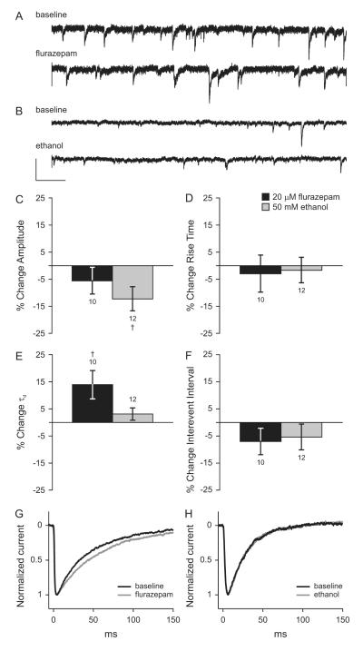
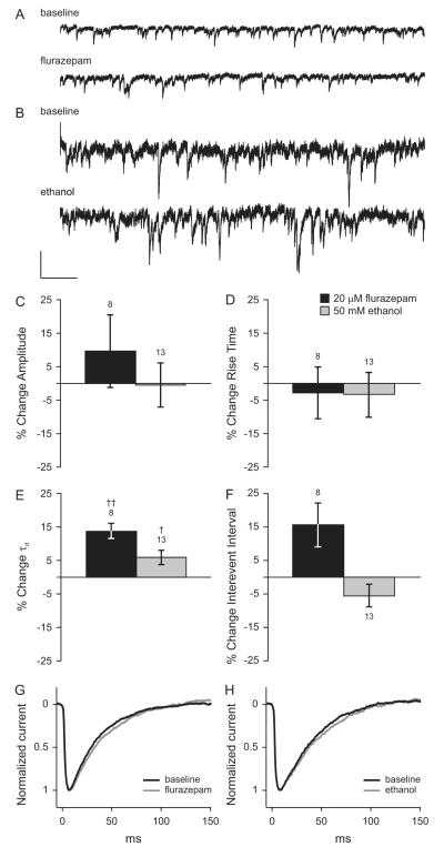
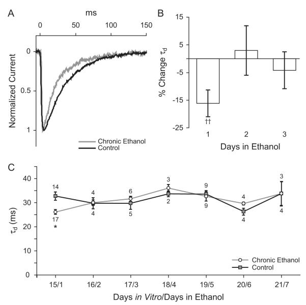
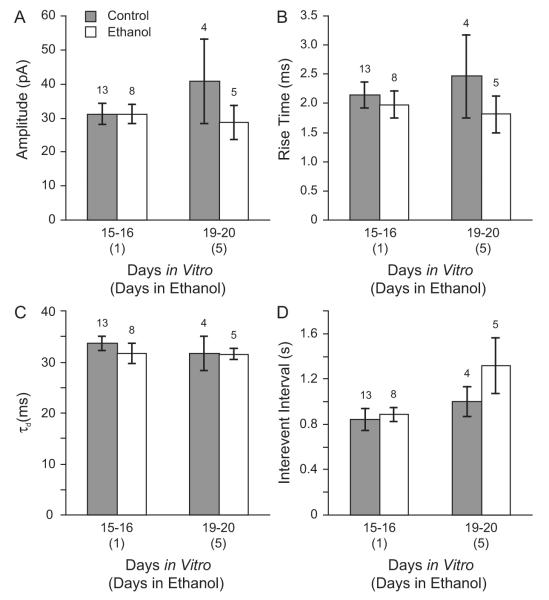
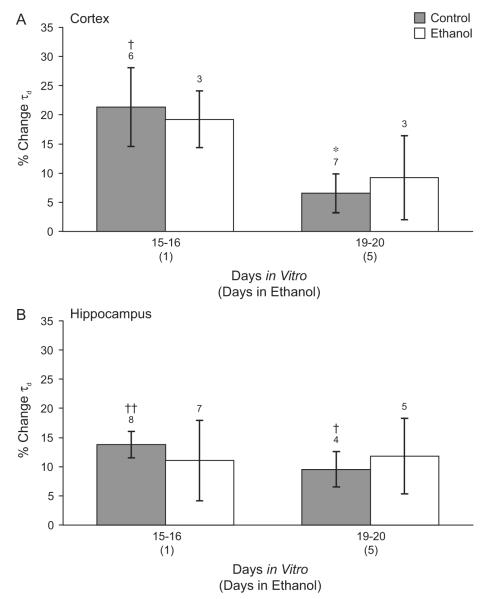
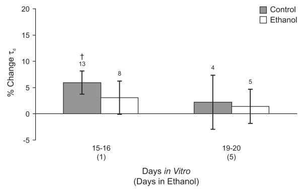
Decades after ethanol was first described as a gamma-aminobutyric acid (GABA) mimetic, the precise mechanisms that produce the acute effects of ethanol and the physiological adaptations that underlie ethanol tolerance and dependence remain unclear. Although a substantial body of evidence suggests that ethanol acts on GABAergic neurotransmission to enhance inhibition in the central nervous system, the precise mechanisms underlying the physiological effects of both acute and chronic ethanol exposure are still under investigation. We have used in vitro ethanol exposure followed by recording of miniature inhibitory postsynaptic currents (mIPSCs) to determine whether acute or chronic ethanol exposure directly alters synaptic GABA(A) receptor (GABA(A)R) function or GABA release in cultured cortical and hippocampal neurons. Acute ethanol exposure slightly increased the duration of mIPSCs in hippocampal neurons but did not alter mIPSC kinetics in cortical neurons. Acute ethanol exposure did not change mIPSC frequency in either hippocampal or cortical neurons. One day of chronic ethanol exposure produced a transient decrease in mIPSC duration in cortical neurons but did not alter mIPSC kinetics in hippocampal neurons. Chronic ethanol exposure did not change mIPSC frequency in either hippocampal or cortical neurons. Chronic ethanol exposure also did not produce substantial cross-tolerance to a benzodiazepine in either hippocampal or cortical neurons. The results suggest that ethanol exposure in vitro has limited effects on synaptic GABA(A)R function and action potential-independent GABA release in cultured neurons and that ethanol exposure in cultured cortical and hippocampal neurons may not reproduce all the effects that occur in vivo and in acute brain slices.


Figures

Figure 1
Figure 1. Effects of flurazepam and ethanol on mIPSCs in cultured cortical neurons
Representative tracings of voltage-clamp recordings from cultured cortical neurons under baseline conditions and during acute exposure to 20 μM flurazepam (A) or 50 mM ethanol (B) in the extracellular solution. A and B are shown at the same scale. Scale bars: x = 1s, y = 10 pA. Mean changes in mIPSC amplitude (C), rise time (D), decay time constant (E), and interevent interval (F) during exposure to 20 μM flurazepam (black bars) or 50 mM ethanol (gray bars) relative to the no-drug baseline. † p < 0.05 by paired t-test. Scaled and averaged mIPSCs under baseline conditions and during exposure to flurazepam (G) or ethanol (H).
Figure 2
Figure 2. Effects of flurazepam and ethanol on mIPSCs in cultured hippocampal neurons
Representative tracings of voltage-clamp recordings from cultured hippocampal neurons aged 15-16 days in vitro under baseline conditions and during acute exposure to 20 μM flurazepam (A) or 50 mM ethanol (B) in the extracellular solution. A and B are shown at the same scale. Scale bars: x = 1s, y = 10 pA. Mean changes in average mIPSC amplitude (C), rise time (D), decay time constant (E), and interevent interval (F) during exposure to 20 μM flurazepam (black bars) or 50 mM ethanol (gray bars) relative to the no-drug baseline are shown. † p < 0.05, †† p < 0.01 by paired t-test. Scaled and averaged mIPSCs under baseline conditions and during exposure to flurazepam (G) or ethanol (H).
Figure 3
Figure 3. Effect of chronic in vitro ethanol exposure on mIPSCs in cortical neurons
Cultured neurons aged 14 days in vitro were exposed to 50 mM ethanol in the tissue culture medium for 1 to 7 days. A. Averaged mIPSCs for a control neuron and a neuron exposed to 50 mM ethanol for 24 hours. B. Percent change in average mIPSC decay time constant calculated relative to age and culture matched controls for neurons exposed to 50 mM ethanol for 1,2, or 3 days. †† p < 0.005 compared to controls by paired t-test. C. Absolute values for mIPSC decay time constants in ethanol treated neurons and controls. One day of ethanol treatment decreased mIPSC τd from 32.7±1.7 ms in control cells to 26.2±1.0 ms in ethanol treated cells. † p < 0.05 by Holm-Sidak posthoc test.
Figure 4
Figure 4. Chronic ethanol treatment does not alter synaptic GABAAR function in cultured hippocampal neurons
Beginning at 14 or 15 days in vitro, hippocampal neurons were cultured in the absence (gray bars) or presence (open bars) of 50 mM ethanol in the tissue culture medium. Average mIPSC amplitude (A), rise time (B), decay time constant (C), and interevent interval (D) are shown for age-matched controls and neurons treated with ethanol for 1 to 5 days. Ethanol treatment did not alter mIPSC amplitude, kinetics, or frequency.
Figure 5
Figure 5. Effect of chronic ethanol treatment on the acute effect of benzodiazepine
Both cortical (A) and hippocampal neurons (B) were cultured in the absence (gray bars) or presence (open bars) of 50 mM ethanol in the tissue culture medium. The increase in mIPSC τd during acute application of 20 μM flurazepam in the external solution is shown. (A) Cultured cortical cells 19-20 days in vitro were less sensitive to flurazepam than control neurons 15-16 days in vitro. Chronic ethanol exposure did not alter mIPSC sensitivity to flurazepam at either timepoint. p < 0.05 ANOVA. * p < 0.05 compared to control 15-16 DIV by Holm-Sidak post-hoc test, † p < 0.05 compared to pre-drug baseline by paired t-test. (B) In cultured hippocampal neurons, flurazepam significantly increased mIPSC τd in control cells 15-16 and 19-20 days in vitro. Ethanol treatment for 1 or 5 days did not alter mIPSC flurazepam sensitivity. † p < 0.05, †† p < 0.01 compared to pre-drug baseline by paired t-test.
Figure 6
Figure 6. Effect of chronic ethanol treatment on acute effects of ethanol in cultured hippocampal neurons
Changes in mIPSC τd during acute application of 50 mM ethanol are shown for age-matched control neurons (gray bars) and for hippocampal neurons cultured in the presence of 50 mM ethanol for 1 or 5 days (open bars). Acute application of ethanol increased mIPSC τd in control neurons 15-16 days in vitro but not in older control neurons or ethanol treated neurons. † p < 0.05 by paired t-test comparing acute ethanol exposure to pre-drug baseline.

Similar articles

Cited by

References

    1. Aguayo LG. Ethanol potentiates the GABAA-activated Cl− current in mouse hippocampal and cortical neurons. Eur. J. Pharmacol. 1990;187:127–130. - PubMed
    1. Ariwodola OJ, Weiner JL. Ethanol potentiation of GABAergic synaptic transmission may be self-limiting: role of presynaptic GABA-B receptors. J. Neurosci. 2004;24:10679–10686. - PMC - PubMed
    1. Bailey CD, Brien JF, Reynolds JN. Chronic prenatal ethanol exposure increases GABA(A) receptor subunit protein expression in the adult guinea pig cerebral cortex. J. Neurosci. 2001;21:4381–4389. - PMC - PubMed
    1. Bouron A. Modulation of spontaneous quantal release of neurotransmitters in the hippocampus. Prog. Neurobiol. 2001;63:613–635. - PubMed
    1. Cagetti E, Liang J, Spigelman I, Olsen RW. Withdrawal from chronic intermittent ethanol treatment changes subunit composition, reduces synaptic function, and decreases behavioral responses to positive allosteric modulators of GABAA receptors. Mol. Pharmacol. 2003;63:53–64. - PubMed

Publication types