. 2010 Feb 20;397(2):379-88.
doi: 10.1016/j.virol.2009.11.030. Epub 2009 Dec 9.

The phosphatase Shp2 is required for signaling by the Kaposi's sarcoma-associated herpesvirus viral GPCR in primary endothelial cells

Affiliations

The phosphatase Shp2 is required for signaling by the Kaposi's sarcoma-associated herpesvirus viral GPCR in primary endothelial cells

Thomas Bakken et al. Virology. .

Abstract

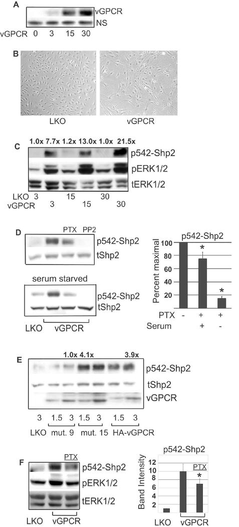
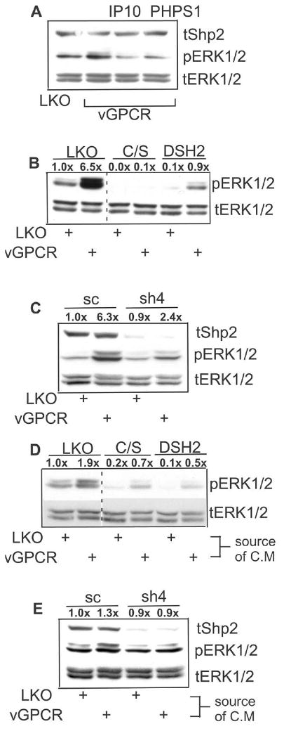
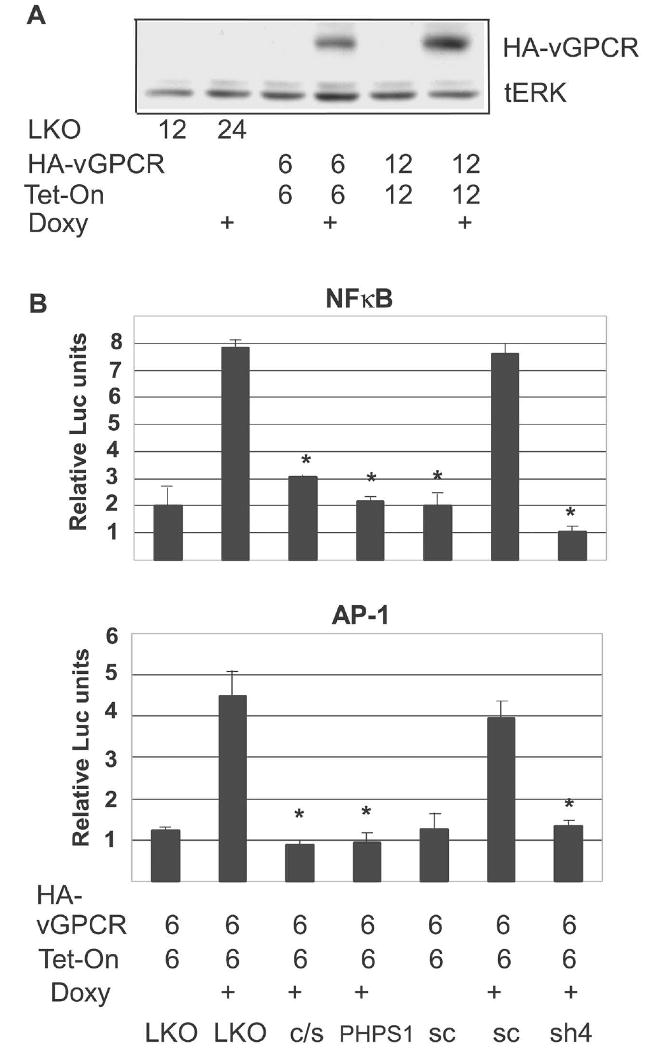
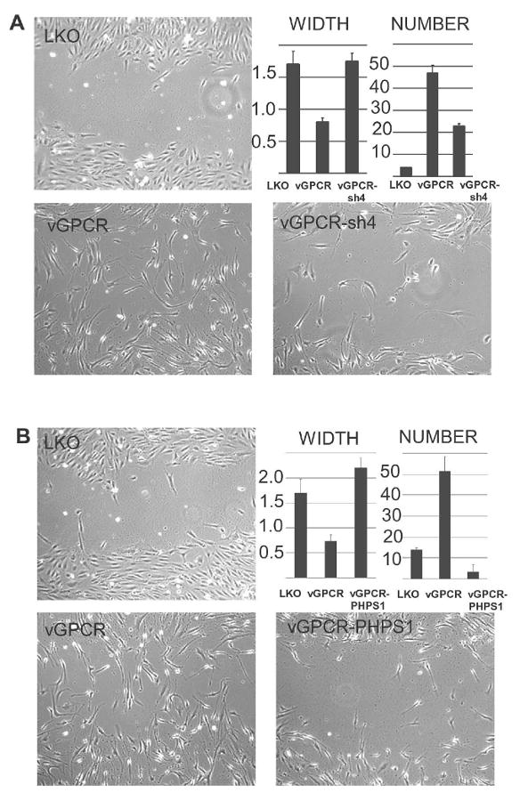
Kaposi's sarcoma-associated herpesvirus (KSHV) is the causative agent of Kaposi's sarcoma (KS), an AIDS-related endothelial cell malignancy that is the most common cancer in central and southern Africa. The KSHV viral G protein-coupled receptor (vGPCR) is a viral oncogene that conveys a survival advantage to endothelial cells and causes KS-like tumors in mouse models. In this study we investigate the role of Shp2, a protein tyrosine phosphatase in vGPCR signaling. Shp2 is vital to many cytokine-induced signaling pathways and is dysregulated in various infections and malignancies. It has also recently been implicated in angiogenesis. We find that vGPCR activity results in phosphorylation of regulatory tyrosines in Shp2 and that in turn, Shp2 is required for vGPCR-mediated activation of MEK, NFkappaB, and AP-1. Furthermore, both genetic and chemical inhibition of Shp2 abrogate vGPCR-induced enhancement of endothelial cell migration. This establishes Shp2 as an important point of convergence of KSHV vGPCR signaling and a potential molecular target in the design of an anti-KSHV therapeutic regimen.


Figures

Figure 1
Figure 1
vGPCR-induced Shp2 phosphorylation is Gαi-independent and Src-dependent, but Erk1/2 independent. HEK 293 cells were transfected with pcDNA3.1+ as control plasmid or with pcKSHV-vGPCR (A,C) or Gq(209L) plasmid (B) as shown. (A-C) After 48 hours protein lysates were Western blotted for phospho-542-Shp2; membranes were then stripped and re-probed for total Shp2. (A,B) Inhibitors were added 36 hours post-transfection where indicated. PTX (pertussis toxin, 100 ng/ml), PP2 (5 mM), PD (PD98059, 25 μM). (C) Cells were co-transfected with Src-DN plasmid where noted. (D) Cells were transfected with scrambled shRNA (sc) or shRNA targeted to Src (shSrc). After 48 hours, cells were transfected with vGPCR and protein harvested after 48 additional hours. Western blot was performed for p542-Shp2, stripped and re-probed for total Shp2. Total levels of Src are also shown to verify knockdown.
Figure 2
Figure 2
Shp2 is required for vGPCR activation of MEK, NFκB and AP-1. (A) HEK 293 cells were transfected with pcDNA3.1+ or pKSHV-vGPCR and incubated with NSC-87877 at doses shown overnight. Western blot for phospho-ERK1/2 was then done followed by blot for total ERK1/2. Numbers above bands represent density normalized to control, lane 1. (B) Cells were transfected with pcDNA3.1+ or vGPCR along with the indicated luciferase reporter construct and a constant amount of β-Gal plasmid. 25 μm NSC-87877 (grey bars) was added 5 hours later. Luciferase activity was assessed 24 hours post-transfection. Results were normalized to β–GAL levels. (C) HEK 293 were transfected with empty pcDNA3.1+, scrambled shRNA (sc) or one of three different candidate shRNA plasmid constructs (Open Biosystems) to knock down Shp2 which was then assessed by Western blot. (D) 48 hours after transfection with Shp2 shRNA (sh3), cells were transfected with vGPCR or empty plasmid. After 24 hours protein was harvested, transferred to PVDF which was then cut into three sections to immunoblot for total Shp2, total vGPCR, and phospho-ERK1/2; the latter was stripped and re-probed for total ERK1/2. (E) Shp2 was knocked down with shRNA as in (C), followed by transfection with vGPCR or control plasmid along with the indicated luciferase reporter plasmid and a β–GAL construct for normalization. (F) Cells stably expressing either scrambled shRNA (sc) or Shp2 shRNA (sh3) were subjected to a sub-maximal dose (10 μM) of NSC-87877 overnight and Western blot performed as above. (G) Density of phospho-ERK1/2 bands from 2F was normalized to corresponding total ERK1/2 band; each experimental point was then normalized to the control, lane 1. Bars = S.D. * denotes p≤0.05 by student's t –test.
Figure 3
Figure 3
Lentiviral transduction of vGPCR into BOEC results in a Gαq- and Gαi-dependent increase in phosphorylated form of Shp2. (A) BOEC cells were transduced with lenti-vGPCR at MOI shown. After 48 hours, RIPA lysates were immunoblotted for vGPCR. ‘NS’ designates a non-specific band. (B) Micrograph showing lenti-vGPCR induced change in morphology from cobblestone to spindled. (C) BOEC were transduced with control lentivirus (LKO) or lenti-vGPCR at MOI shown. At 48 hours RIPA lysates were transferred to PVDF which was cut and simultaneously immunoblotted for p542-Shp2, and pERK1/2; blot was stripped and re-probed for total ERK1/2 as a loading control. Numbers above bands represent p542-Shp2 band density normalized to corresponding loading control and then normalized to control, lane 1. (D) BOEC were lenti-transduced with control virus (LKO) or vGPCR at an MOI of 3. Forty eight hours later, cells were serum-starved (bottom panel) or not (top panel). At 60 hours post-transduction PTX (100 nM), or the Src inhibitor PP2 (5 μM) were added and protein lysates harvested and immunoblotted for p542-Shp2 at 72 hours post-transduction. Blots were stripped and re-probed for total Shp2. Graph to right represents p542-Shp2 band densities normalized to loading control and then normalized to peak levels in the corresponding lane 2. Bands were undetectable in PP2 lanes and not represented graphically. (E) BOEC were lenti-transduced with LKO, HA-vGPCR, vGPCR mutant #9 (inactive) or vGPCR mutant #15 (Gq-only), each at two different MOI as shown. After 72 hours cells were harvested for protein and immunoblotting done for p542-Shp2, total Shp2 and vGPCR. Lanes 3, 4 and 7 had most similar amounts of vGPCR construct and were used for densitometry in which p542-Shp2 and vGPCR bands were normalized to tShp2 loading control and then p542-Shp2 normalized to corresponding vGPCR band. Densities were normalized to inactive mutant, lane 3, and fold change shown numerically above bands. (F) Lenti-vGPCR infected BOEC were serum- starved overnight 60 hours post-transduction. 30 minutes before harvesting protein, 100 nM PTX was added where indicated. p542-Shp2 bands were quantified and normalized to tERK1/2 loading control and then to control, lane1, and represented graphically. PTX, pertussis toxin. Bars= S.D. *= p≤0.05
Figure 4
Figure 4
KSHV vGPCR-induced MEK activation in BOEC requires intact Shp2. BOEC cells were lenti-transduced with control virus (LKO) or lenti-vGPCR (MOI 3). At 48 hours RIPA lysates were immunoblotted for phospho-ERK1/2, stripped and re-probed for total ERK1/2. (A) At 36 hours post-transduction, IP-10 (100 nM) or the Shp2 inhibitor PHPS1 (10 μM) was added. (B) 48 hours prior to lenti-vGPCR transduction, BOEC were transduced where shown with control lenti-LKO, lenti-Shp2-c/s, or -DSH2, dominant negatives of Shp2. (C) Experiment performed as in (B) but using scrambled (sc) control versus lenti-sh4. (D) BOEC were transduced with control lentivirus, lenti-Shp2-c/s, or lenti-DSH2 as shown. After 48 hours cells were serum-starved for 17 hours in M199 (0.5% FBS), then exposed for 20 minutes to conditioned medium from BOEC that had been previously transfected with either control LKO lentivirus or lenti-vGPCR. Protein lysates were immunoblotted for pERK1/2 and total ERK1/2. (E) Experiment performed as in (D) but using lenti-sc as control versus lenti-sh4. Densitometry was performed and numbers over bands indicate fold intensity changes as normalized to loading controls and then to control bands.
Figure 5
Figure 5
Shp2 is required for vGPCR-mediated activation of NFκB and AP-1 in BOEC. (A) BOEC were transfected with control virus or with pLVX-Tight-HA-vGPCR (HA-vGPCR) and pLVX-Tet-On (Tet-On) at MOI shown. After 48 hours cells were split equally and doxycycline (2 μg/ml) was added where indicated for an additional 48 hours. Protein lysates were harvested and immunoblotted for HA-vGPCR total ERK1/2 as a loading control. (B) BOEC were co-transduced with lenti-NFκB-luc reporter (top panel) along with pLVX-Tight-HA-vGPCR and pLVX-Tet-On. After 24 hours, cells were split, transduced again with control LKO versus Shp2 dominant-negative lentiviruses, or lenti-scrambled versus lenti-sh4. Doxycycline (doxy) was added to cause vGPCR expression where indicated for 48 hours, after which luciferase assay was performed. PHPS1 (10 μM) was added 17 hours before harvesting where shown. Bottom Panel: Identical to top except lenti-AP1-luc reporter was used. Values were normalized to protein concentration. Bars= S.D. * denotes p≤0.05 compared to maximal activation.
Figure 6
Figure 6
Shp2 is required for vGPCR-induced migration in BOEC. (A) BOEC were lenti- transduced with control virus LKO or vGPCR with and without lenti-sh4 to knockdown Shp2. After 48 hours cells reached confluence and a scratch “wound” was made with a pipette tip and medium changed to low-serum (0.1% FBS) without additional supplements. The width of the scratch and the number of cells migrating back into the wound were quantified using NIH ImageJ, 10 hours post-wounding (width measured in arbitrary units). (B) Shp2 was inhibited by PHPS1 (10 μM) where indicated for 12 hours prior to wounding. Error bars represent S.D. One representative of 3 experiments is shown.

Similar articles

Cited by

References

    1. Adachi M, Fischer EH, Ihle J, Imai K, Jirik F, Neel B, Pawson T, Shen S, Thomas M, Ullrich A, Zhao Z. Mammalian SH2-containing protein tyrosine phosphatases. Cell. 1996;85(1):15. - PubMed
    1. Alonso A, Sasin J, Bottini N, Friedberg I, Osterman A, Godzik A, Hunter T, Dixon J, Mustelin T. Protein tyrosine phosphatases in the human genome. Cell. 2004;117(6):699–711. - PubMed
    1. Arvanitakis L, Geras-Raaka E, Varma A, Gershengorn MC, Cesarman E. Human herpesvirus KSHV encodes a constitutively active G-protein-coupled receptor linked to cell proliferation. Nature. 1997;385(6614):347–50. - PubMed
    1. Bais C, Santomasso B, Coso O, Arvanitakis L, Raaka EG, Gutkind JS, Asch AS, Cesarman E, Gershengorn MC, Mesri EA. G-protein-coupled receptor of Kaposi's sarcoma-associated herpesvirus is a viral oncogene and angiogenesis activator. Nature. 1998;391(6662):86–9. - PubMed
    1. Burshtyn DN, Scharenberg AM, Wagtmann N, Rajagopalan S, Berrada K, Yi T, Kinet JP, Long EO. Recruitment of tyrosine phosphatase HCP by the killer cell inhibitor receptor. Immunity. 1996;4(1):77–85. - PMC - PubMed

Publication types

MeSH terms

Substances