. 2010 Feb 20;397(2):358-68.
doi: 10.1016/j.virol.2009.11.010. Epub 2009 Dec 9.

Ubiquitination of tombusvirus p33 replication protein plays a role in virus replication and binding to the host Vps23p ESCRT protein

Affiliations

Ubiquitination of tombusvirus p33 replication protein plays a role in virus replication and binding to the host Vps23p ESCRT protein

Daniel Barajas et al. Virology. .

Abstract

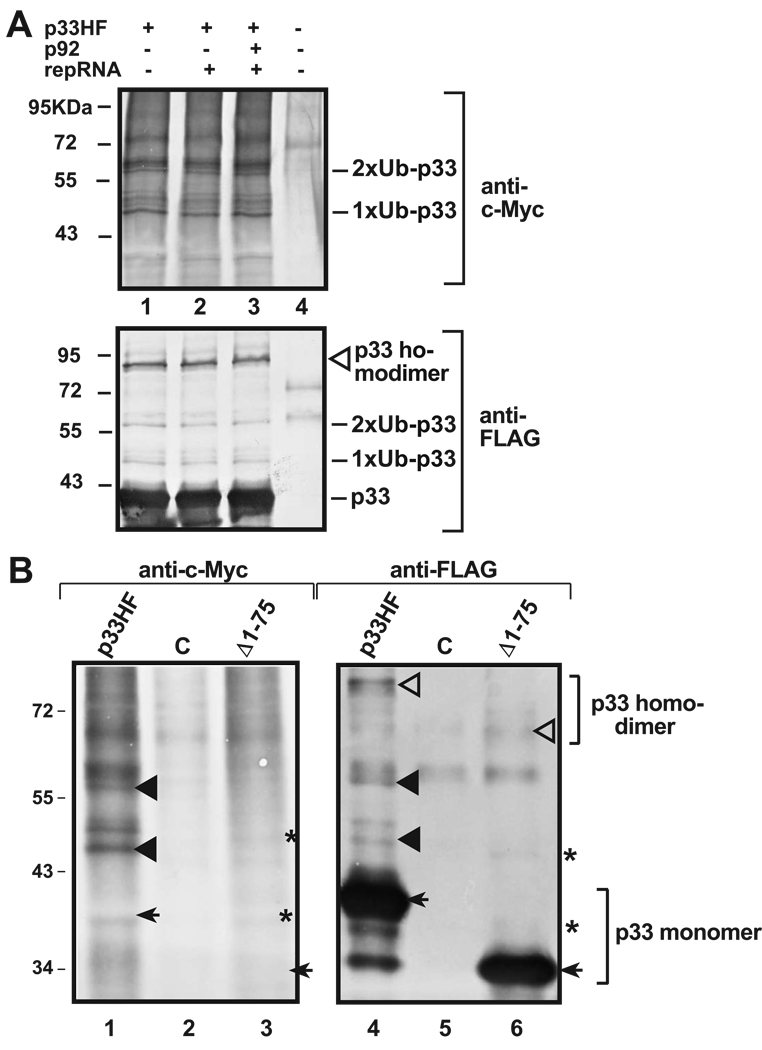
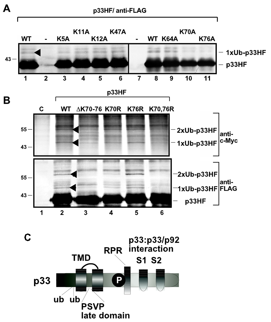
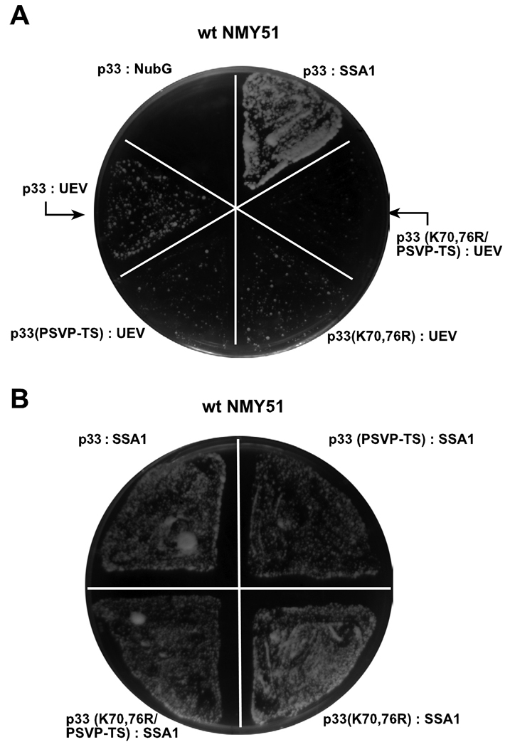
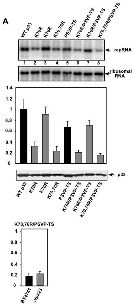
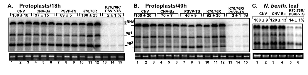
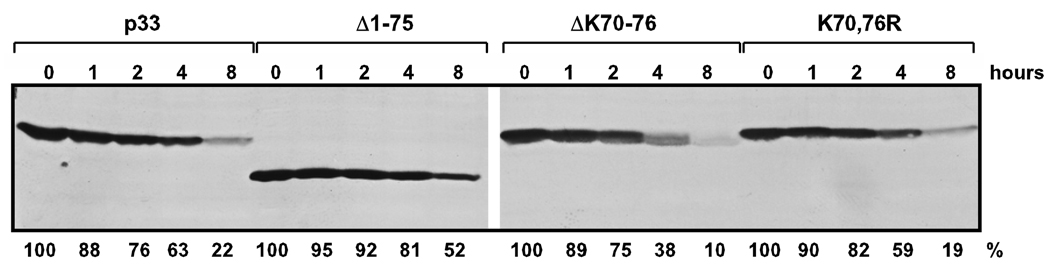
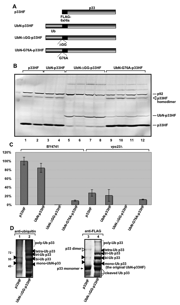
Post-translational modifications of viral replication proteins could be widespread phenomena during the replication of plus-stranded RNA viruses. In this article, we identify two lysines in the tombusvirus p33 replication co-factor involved in ubiquitination and show that the same lysines are also important for the p33 to interact with the host Vps23p ESCRT-I factor. We find that the interaction of p33 with Vps23p is also affected by a "late-domain"-like sequence in p33. The combined mutations of the two lysines and the late-domain-like sequences in p33 reduced replication of a replicon RNA of Tomato bushy stunt virus in yeast model host, in plant protoplasts, and plant leaves, suggesting that p33-Vps23p ESCRT protein interaction affects tombusvirus replication. Using ubiquitin-mimicking p33 chimeras, we demonstrate that high level of p33 ubiquitination is inhibitory for TBSV replication. These findings argue that optimal level of p33 ubiquitination plays a regulatory role during tombusvirus infections.


Figures

Fig. 1
Fig. 1
Ubiquitination of p33 replication protein in yeast. (A) The effect of co-expression of p92pol and repRNA on p33 ubiquitination. The membrane-bound p33HF (tagged with 6×His and FLAG) was purified via FLAG-affinity chromatography after membrane solubilization from yeast co-expressing c-Myc-tagged ubiquitin from a plasmid. The ubiquitinated p33 was detected with Western blot using anti-c-Myc (top panel) or anti-FLAG antibodies (bottom panel), based on the 8 and 16 KDa increase of MW for mono (1×Ub)- and bi (2×Ub)-ubiquitination products of p33. Note that the additional p33-specific bands near the mono- and bi-ubiquitinated bands could be phosphorylated forms (Shapka, Stork, and Nagy, 2005) of the ubiquitinated p33HF. The origin of smear detected with anti-c-Myc antibody is unknown, though they are probably co-purified ubiquitinated host proteins, since the p33-derived products would also be detected by the anti-FLAG antibody (bottom panel). Also, these products are missing in the control sample from yeast expressing 6× His-FLAG peptide from pYC-HF (lane 4). The p33 homodimer is marked with an empty arrowhead. Note that co-expression of TBSV repRNA and/or p92 RdRp protein did not affect the ubiquitination status of p33. (B) Testing the ubiquitination status of an N-terminal deletion mutant of p33 replication protein in yeast. The positions of the deleted amino acids are indicated and the expected FLAG-purified p33HF products are marked with arrows. The ubiquitinated p33 was detected with Western blot using anti-c-Myc (left panel) or anti-FLAG antibodies (right panel). The mono- and biubiquitinated p33HF products are shown with black arrowheads, while the p33HF-homodimer is marked with an open arrowhead. The calculated sizes of the expected, but not detected, mono (1×Ub)- and bi (2×Ub)-ubiquitination products of the p33 mutant are marked with asterisks. Note that the N-terminal deletion in p33 eliminated p33 mono- and biubiquitination in yeast. See further details in panel A.
Fig. 2
Fig. 2
Identification of the ubiquitinated lysines in p33 replication protein in yeast. (A) Testing mono-ubiquitination of the first seven N-terminal lysines in the p33 replication protein. The positions of the lysines mutated to alanines are indicated and the expected FLAG-purified p33HF products are marked. The purified p33HF was detected with Western blot using anti-FLAG antibody. See further details in Fig. 1A. (B) The positions of lysine to arginine mutations or deletion are indicated. The membrane-bound p33HF was purified via FLAG-affinity chromatography. The ubiquitinated p33 was identified via anti-c-Myc antibody (top panel) or via anti-FLAG antibody (bottom panel). Note that single K76R mutation in p33HF did not have effect on ubiquitination of p33, likely due to ubiquitination of an alternative lysine in this mutant. See further details in Fig. 1A. (C) Schematic representation of the ubiquitinated lysines and known functional motifs/domains in p33 replication protein. TMD, transmembrane domain; ub, ubiquitinated lysines; P, phosphorylated serine/threonine; RPR, proline/arginine-rich RNA binding region; S1 and S2 subdomains involved in p33:p33/p92 interaction.
Fig. 3
Fig. 3
Interaction between Vps23p ESCRT protein and p33 replication protein. (A) Split ubiquitin assay was used to test binding between p33 or its mutants and the N-terminal UEV domain of Vps23p in yeast. The bait p33 or its mutant derivatives were co-expressed with the shown prey proteins. Ssa1p (HSP70 chaperone), and the empty prey vector (NubG) were used as positive and negative controls, respectively. (B) A control split ubiquitin assay was based on using the documented interaction between p33 and Ssa1p. Note that the p33 mutants showed comparable level of interaction with Ssa1p to that of wt p33, suggesting that the p33 mutants were expressed and maintained stably in yeast.
Fig. 4
Fig. 4
Mutations of lysines 70 and 76 and the 'late domain'-like motif in p33 inhibit TBSV accumulation in yeast. (A) Top panel: Northern blot analysis of total RNA extracts obtained from yeast 48 hours after induction of repRNA replication. The given mutated p33 is shown on the top. Middle panel shows the ribosomal RNAs as loading controls for the Northern blot. The graph shows the level of TBSV repRNA accumulation in comparison with wt p33 (100%). Each mutant was tested in eight repeats. Bottom panel shows a Western blot analysis of total protein extracts for measuring p33HF levels in yeast using anti-6×His antibody. (B) The level of TBSV repRNA accumulation in the presence of K70, 76R/PSVP-TS p33 mutant and wt p92 in the parental and Δvps23 yeast. Note that the accumulation level of TBSV repRNA in the presence of wt p33/p92 in the parental yeast was chosen as 100%.
Fig. 5
Fig. 5
Inhibition of tombusvirus RNA accumulation in plant protoplasts and plants by mutations in p33 known to affect its interaction with Vps23p. (A–B) The accumulation of CNV genomic and subgenomic RNAs is measured by Northern blotting in N. benthamiana protoplasts incubated for 18 (panel A) or 40 hours (panel B). The CNV genomic RNA had mutations in lysines 70 and 76 and/or within the late-domain PSVP region of p33 ORF as shown. CNV-Ba carries a BamHI site within the 5' noncoding region that was used to engineer the other mutations in p33. Ribosomal RNA (bottom panels) was used as a loading control. The experiments were repeated two times. (C) The accumulation of CNV RNAs is measured by Northern blotting in N. benthamiana leaves. CNV infection was launched from the 35S promoter in an Agrobacterium plasmid (introduced to the leaves via agroinfiltration). Samples were taken from the infiltrated leaves 3.5 days after infiltration.
Fig. 6
Fig. 6
Stability of p33 is not changed due to N-terminal deletions or mutations of lysines 70 and 76 involved in ubiquitination. Yeasts expressing wt p33HF or its mutants from plasmids with the inducible /repressible GAL1 promoter were grown in 2% galactose minimal media for 24 hours at 29°C to produce p33. Then, yeasts were subsequently transferred to 2% glucose minimal media and grown at 29°C to shut down p33 production. Total protein samples were taken at 0, 1, 2, 4 and 8 hours after the transfer to glucose and p33 accumulation was measured with Western blotting using anti-FLAG antibody. The data with K70,76R indicate that lysines 70 and 76 are unlikely to be involved in p33 stability/degradation.
Fig. 7
Fig. 7
The effect of UbN-p33 chimeras on p33 accumulation, polyubiquitination and TBSV repRNA accumulation. (A) Schematic representation of the UbN-p33 chimeras used in this study. We have expressed p33HF derivatives carrying (i) a single ubiquitin-tag at the N-terminus that had the cleavable wt Ub sequence, (ii) two glycine deletions to inhibit its cleavage, (iii) or a G76A mutation to partly inhibit cleavage by DUBs in yeast. (B) Western-blot analysis of p33HF and UbN-p33 chimeras expressed in yeast using anti-FLAG antibody and total protein extract. (C) Accumulation of TBSV repRNA in wt or vps23Δ yeast co-expressing the shown p33HF derivative, p92pol and repRNA based on Northern blot analysis. (D) Possible polyubiqitination of the cleavage-deficient derivative of UbN-p33 chimera in yeast cells. Western blotting of FLAG-affinity purified p33HF was done with either anti-ubiquitin (left panel) or anti-FLAG (right panel) antibodies. Unlike the original p33HF (lane 1), the cleavage-deficient UbN-HFp33 chimera showed many different ubiquitinated p33 forms (marked with arrowheads), including possibly polyubiquitinated forms (bracketed) as shown. The lack of these higher MW forms in the control p33HF, as seen with the anti-FLAG antibody on the right, suggests that p33 does not efficiently accumulate the higher MW forms, but mostly produces the mono- and bi-ubiquitinated forms at detectable levels. Note that these data indicate that the mono- and bi-ubiquitinated forms of p33HF ubiquitinated at K70 and K76 are unlikely to be intermediates for polyubiquitinated forms.

Similar articles

Cited by

References

    1. Bamezai S, Tate S, Breslow E. Inhibition of ubiquitin-dependent proteolysis by des-Gly-Gly-ubiquitin: implications for the mechanism of polyubiquitin synthesis. Biochem Biophys Res Commun. 1989;162(1):89–94. - PubMed
    1. Barajas D, Li Z, Nagy PD. The Nedd4-Type Rsp5p Ubiquitin Ligase Inhibits Tombusvirus Replication by Regulating Degradation of the p92 Replication Protein and Decreasing the Activity of the Tombusvirus Replicase. J Virol. 2009;83(22):11751–11764. - PMC - PubMed
    1. Bossis G, Malnou CE, Farras R, Andermarcher E, Hipskind R, Rodriguez M, Schmidt D, Muller S, Jariel-Encontre I, Piechaczyk M. Down-regulation of c-Fos/c-Jun AP-1 dimer activity by sumoylation. Mol Cell Biol. 2005;25(16):6964–6979. - PMC - PubMed
    1. Bowers K, Stevens TH. Protein transport from the late Golgi to the vacuole in the yeast Saccharomyces cerevisiae. Biochim Biophys Acta. 2005;1744(3):438–454. - PubMed
    1. Brigati C, Giacca M, Noonan DM, Albini A. HIV Tat, its TARgets and the control of viral gene expression. FEMS Microbiol Lett. 2003;220(1):57–65. - PubMed

Publication types

Substances