. 2009 Dec;124(6):1289-302.e4.
doi: 10.1016/j.jaci.2009.10.038.

Large deletions and point mutations involving the dedicator of cytokinesis 8 (DOCK8) in the autosomal-recessive form of hyper-IgE syndrome

Affiliations

Large deletions and point mutations involving the dedicator of cytokinesis 8 (DOCK8) in the autosomal-recessive form of hyper-IgE syndrome

Karin R Engelhardt et al. J Allergy Clin Immunol. 2009 Dec.

Erratum in

  • J Allergy Clin Immunol. 2010 Mar;125(3):743. Kutuculer, Necil [corrected to Kutukculer, Necil]

Abstract

Background: The genetic etiologies of the hyper-IgE syndromes are diverse. Approximately 60% to 70% of patients with hyper-IgE syndrome have dominant mutations in STAT3, and a single patient was reported to have a homozygous TYK2 mutation. In the remaining patients with hyper-IgE syndrome, the genetic etiology has not yet been identified.

Objectives: We aimed to identify a gene that is mutated or deleted in autosomal recessive hyper-IgE syndrome.

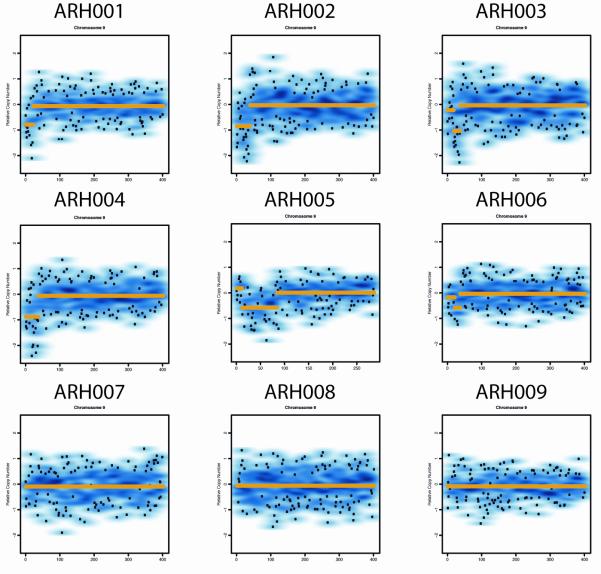
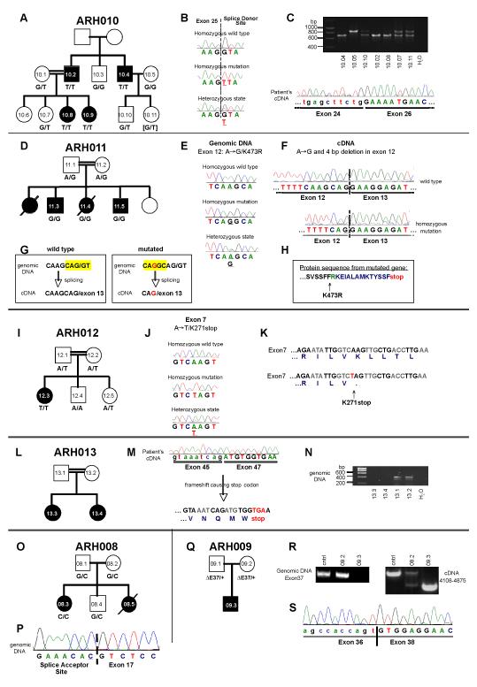
Methods: We performed genome-wide single nucleotide polymorphism analysis for 9 patients with autosomal-recessive hyper-IgE syndrome to locate copy number variations and homozygous haplotypes. Homozygosity mapping was performed with 12 patients from 7 additional families. The candidate gene was analyzed by genomic and cDNA sequencing to identify causative alleles in a total of 27 patients with autosomal-recessive hyper-IgE syndrome.

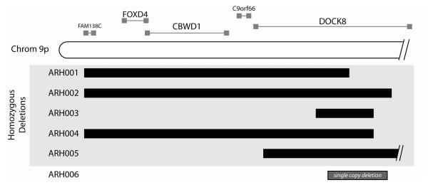
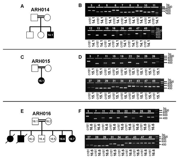
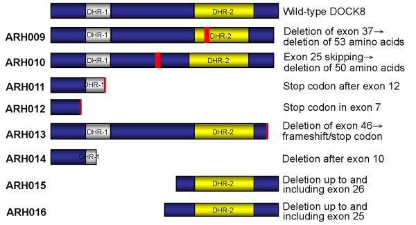
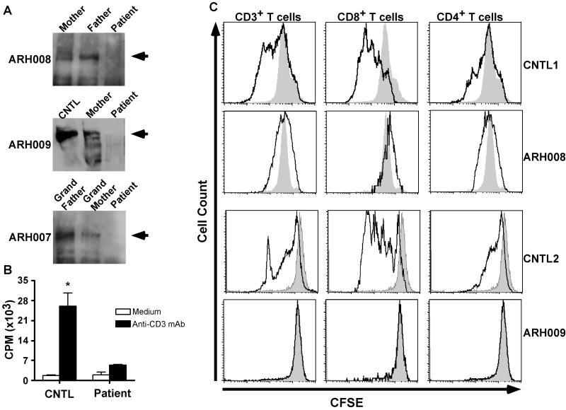
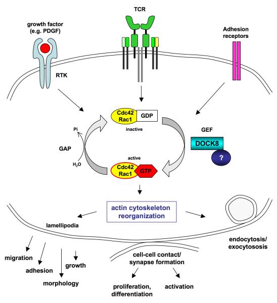
Results: Subtelomeric biallelic microdeletions were identified in 5 patients at the terminus of chromosome 9p. In all 5 patients, the deleted interval involved dedicator of cytokinesis 8 (DOCK8), encoding a protein implicated in the regulation of the actin cytoskeleton. Sequencing of patients without large deletions revealed 16 patients from 9 unrelated families with distinct homozygous mutations in DOCK8 causing premature termination, frameshift, splice site disruption, and single exon deletions and microdeletions. DOCK8 deficiency was associated with impaired activation of CD4+ and CD8+T cells.

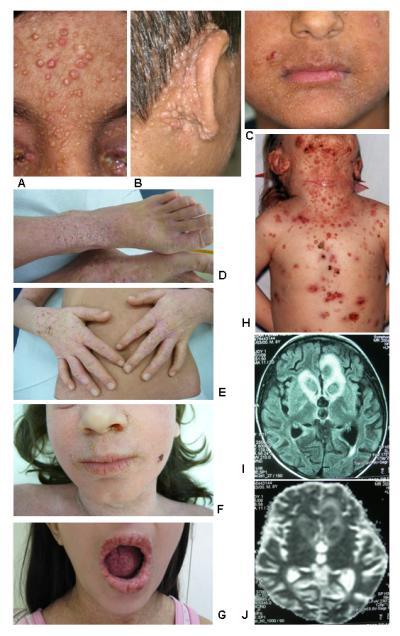
Conclusion: Autosomal-recessive mutations in DOCK8 are responsible for many, although not all, cases of autosomal-recessive hyper-IgE syndrome. DOCK8 disruption is associated with a phenotype of severe cellular immunodeficiency characterized by susceptibility to viral infections, atopic eczema, defective T-cell activation and T(h)17 cell differentiation, and impaired eosinophil homeostasis and dysregulation of IgE.


Figures

Figure 1
Figure 1
Clinical findings in patients with DOCK8 mutations. Panels A-C show the severe Molluscum contagiosum burden of patient ARH014; panel H illustrates the Molluscum infection of patient ARH012; panels D-F show the severe dermatitis in patients ARH010.8 and ARH010.9; and panel G demonstrates the severe oral papilloma virus infection of patient ARH009. Panels F (MRI) and G (diffusion scan) document the cause of death in patient ARH003 who developed an undefined form of encephalitis.
Figure 2
Figure 2
A. ROMA data demonstrating copy number abnormalities consistent with subtelomeric deletions of 9p in AR-HIES. Individuals ARH001-ARH004 have homozygous deletions, ARH005 has a compound heterozygous deletion, and ARH006 has a heterozygous deletion. The remaining subjects do not have demonstrable deletions. Genome-wide SNP Nsp 250k arrays were used for subjects ARH001-ARH009. B. HIES patient deletions and homozygous intervals and known and predicted genes at the terminus of chromosome 9p. C9orf66 is an open reading frame, and FAM138C is a noncoding RNA gene. FOXD4 is a transcription factor, and CBWD has a cobalamin binding domain and nuclease function. DOCK8 is described in the text.
Figure 2
Figure 2
A. ROMA data demonstrating copy number abnormalities consistent with subtelomeric deletions of 9p in AR-HIES. Individuals ARH001-ARH004 have homozygous deletions, ARH005 has a compound heterozygous deletion, and ARH006 has a heterozygous deletion. The remaining subjects do not have demonstrable deletions. Genome-wide SNP Nsp 250k arrays were used for subjects ARH001-ARH009. B. HIES patient deletions and homozygous intervals and known and predicted genes at the terminus of chromosome 9p. C9orf66 is an open reading frame, and FAM138C is a noncoding RNA gene. FOXD4 is a transcription factor, and CBWD has a cobalamin binding domain and nuclease function. DOCK8 is described in the text.
Figure 3
Figure 3
Exonic deletions in DOCK8. Pedigrees are shown in panels A, C and E. Squares: males; circles: females. Filled symbols: patients; slash: deceased individuals. The lack of PCR products from patients’ DNA compared to control DNA suggests exonic deletions (B, D, F).
Figure 4
Figure 4
Mutations in DOCK8. Pedigrees are shown in panels A, D, I, L, O, and Q. Squares: males; circles: females. Filled symbols: patients; slash: deceased individuals. Point mutations in the splice site (B, P) or within exons (E, J). Stop codon caused by point mutation (K) or frameshift (H, M). Generation of a cryptic splice site leading to a 4 bp deletion (F, G). Exon skipping shown by cDNA sequencing (C, M, S) and PCR (C, R). Lack of PCR products from genomic DNA suggesting exonic deletions (N, R).
Figure 5
Figure 5
Cartoon showing the predicted impact of the mutations on DOCK8 protein expression. In three families, the mutation results in a truncated protein affecting both DHR domains, whereas in two families the truncated protein lacks the DHR-1 domain. In family ARH009, 53 amino acids are missing within the DHR-2 domain, and in family ARH0010, 50 amino acids are missing in-between the two DHR domains. In family ARH013, DOCK8 is truncated at the C-terminus.
Figure 6
Figure 6
DOCK8 deficiency impairs T cell activation. A. DOCK8 protein expression in probands, family members and control samples. Lysates from PBMC, normalized for protein content, were analyzed by immunoblotting with an anti-human DOCK8 antibody. A dominant band of about 180 KDa (arrowhead) and several smaller isoforms were detected in control and family members samples but not in those of the probands. B. Proliferative responses of PBMC to anti-CD3 mAb treatment (n=2-5/group; *p=0.02). C. DOCK8 deficiency impairs the activation of both CD4+ and CD8+ T cells. PBMC were loaded with carboxy-fluorescein succinimidyl ester (CFSE) and stimulated with anti-CD3 + anti-CD28 mAb for 3 days. Gated populations of mAb-stimulated CD3+, CD4+ and CD8+ T cells were analyzed for CFSE fluorescence intensity (solid lines), and compared to the respective unstimulated cell population (shaded area).
Figure 7
Figure 7
Hypothetical function of DOCK8. DOCK8 is likely to function as a guanine-nucleotide exchange factor (GEF) for the Rho-GTPases Cdc42 and Rac1, turning them into the active, GTP-bound form upon receptor engagement (e.g. receptor tyrosine kinases, antigen receptors and adhesion receptors). An unknown protein possibly stabilizes the interaction of DOCK8 with Cdc42 and Rac1. GTPase activation induces dynamic filamentous actin rearrangements and lamellipodia formation, leading to cell growth, migration and adhesion. Given the clinical phenotype of the AR-HIES patients with DOCK8 deficiency, we propose an important role of DOCK8 in T-cell actin dynamics, which might be important for the formation of the immunological synapse, leading to T cell activation, proliferation, and differentiation.

Similar articles

Cited by

References

    1. Grimbacher B, Holland SM, Gallin JI, Greenberg F, Hill SC, Malech HL, et al. Hyper-IgE syndrome with recurrent infections--an autosomal dominant multisystem disorder. N Engl J Med. 1999;340:692–702. - PubMed
    1. Grimbacher B, Holland SM, Puck JM. Hyper-IgE syndromes. Immunol Rev. 2005;203:244–50. - PubMed
    1. Holland SM, DeLeo FR, Elloumi HZ, Hsu AP, Uzel G, Brodsky N, et al. STAT3 mutations in the hyper-IgE syndrome. N Engl J Med. 2007;357:1608–19. - PubMed
    1. Grimbacher B, Schaffer AA, Holland SM, Davis J, Gallin JI, Malech HL, et al. Genetic linkage of hyper-IgE syndrome to chromosome 4. Am J Hum Genet. 1999;65:735–44. - PMC - PubMed
    1. Woellner C, Gertz EM, Schäffer AA, Lagos M, Perro M, Glocker EO, et al. Mutations in the signal transducer and activator of transcription 3 (STAT3) and diagnostic guidelines for the Hyper-IgE Syndrome. J Allergy Clin Immunol. 2009 accepted for publication. - PMC - PubMed

Publication types

MeSH terms

Substances