

Defining the borders of splenic marginal zone lymphoma: a multiparameter study
- PMID: 20004934
- PMCID: PMC2879876
- DOI: 10.1016/j.humpath.2009.09.007
Defining the borders of splenic marginal zone lymphoma: a multiparameter study
Abstract
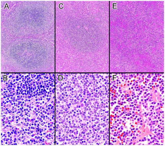
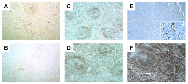
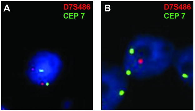
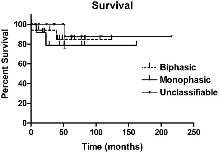
Classic splenic marginal zone lymphomas are CD5-, CD10-, CD23-, CD43-, and usually IgD+ with biphasic white pulp nodules. However, the 2008 World Health Organization classification accepts splenic marginal zone lymphomas with monophasic marginal zone-like white pulp nodules and recognizes a group of unclassifiable splenic small B-cell lymphomas. To explore the relationship of classic splenic marginal zone lymphomas to these other less well-defined splenic lymphomas, a multiparameter study of 47 splenic marginal zone lymphomas and unclassifiable splenic small B-cell lymphomas was performed. Seventeen of 31 splenic marginal zone lymphomas were biphasic, and 14 were monophasic (90%-100% marginal zone-like white pulp nodules). Sixteen cases were unclassifiable splenic small B-cell lymphomas, most lacking a marginal zone-type component. There were many clinical similarities between the 3 groups, including similar survivals. Monophasic and unclassifiable cases were less likely to have a typical splenic marginal zone lymphoma phenotype (28.6%, 23.1%) compared with biphasic cases (86.7%), usually because of IgD negativity (P < .003). Thirty-four of 42 (81%) cases had cytogenetic abnormalities by fluorescence in situ hybridization; and 17 of 20 (85%), by classical cytogenetics. The most frequent fluorescence in situ hybridization abnormalities among the splenic marginal zone lymphomas were del(7)(q31) (26%), +12 (25%), and +3q27 (27%); and among the unclassifiable cases, +12 (50%) and +3q27 (36%). Five of 6 unclassifiable cases with exclusively small non-marginal zone-like lymphocytes involving both white and red pulp had +12 compared with 9 of 34 other cases (P < .02). CDK6 (2 cases) and BCL3 (1 case) rearrangements were only seen in the unclassifiable group. These results support including both biphasic and monophasic cases as splenic marginal zone lymphomas, but suggest that the lack of a non-marginal zone-like population in the monophasic group is associated with some biologic differences. They also demonstrate a relatively large proportion of unclassifiable cases, including a group with frequent +12.
Copyright 2010 Elsevier Inc.
Figures
Similar articles
-
A marginal zone pattern may be found in different varieties of non-Hodgkin's lymphoma: the morphology and immunohistology of splenic involvement by B-cell lymphomas simulating splenic marginal zone lymphoma.Histopathology. 1998 Sep;33(3):230-9. doi: 10.1046/j.1365-2559.1998.00478.x. Histopathology. 1998. PMID: 9777389
-
Splenic marginal zone lymphoma.Semin Diagn Pathol. 2003 May;20(2):121-7. doi: 10.1016/s0740-2570(03)00012-1. Semin Diagn Pathol. 2003. PMID: 12945935 Review.
-
Primary nodal marginal zone lymphomas of splenic and MALT type.Am J Surg Pathol. 1999 Jan;23(1):59-68. doi: 10.1097/00000478-199901000-00006. Am J Surg Pathol. 1999. PMID: 9888704
-
Clinical and biological features of B-cell neoplasms with CDK6 translocations: an association with a subgroup of splenic marginal zone lymphomas displaying frequent CD5 expression, prolymphocytic cells, and TP53 abnormalities.Br J Haematol. 2021 Apr;193(1):72-82. doi: 10.1111/bjh.17141. Epub 2020 Dec 13. Br J Haematol. 2021. PMID: 33314017
-
[Marginal zone lymphomas: extranodal MALT type, nodal and splenic].Pathologe. 2000 Mar;21(2):162-77. doi: 10.1007/s002920050383. Pathologe. 2000. PMID: 10840821 Review. German.
Cited by
-
TLR-mediated activation of Waldenström macroglobulinemia B cells reveals an uncoupling from plasma cell differentiation.Blood Adv. 2020 Jun 23;4(12):2821-2836. doi: 10.1182/bloodadvances.2019001279. Blood Adv. 2020. PMID: 32574366 Free PMC article.
-
Follicular lymphoma and marginal zone lymphoma: how many diseases?Virchows Arch. 2023 Jan;482(1):149-162. doi: 10.1007/s00428-022-03432-2. Epub 2022 Nov 17. Virchows Arch. 2023. PMID: 36394631 Free PMC article. Review.
-
A Review on Splenic Diffuse Red Pulp Small B-Cell Lymphoma.Curr Oncol. 2021 Dec 6;28(6):5148-5154. doi: 10.3390/curroncol28060431. Curr Oncol. 2021. PMID: 34940070 Free PMC article. Review.
-
Improved classification of leukemic B-cell lymphoproliferative disorders using a transcriptional and genetic classifier.Haematologica. 2017 Sep;102(9):e360-e363. doi: 10.3324/haematol.2016.160374. Epub 2017 May 18. Haematologica. 2017. PMID: 28522579 Free PMC article. No abstract available.
-
New developments in the pathology of malignant lymphoma: a review of the literature published from October 2009 to January 2010.J Hematop. 2010 Jun;3(1):47-58. doi: 10.1007/s12308-010-0060-x. Epub 2010 Jun 2. J Hematop. 2010. PMID: 21633487 Free PMC article. No abstract available.
References
-
- Isaacson PGPM, Catovsky D, Swerdlow S, Montserrat E, Berger F, Muller-Hermelink HK, Nathwani B, Harris NL. Splenic marginal zone lymphoma. In: Jaffe ESHN, Stein H, Vardiman JW, editors. Pathology and Genetics of Tumors of Haematopoietic and Lymphoid Tissues. Lyon: IARC Press; 2001. pp. 135–137.
-
- Hammer RD, Glick AD, Greer JP, Collins RD, Cousar JB. Splenic marginal zone lymphoma. A distinct B-cell neoplasm. Am J Surg Pathol. 1996;20:613–626. - PubMed
-
- Isaacson PGPM, Berger F, Swerdlow SH, Thieblemont C, Pittaluga S, Harris NL. Splenic Marginal Zone Lymphoma. In: Swerdlow SHCE, Harris NL, Jaffe ES, Pileri SA, Stein H, Jurgen T, Vardiman JW, editors. WHO Classification of Tumours of Haematopoietic and Lymphoid Tissues. Lyon: IARC Press; 2008. pp. 185–187.
-
- Boonstra R, Bosga-Bouwer A, van Imhoff GW, Krause V, Palmer M, Coupland RW, Dabbagh L, van den Berg E, van den Berg A, Poppema S. Splenic marginal zone lymphomas presenting with splenomegaly and typical immunophenotype are characterized by allelic loss in 7q31-32. Mod Pathol. 2003;16:1210–1217. - PubMed
-
- Matutes E, Oscier D, Montalban C, Berger F, Callet-Bauchu E, Dogan A, Felman P, Franco V, Iannitto E, Mollejo M, Papadaki T, Remstein ED, Salar A, Sole F, Stamatopoulos K, Thieblemont C, Traverse-Glehen A, Wotherspoon A, Coiffier B, Piris MA. Splenic marginal zone lymphoma proposals for a revision of diagnostic, staging and therapeutic criteria. Leukemia. 2008;22:487–495. - PubMed
Publication types
MeSH terms
Grants and funding
LinkOut - more resources
Full Text Sources
