. 2009 Dec 24;139(7):1353-65.
doi: 10.1016/j.cell.2009.11.034. Epub 2009 Dec 10.

Sensory neuron-specific GPCR Mrgprs are itch receptors mediating chloroquine-induced pruritus

Affiliations

Sensory neuron-specific GPCR Mrgprs are itch receptors mediating chloroquine-induced pruritus

Qin Liu et al. Cell. .

Abstract

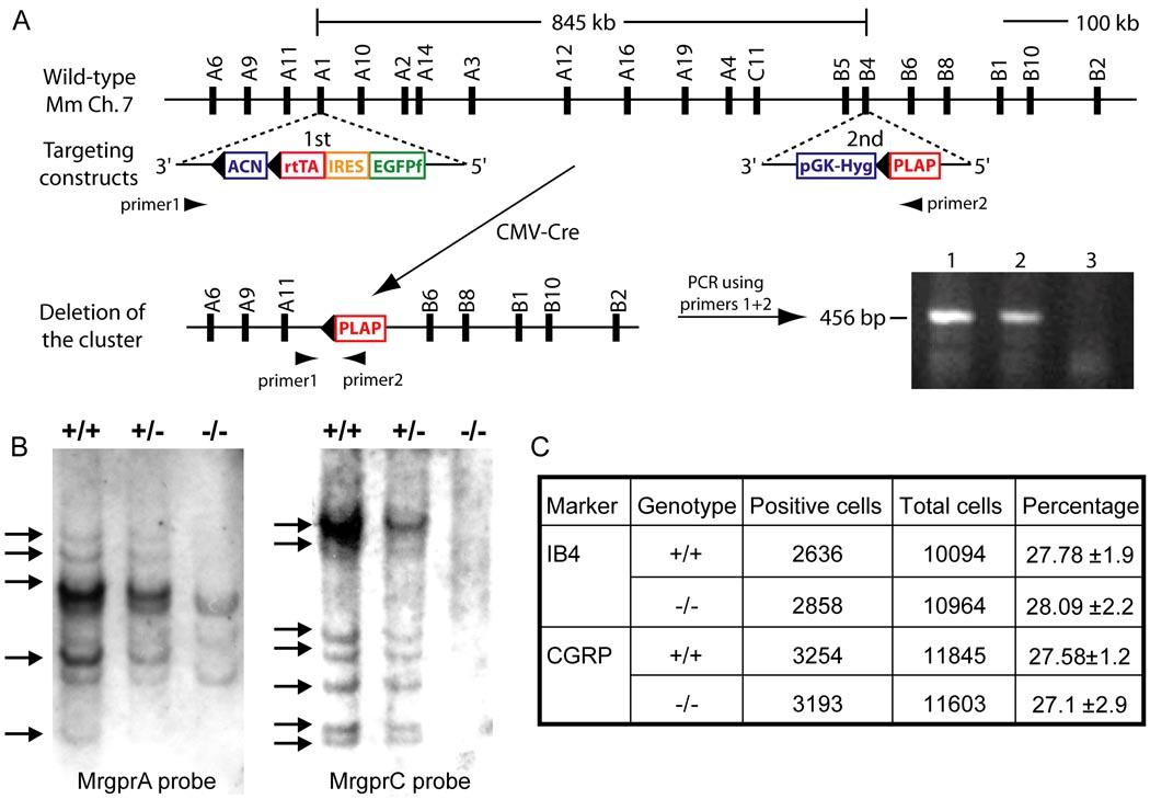
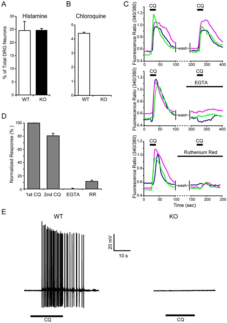
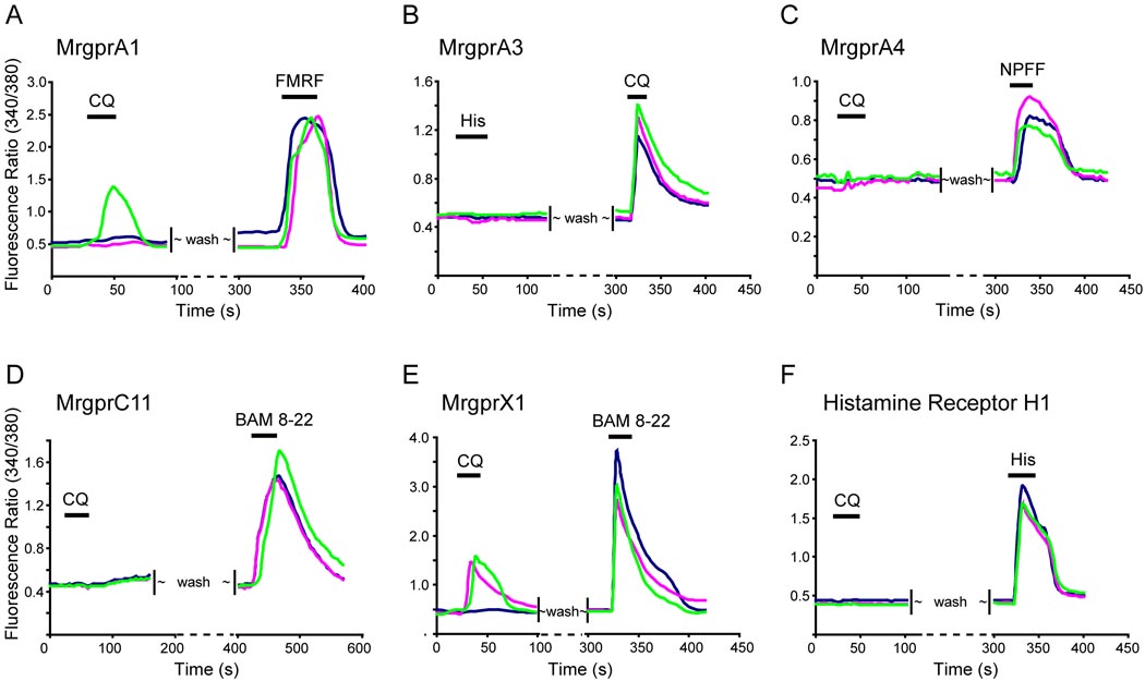
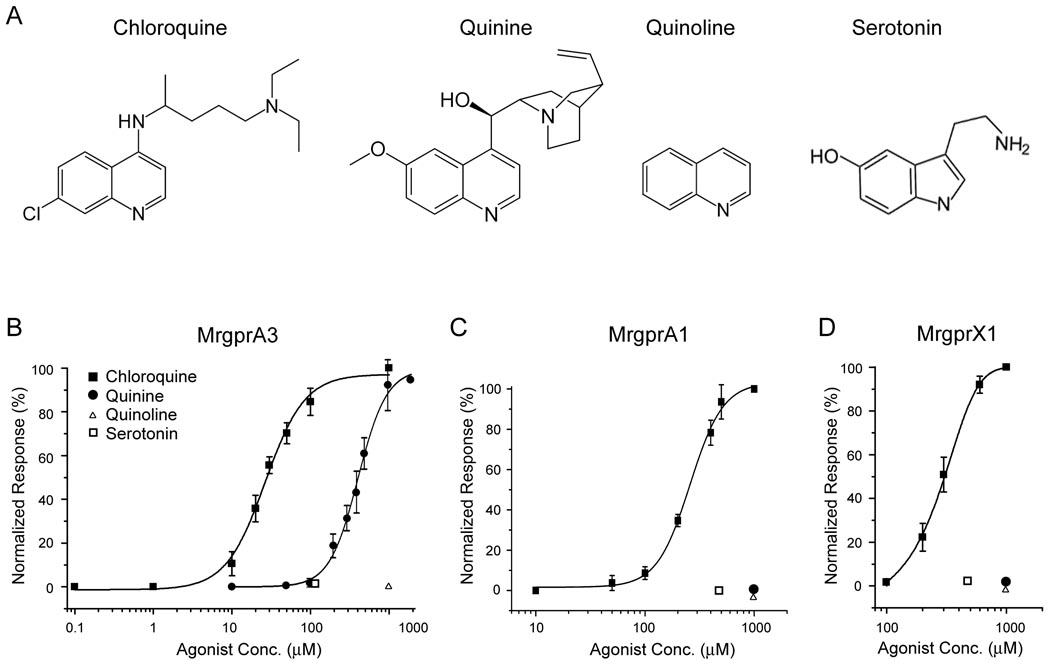
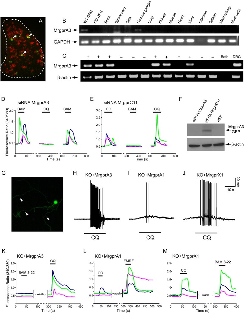
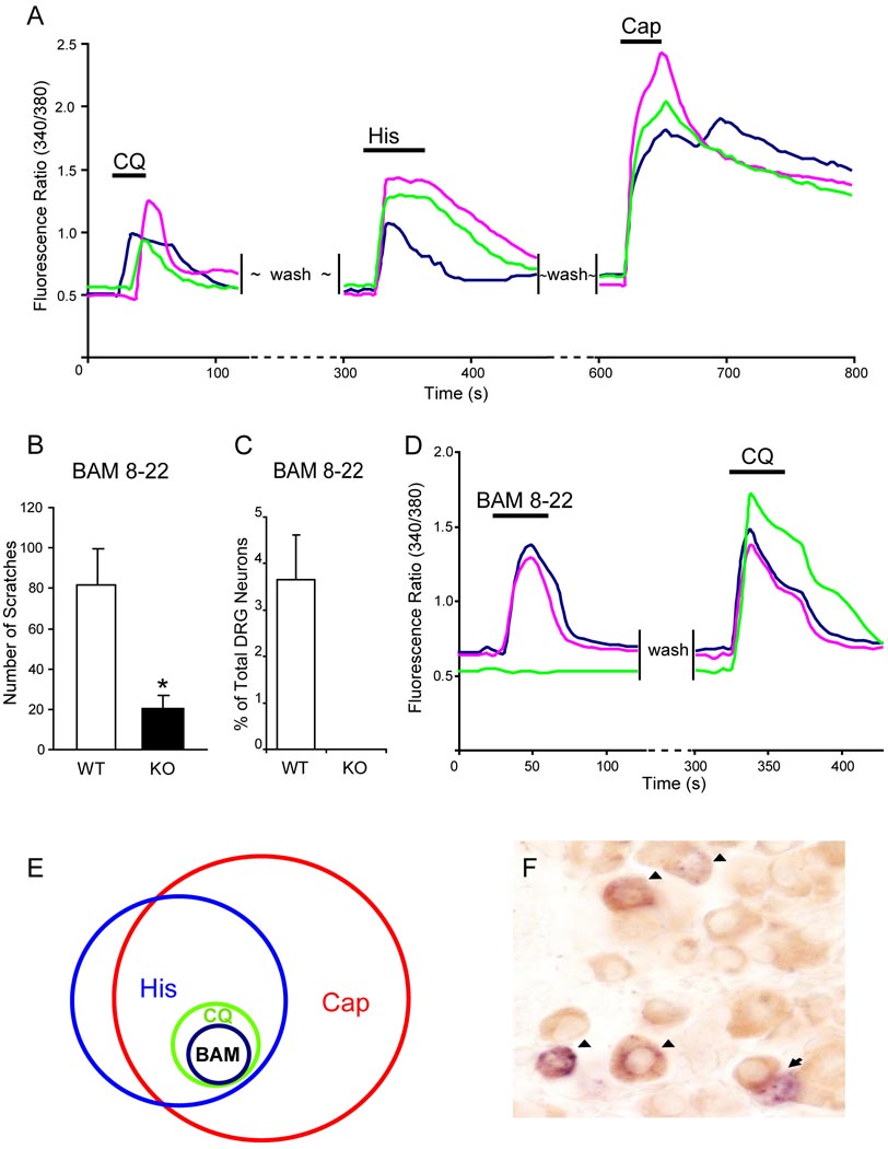
The cellular and molecular mechanisms mediating histamine-independent itch in primary sensory neurons are largely unknown. Itch induced by chloroquine (CQ) is a common side effect of this widely used antimalarial drug. Here, we show that Mrgprs, a family of G protein-coupled receptors expressed exclusively in peripheral sensory neurons, function as itch receptors. Mice lacking a cluster of Mrgpr genes display significant deficits in itch induced by CQ but not histamine. CQ directly excites sensory neurons in an Mrgpr-dependent manner. CQ specifically activates mouse MrgprA3 and human MrgprX1. Loss- and gain-of-function studies demonstrate that MrgprA3 is required for CQ responsiveness in mice. Furthermore, MrgprA3-expressing neurons respond to histamine and coexpress gastrin-releasing peptide, a peptide involved in itch sensation, and MrgprC11. Activation of these neurons with the MrgprC11-specific agonist BAM8-22 induces itch in wild-type but not mutant mice. Therefore, Mrgprs may provide molecular access to itch-selective neurons and constitute novel targets for itch therapeutics.


Figures

Figure 1
Figure 1
Targeted deletion of a cluster of Mrgpr genes. (A) Top horizontal line represents Mrgpr gene cluster on WT mouse chromosome 7. The distance between MrgprA1 and MrgprB4 is 845 kilobases, which contains 12 intact Mrgprs (each represented by a black bar with its name on top). Targeting constructs containing loxP sites (black triangles) and the selection marker genes were introduced to the MrgprA1 and MrgprB4 loci in ES cells by two rounds (1st and 2nd) of electroporation and homologous recombination. Positive ES clones with correct targeting in the two loci underwent a third round of electroporation with CMV-Cre construct. Cre-mediated recombination resulted in deletion of an Mrgpr cluster between loxP sites. The deletion event in ES cells (lane 1 and 2; lane 3 as negative control using WT ES cells) was detected by PCR amplification using primers 1 and 2 flanking the cluster (shown as arrowheads). The PCR product (456 bp) was further confirmed by sequencing. (B) Southern blot of genomic DNA with an MrgprA or MrgprC probe. The genomic DNA was digested with BglII. Due to cross-hybridization, a single MrgprC or MrgprA probe can label multiple members of the MrgprC or MrgprA subfamily in WT (+/+) and cluster heterozygous mice (+/−) DNA. In homozygous mice (−/−), most of the positive bands are absent (arrows). (C) The deletion of Mrgpr genes does not affect cell fate determination of small-diameter sensory neurons. The proportion of nonpeptidergic (IB4+) and peptidergic (CGRP+) small-diameter sensory neurons does not differ between WT and Mrgpr-clusterΔ−/− mice (n = 3).
Figure 2
Figure 2
Mrgpr-clusterΔ−/− mice show severe deficiency in CQ-induced itch. (A–D) Mrgpr-clusterΔ−/− mice respond normally to noxious acute thermal stimuli. Response latencies in tail immersion (50°C, n = 12 per genotype, A), hot plate (50°C, n = 11 per genotype, B), Hargreaves (n = 24 per genotype, C) and cold plate (0 °C, WT n = 13, KO n = 9, D) tests did not differ between WT and Mrgpr-clusterΔ−/− mice. (E) The paw withdrawal threshold of Mrgpr-clusterΔ−/− mice to punctate mechanical stimuli (Von Frey) was comparable to that of WT mice (n = 12 per genotype). (F) Mrgpr-clusterΔ−/− mice responded normally to noxious acute chemical stimuli. The writhing responses to intraperitoneal injection of acetic acid (0.6%, 15 ml/kg) were indistinguishable between WT and Mrgpr-clusterΔ−/− mice (n = 12 per genotype). (G and H) Mrgpr-clusterΔ−/− mice displayed normal histamine-dependent itch. The total scratching bouts were not significantly different between WT and Mrgpr-clusterΔ−/− mice during the first 30 min after subcutaneous injection of histamine (10 µmol; WT n = 7, KO n = 10, G) or compound 48/80 (100 µg/50 µl; WT n = 8, KO n = 7, H). (I) Mrgpr-clusterΔ−/− mice showed deficiency in CQ-induced itch. The total scratching bouts during the first 30 min after CQ injection (200 µg/50 µl, 8 mM) were significantly decreased in Mrgpr-clusterΔ−/− mice (n = 9) than in WT littermates (n = 8). The time course shows bouts of scratching at 5 min intervals. (J) Quinoline (QN) failed to induce itch in both WT and Mrgpr-clusterΔ−/− mice. However, subsequent injection of CQ induced a strong scratch response in WT and a much weaker response in Mrgpr-clusterΔ−/− mice (n = 5 per genotype). (K) SASH mice showed a mild but significant reduction in CQ-induced itch compared with WT mice (SASH n = 8, WT n = 7). (L) SASH mice released significantly less histamine than WT mice after IgE stimulation of the skin (n = 5 per genotype), which provides strong evidence for the mast cell deficiency in SASH mice. The data are presented as mean ± SEM. *, P<0.05; **, P<0.01; ***, P<0.005; two-tailed unpaired t-test or two-way ANOVA.
Figure 3
Figure 3
The response of DRG neurons to CQ is Mrgpr-dependent. (A) The response to histamine was not impaired in Mrgpr-deficient DRG neurons. Calcium imaging showed that the percentage of Mrgpr-clusterΔ−/− DRG neurons responding to histamine (50 µM) was similar to that of WT neurons (n = 3 per genotype). (B) ~4.4% of WT DRG neurons responded to CQ (1 mM) with increased [Ca2+]i whereas Mrgpr-clusterΔ−/− DRG neurons failed to respond to the drug (n = 3 per genotype). (C–D) Extracellular calcium was required for the CQ-induced [Ca2+]i increase in DRG neurons. (C) shows representative traces from 3 different DRG neurons in calcium imaging assays. (D) The CQ-induced increase in [Ca2+]i was almost completely blocked with EGTA treatment. Ruthenium red (RR) also significantly attenuated [Ca2+]i increase evoked by CQ. As a control, sequential treatment of CQ only caused a ~20% reduction in [Ca2+]i increase. (E) CQ (1 mM) induced APs in DRG neurons. In WT DRG neurons, all CQ-sensitive neurons (as determined by calcium imaging, n=5) elicited a train of APs evoked by subsequent CQ treatment. In contrast, none of the neurons tested (n=11) from Mrgpr-clusterΔ−/− mice showed any response to the drug.
Figure 4
Figure 4
Mouse MrgprA3 and human MrgprX1 are the predominant receptors for CQ. HEK293 cells were transfected with expression constructs for Mrgprs and histamine H1 receptor. The effects of different agonists on these transfected cells were tested via calcium imaging. Each figure shows a typical response from three different cells. (A) Fewer than half of MrgprA1-tranfected cells responded to CQ (1 mM) with increased [Ca2+]i whereas all transfected cells responded to FMRF (2 µM). (B) All MrgprA3-expressing cells responded to CQ but not histamine. (C) MrgprA4-expressing cells respond to NPFF (2 µM) but not CQ. (D) MrgprC11-expressing cells failed to respond to CQ whereas they responded to BAM8-22 (2 µM). (E) Human MrgprX1 responded to both CQ and BAM8-22 (2 µM). (F) Cells expressing the histamine H1 receptor exhibited a strong response to histamine (50 µM) but failed to respond to CQ.
Figure 5
Figure 5
Mrgprs are selectively activated by CQ. (A) Molecules with structures related to CQ. Dose-response curves for MrgprA3 (B), MrgprA1 (C), and MrgprX1 (D) expressed in HEK 293 cells to the molecules in (A). Each data point represents the mean ± SEM of at least three independent experiments and at least 50 GFP+ cells were analyzed each time. Calcium responses at each ligand concentration were normalized to the maximal response subsequently elicited.
Figure 6
Figure 6
MrgprA3 is required for CQ responsiveness in mouse DRG neurons. (A) Fluorescent in situ hybridization of DRG sections with MrgprA3 (green, arrowheads) and MrgprD (red). The white dashed line outlines the DRG. (B) RT-PCR analysis of 14 mouse tissues or cell types for expression of MrgprA3. The only tissues containing MrgprA3 are WT DRG and nodose ganglia. Notably no band was found in Mrgpr-clusterΔ−/− DRG, confirming MrgprA3 was deleted in Mrgpr-clusterΔ−/− mice. (C) Single cell RT-PCR was performed on individual DRG neurons with the responsiveness to CQ (1 mM) established by calcium imaging (shown here are 12 representative neurons). MrgprA3 mRNA was detected in 8/9 CQ-responsive neurons (+), but was not detected in any of 11 CQ-unresponsive neurons (−). For a negative control, sample of bath solution was used (Bath); Diluted total DRG cDNA was used as positive control (DRG). Arrows indicate predicted product size for MrgprA3 (150 bp) and β-actin (302 bp). No product was detected in RT- controls from MrgprA3-expressing cells (n=8). (D) and (E) show representative traces from 3 different WT DRG neurons electroporated with siRNAs in calcium imaging assays. (D) CQ-induced increase in [Ca2+]i was completely lost in WT neurons electroporated with MrgprA3 siRNA. However, these neurons (normally express both MrgprA3 and MrgprC11) are still sensitive to BAM8-22 (BAM). 24 BAM8-22 sensitive neurons were analyzed. (E) As a control, CQ responsiveness in WT neurons electroporated with MrgprC11 siRNA remained intact (10 CQ sensitive neurons analyzed). But MrgprC11 siRNA completely abolished BAM8-22 sensitivity. (F) The efficiency and specificity of MrgprA3 siRNA were tested by co-transfecting HEK293 cells with MrgprA3 siRNA and expression constructs of MrgprA3 or MrgprC11. Western blot shows that MrgprA3 siRNA specifically knocked-down the expression of MrgprA3, but not MrgprC11. (G–M) Mrgpr A3 and MrgprX1 selectively rescued CQ responsiveness in Mrgpr-clusterΔ−/− DRG neurons. (G) Visualization of Mrgpr-clusterΔ−/− DRG neurons that express MrgprA3-GFP protein. Note the membrane and axon localization (arrowheads) of MrgprA3-GFP in DRG neurons. (H) All Mrgpr-clusterΔ−/− neurons electroporated with MrgprA3 fired a train of APs upon CQ treatment (n=6). (I) Fewer than half of MrgprA1-electroporated neurons (3 out of 7) elicited a few APs upon CQ treatment. (J) Most Mrgpr-clusterΔ−/− neurons electroporated with MrgprX1 (5 out of 7 GFP-positive neurons recorded) also generated a train of APs in response to CQ. (K–M) Each figure shows typical calcium traces from three different neurons. (K) All MrgprA3-expressing Mrgpr-clusterΔ−/− neurons showed increased [Ca2+]i in response to CQ (1 mM) but not BAM8-22 (2 µM). (L) All MrgprA1-electroporated mutant neurons showed a strong response to FMRF (2 µM) whereas only a small portion responded to CQ. (M) Electroporation of MrgprX1 rendered Mrgpr-clusterΔ−/− DRG neurons sensitivity to both CQ and BAM8-22.
Figure 7
Figure 7
CQ-responsiveness defines a specific subpopulation of DRG neurons. (A) CQ-responsive neurons represented a small population of DRG neurons that also responded to histamine (50 µM) and capsaicin (1 µM) with increased [Ca2+]i monitored by calcium imaging. (B) The total scratching bouts during the first 30 min after BAM8-22 intradermal injection (50 µI of 1 mM). WT mice exhibited significantly stronger scratching responses after injection than Mrgpr-clusterΔ−/− littermates did (n=8 per genotype; * p<0.05). (C) As determined by calcium imaging, 3.6% of WT DRG neurons responded to BAM8-22 (2 µM) with increased [Ca2+]i and all of them are also CQ-sensitive (D), whereas Mrgpr-clusterΔ−/− DRG neurons failed to respond to the drug (n=3 per genotype) (C). (E) The Venn diagram illustrates the relationships of histamine- (His), capsaicin- (Cap), chloroquine- (CQ), and BAM8-22- (BAM) responsive neurons in adult DRG. The sizes of the circles are proportional to the sizes of the cell populations. (F) WT adult DRG sections were doubly stained by in situ hybridization for MrgprA3 (blue) and immunostaining using anti-GRP antibody (brown). Most MrgprA3+ cells (51 out of 55) express GRP. Arrowheads indicate MrgprA3/GRP co-expressing neurons. Arrows indicate MrgprA3+/GRP cells.

Similar articles

Cited by

References

    1. Abila B, Ezeamuzie IC, Igbigbi PS, Ambakederemo AW, Asomugha L. Effects of two antihistamines on chloroquine and histamine induced weal and flare in healthy African volunteers. Afr J Med Med Sci. 1994;23:139–142. - PubMed
    1. Adam ME, Karim EF, Elkadaru AY, Ibrahim KE, Berger BJ, Wiese M, Babiker HA. Imipramine induced complete reversal of chloroquine resistance in plasmodium falciparum infections in Sudan. Saudi Pharmaceutical Journal. 2004;12:130–135.
    1. Ademowo OG, Walker O, Sodeinde O. Prevalence of chloroquine-induced pruritus: evidence for heredofamilial factors. Malaria Infect Dis Afr. 1998;8:14–18.
    1. Ajayi AA, Oluokun A, Sofowora O, Akinleye A, Ajayi AT. Epidemiology of antimalarial-induced pruritus in Africans. Eur J Clin Pharmacol. 1989;37:539–540. - PubMed
    1. Alving K, Sundstrom C, Matran R, Panula P, Hokfelt T, Lundberg JM. Association between histamine-containing mast cells and sensory nerves in the skin and airways of control and capsaicin-treated pigs. Cell Tissue Res. 1991;264:529–538. - PubMed

Publication types