. 2010 Jan 12;20(1):75-9.
doi: 10.1016/j.cub.2009.11.016. Epub 2009 Dec 10.

RalA-exocyst complex regulates integrin-dependent membrane raft exocytosis and growth signaling

Affiliations

RalA-exocyst complex regulates integrin-dependent membrane raft exocytosis and growth signaling

Nagaraj Balasubramanian et al. Curr Biol. .

Abstract

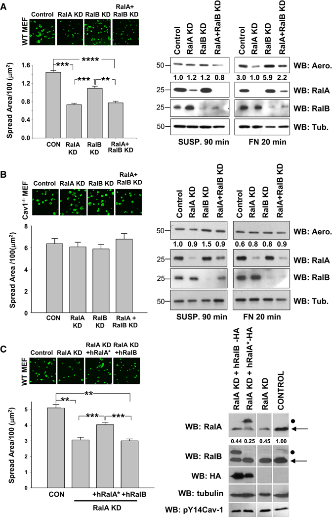
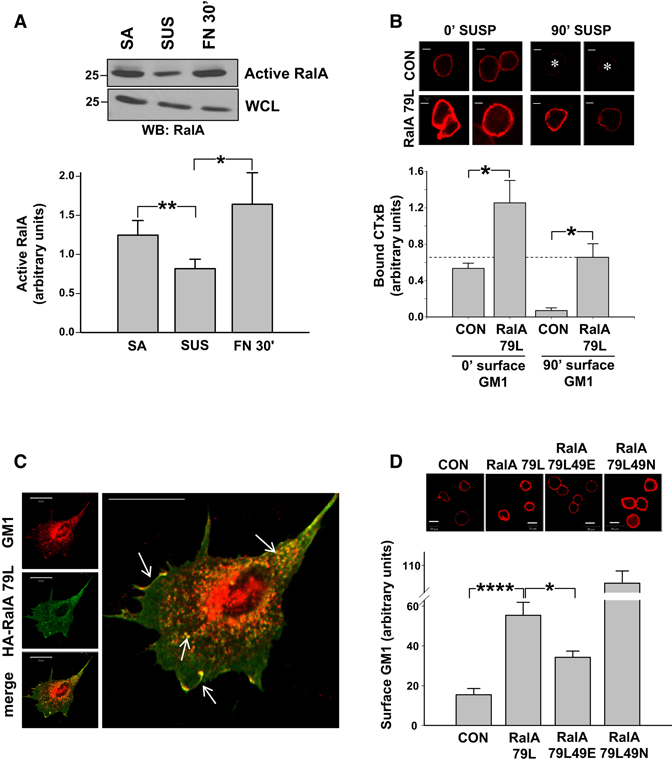
Anchorage dependence of cell growth is a key metastasis-suppression mechanism that is mediated by effects of integrins on growth signaling pathways. The small GTPase RalA is activated in metastatic cancers through multiple mechanisms and specifically induces anchorage independence. Loss of integrin-mediated adhesion triggers caveolin-dependent internalization of cholesterol- and sphingolipid-rich lipid raft microdomains to the recycling endosomes; these domains serve as platforms for many signaling pathways, and their clearance from the plasma membrane (PM) after cell detachment suppresses growth signaling. Conversely, readhesion triggers their return to the PM and restores growth signaling. Activation of Arf6 by integrins mediates exit of raft markers from the recycling endosomes but is not sufficient for return to the PM. We now show that RalA but not RalB mediates integrin-dependent membrane raft exocytosis through the exocyst complex. Constitutively active RalA restores membrane raft targeting to promote anchorage-independent growth signaling. Ras-transformed pancreatic cancer cells also show RalA-dependent constitutive PM raft targeting. These results identify RalA as a key determinant of integrin-dependent membrane raft trafficking and regulation of growth signaling. They therefore define a mechanism by which RalA regulates anchorage dependence and provide a new link between integrin signaling and cancer.


Figures

Figure 1
Figure 1. Ral inhibition delays cell spreading and raft exocytosis
(A) WT and Cav1−/− MEFs expressing Myc-tagged Ral binding domains (RBDs) from Sec5 or RLIP were serum starved overnight, suspended for 90min and replated on FN (2 µg/ml) for 30min. Cells were fixed and stained with phalloidin-Alexa 488 and images taken. Graph: represents spread surface area/100; values are mean ± SE in µm2. (n=95 WT MEFs, n=70 Cav1−/− MEFs). Three experiments gave similar results. Suspended and re-adherent (B) WT and (C) Cav1−/− MEFs were incubated on ice with purified proaerolysin for 30min then rinsed. Bound proaerolysin was detected in cell lysate by Western blotting (WB : Aero) using tubulin as loading control (WB : Tub). RBD constructs were detected by western blotting for myc (WB: Myc). Values listed under the aerolysin blot represent band intensity normalized to tubulin relative to suspended control cells in each cell type. Blots are representative of three independent experiments. **** =p<0.00001 to * =p<0.01.
Figure 2
Figure 2. Effects of Ral knockdown on cell spreading and surface rafts
(A) WT MEFs and (B) Cav1−/− MEFs were transfected with RalA siRNA (RalA KD), RalB siRNA (RalB KD), both (RalA+ RalB KD) or control siRNA (CON). Cells were detached, held in suspension for 90min and replated on FN (2 µg/ml) for 20min. Cells were fixed and stained with phalloidin-Alexa 488 and images taken. Graph: represents spread surface area/100; values are mean ± SE in µm2. (>100 cells were analysed per experiment, 3 independent experiments gave similar results). Surface binding of proaerolysin was measured as in Fig 1 (WB : Aero) using tubulin (WB : Tub) as loading control (right panel). Values represent band intensity normalized to tubulin relative to control 90min suspended cell band intensity. RalA and RalB expression were detected by western blotting (WB : RalA, RalB) (C) RalA-depleted WT MEFs were transfected with vectors for HA tagged siRNA-resistant human RalA (hRalA*) or human RalB. Cells were treated as above and spreading quantitated. Graph: spread surface area/100; values are mean ± SE in µm2. (n=128 cells). Two independent experiments gave similar results. Quantified values represent endogenous RalA (←) band intensity normalized to tubulin. Mutant siRNA-resistant hRalA and RalB are indicated by •. **** =p<0.00001 to * =p<0.01.
Figure 3
Figure 3. Adhesion-dependent RalA activation promotes raft plasma membrane localization
(A) WT MEFs stably adherent (SA), suspended for 90min (SUS), or replated on FN (10 µg/ml) for 30min (FN30’) were assayed for active RalA using GST-Sec5 RBD beads and Western blotting for RalA (WB: RalA). Graph: Active RalA bands were quantified and normalized to WCL. Values are means ± SE, n=5. (** =p<0.01, * =p<0.05) (B) Untransfected (CON) and HA-RalA79L-transfected WT MEFs were serum starved overnight, detached, held in suspension for 0min or 90min, and surface labeled with CTxB-Alexa 594 (GM1-CTxB). Cells were then fixed and images taken. Graph: Values are mean surface intensity ± SE, n=10, representative of two independent experiments. *p < 0.01. Scale bar represents 5 µm. (C) HA tagged RalA79L-expressing WT MEFs surface labeled with CTxB-Alexa594 were suspended and replated on FN (2 µg/ml) for 30min. Cells were fixed and stained for HA. Arrows indicated overlap in vesicles and membrane ruffles. Scale bar represents 20 µm. (D) Untransfected (CON) MEFs and MEFs expressing HA-RalA79L, RalA79L 49E or RalA79L 49N were serum starved overnight, detached and held in suspension for 90min. Cells were then surface labeled with CTxB-Alexa 594 (GM1-CTxB), fixed and images taken. Graph: Values are mean surface intensity ± SE, n=14, representative of two independent experiments. **** =p<0.00001 to * =p<0.01. Scale bar represents 10 µm.
Figure 4
Figure 4. Sec5 and RalA Regulate Membrane Raft Targeting and Signaling
(A) WT MEFs transfected with control siRNA, Sec5 siRNA oligo #1 (Sec5-1) or # 2 (Sec5-2) were suspended for 90 min and replated on FN (2 µg/ml) for 20min. Cells were fixed and stained with phalloidin-Alexa 488. Graph: spread surface area/100; values are mean ± SE in µm2 (n=75 cells), representative of three independent experiments. (B) Re-adherent control and Sec5 siRNA oligo # 2 treated cells (Sec5-2) were incubated on ice with purified proaerolysin for 30min. Bound proaerolysin was detected in cell lysates by Western blotting (WB:Aero). Sec5, RalA and RalB expression was confirmed by western blotting (WB). Values represent band intensity normalized to RalB levels relative to control (C) WT MEFs expressing Sec5 siRNA oligo #2 (Sec5 KD) were transfected with Myc- WT Sec5 (WT Sec5) or Myc -T11A Sec5 (T11A Sec5). Cells were suspended for 90 min, replated on FN (2 µg/ml) for 20min, fixed and stained with phalloidin-Alexa 488. Graph: spread surface area/100; values are mean ± SE in µm2 (n=65 cells), representative of three independent experiments. (D) Re-adherent cells were incubated on ice with purified proaerolysin for 30min. Bound proaerolysin detected in cell lysate by Western blotting (WB : Aero) using tubulin (WB : tub.) as a loading control. Graph: Aerolysin normalized to tubulin, values are mean ± S.E from three independent experiments. (A–D) ****p < 0.00001; ***p < 0.0001. (E) WT Sec5-GFP (Sec5-GFP) expressing WT MEFs were suspended for 90min, and replated on FN (2 µg/ml) for 20min. Cells were chilled, stained with CTxB-Alexa 594 (Surface GM1), and fixed. (F–G) WT MEFs untransfected (CON) or transfected with HA-RalA79L (RalA 79L) were kept suspended or adherent for 8hours without serum then treated with 10% serum for 30min. Cells were lysed and Western blotted for (F) active (pSer473 Akt) and total Akt (Akt) (G) Phospho-p44/p42 and total Erk (Erk). Phosphorylated Akt and Erk were normalized to total protein. Graph: Values are means ± SE, n=4 (pAkt) and n=3 (pErk). (H) MiaPaCa2 cells (CON) in which RalA (RalAi) and RalB (RalBi) were stably depleted were analyzed by western blotting (WB) for the indicated proteins. (I) Control (CON), RalA (RalAi) and RalB (RalBi) knockdown MiaPaCa2 cells were maintained adherent or suspended for 4hours with serum. Cells were chilled, and proaerolysin surface labeling measured as in Fig 1., Bands were quantified and normalized to tubulin; Graph shows proaerolysin binding in suspended cells relative to adherent cells for each condition. Values are means ± SE, n=3. (F–I) **p < 0.01; *p < 0.05.

References

    1. Schwartz MA, Assoian RK. Integrins and cell proliferation: regulation of cyclin-dependent kinases via cytoplasmic signaling pathways. J Cell Sci. 2001;114:2553–2560. - PubMed
    1. Ryu CH, Kim SW, Lee KH, Lee JY, Kim H, Lee WK, Choi BH, Lim Y, Kim YH, Hwang TK, et al. The merlin tumor suppressor interacts with Ral guanine nucleotide dissociation stimulator and inhibits its activity. Oncogene. 2005;24:5355–5364. - PubMed
    1. Lim KH, O'Hayer K, Adam SJ, Kendall SD, Campbell PM, Der CJ, Counter CM. Divergent roles for RalA and RalB in malignant growth of human pancreatic carcinoma cells. Curr Biol. 2006;16:2385–2394. - PubMed
    1. Lim KH, Baines AT, Fiordalisi JJ, Shipitsin M, Feig LA, Cox AD, Der CJ, Counter CM. Activation of RalA is critical for Ras-induced tumorigenesis of human cells. Cancer Cell. 2005;7:533–545. - PubMed
    1. del Pozo MA, Balasubramanian N, Alderson NB, Kiosses WB, Grande-Garcia A, Anderson RG, Schwartz MA. Phospho-caveolin-1 mediates integrin-regulated membrane domain internalization. Nat Cell Biol. 2005;7:901–908. - PMC - PubMed

Publication types