. 2010 Feb;221(2):346-52.
doi: 10.1016/j.expneurol.2009.12.004. Epub 2009 Dec 11.

Acute glial activation by stab injuries does not lead to overt damage or motor neuron degeneration in the G93A mutant SOD1 rat model of amyotrophic lateral sclerosis

Affiliations

Acute glial activation by stab injuries does not lead to overt damage or motor neuron degeneration in the G93A mutant SOD1 rat model of amyotrophic lateral sclerosis

Masatoshi Suzuki et al. Exp Neurol. 2010 Feb.

Abstract

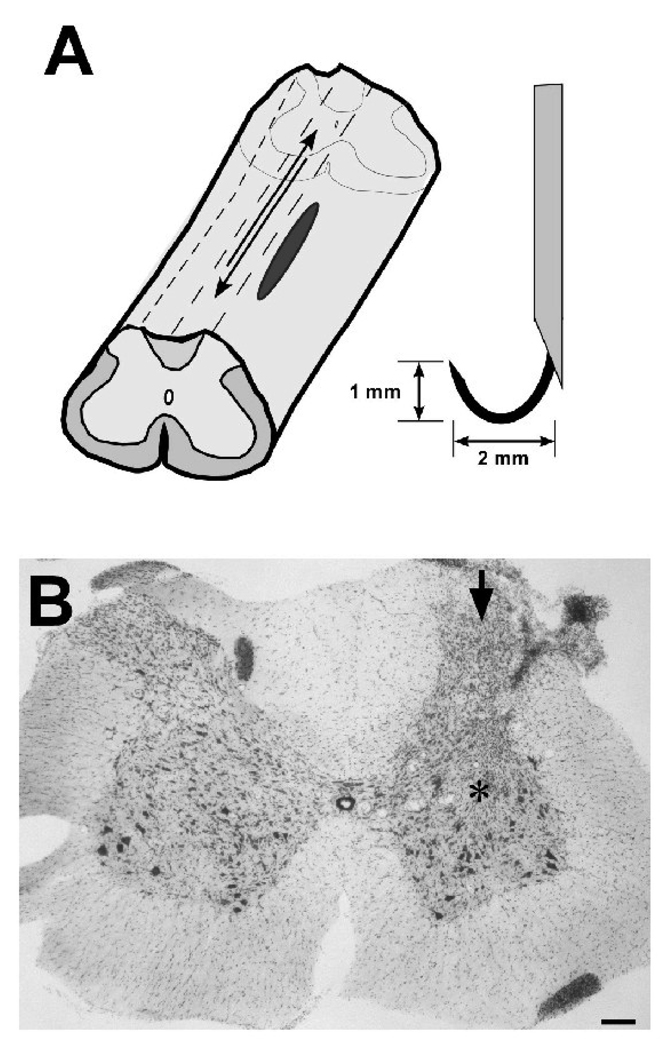
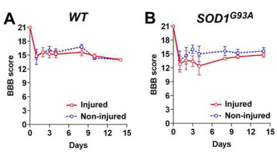
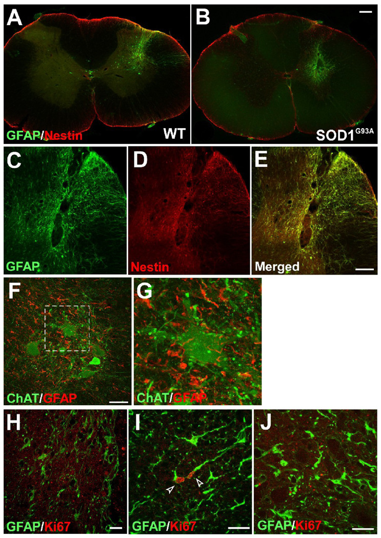
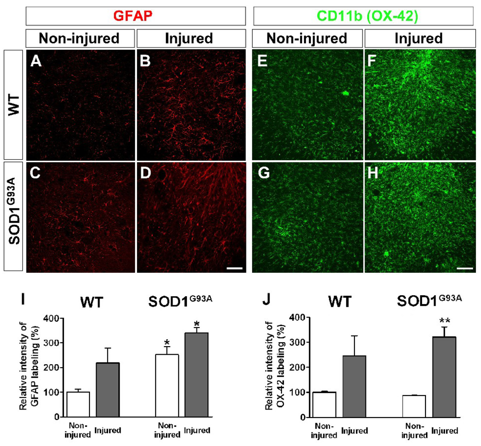
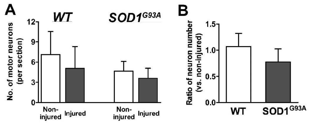
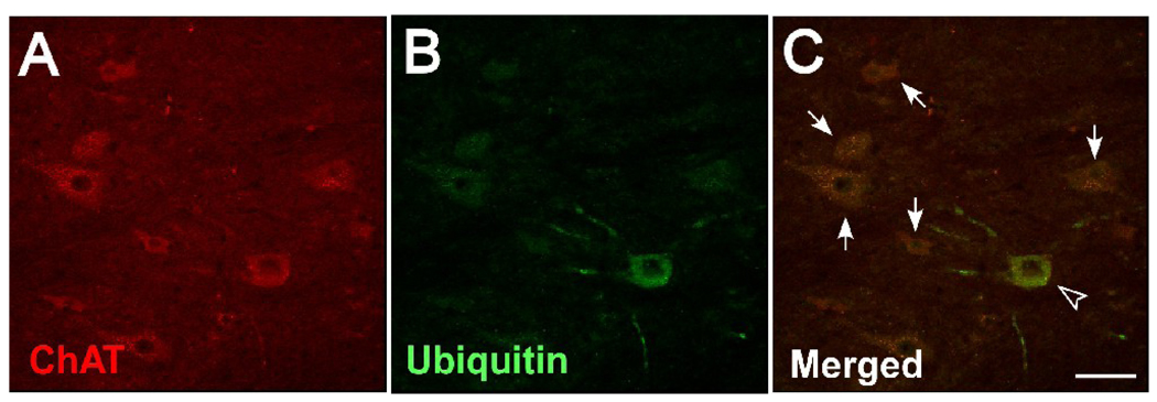
Amyotrophic lateral sclerosis (ALS) is a neurodegenerative disease where motor neurons within the brain and spinal cord are lost, leading to paralysis and death. Recently, a correlation between head trauma and the incidence of ALS has been reported. Furthermore, new invasive neurosurgical studies are being planned which involve inserting needles directly to the spinal cord. We therefore tested whether acute trauma to the spinal cord via a knife wound injury would lead to accelerated disease progression in rodent models of ALS (SOD1(G93A) rats). A longitudinal stab injury using a small knife was performed within the lumbar spinal cord region of presymptomatic SOD1(G93A) rats. Host glial activation was detected in the lumbar area surrounding a micro-knife lesion at 2 weeks after surgery in both wild type and SOD1(G93A) animals. However, there was no sign of motor neuron loss in the injured spinal cord of any animal and normal motor function was maintained in the ipsilateral limb. These results indicate that motor neurons in presymptomatic G93A animals are not affected by an invasive puncture wound injury involving reactive astrocytes. Furthermore, acute trauma alone does not accelerate disease onset or progression in this ALS model which is important for future strategies of gene and cell therapies directly targeting the spinal cord of ALS patients.


Figures

Figure 1
Figure 1. A stab injury model using a micro wire-knife in a rat spinal cord
(A) Schematic illustrations of stab injury model in the lumbar region of rat spinal cords. The stab injuries ran parallel to the long ascending and descending tracts and passed primarily through gray matter at the level of L2-4. The longitudinal stab injuries were 2 mm in length and 1 mm depth and were made with a retractable wire knife. (B) Representative photomicrograph showing tissue adjacent to the stab injury. A micro wire was inserted on the dorsal side of the spinal cord (arrow) and affected gray matter (asterisk). Scale bar: 200 µm.
Figure 2
Figure 2. Ipsilateral limb function is not affected by stab injuries in SOD1G93A rats
Basso-Beattie-Bresnahan (BBB) locomotor rating scales in the WT (A) or SOD1G93A rats (B). A significant difference was not observed between the injured side and non injured side in any group.
Figure 3
Figure 3. Host glial activation following stab injuries in SOD1G93A rats
Immunostaining with anti-GFAP and nestin in the lumbar spinal cord of WT (A) and SOD1G93A rats (B). Significant astrocyte activation in the injured side of the spinal cord, while there were few positive signals in the non-injured side. Particularly, high expression of GFAP (C) and nestin (D) was observed in the injured core. (F) Double labeling with GFAP and ChAT revealed astrocyte activation within regions of motor neurons. (G) A photomicrograph using higher magnification in the area represented by a broken-lined box in F. Double staining with GFAP and proliferation marker Ki67 in the injured core (H, I) and the ventral horn (J). While a few GFAP+/Ki67+ cells (designated by arrow heads) were found in the injured core, there is no double positive cell in the ventral horn. Scale bars: 200 µm in A, B; 100 µm in C-E; 50 µm in F; 20 µm in H-J.
Figure 4
Figure 4. Analysis of glial activation following a stab injury in the spinal cord of SOD1G93A rats
Photomicrographs of the non-injured (A) and injured (B) WT rats and SOD1G93A rats (C, D) indicate astroglial activation in the ventral horn of lumbar spinal cord. Microglial activation was compared between non-injured and injured spinal cord by immunostaining for microglial marker CD11b (OX-42) in WT (E, F) or SOD1G93A animals (G, F). Densitometrical results of the relative intensity of GFAP (I) or OX-42 (J) –labeling. Scale bars: 100 µm in A-H. *: P<0.05 vs. non-injured WT; **: P<0.05 vs. non-injured WT and SOD1G93A.
Figure 5
Figure 5. Motor neurons are maintained following a stab injury in the lumbar spinal cord of SOD1G93A rats
(A) Motor neuron counts showed that a significant difference was not observed in the number of lumbar motor neurons (L2-4) between the WT and SOD1G93A animals. (B) The ratio of motor neurons compared to the non-injured side. There was no obvious difference in the number of large motor neurons in the non-injured side of the spinal cord in both WT and SOD1G93A rats.
Figure 6
Figure 6. Ubiquitin expression in motor neurons of SOD1G93A rats
Even though spinal cord injury did not change motor function of SOD1G93A rats at 129 days of age, double labeled sections for both ChAT (A) and ubiquitin (B) revealed that there is undergoing degeneration in the lumbar spinal cord. Most of the large surviving ChAT+ motor neurons within chimeric regions of the transplanted spinal cord did not express ubiquitin (designated by arrows). A few ChAT+/ubiquitin+ neurons (designated by an arrow head), which represent early degenerating motor neurons, were found in the lumbar spinal cord of SOD1G93A rats but not in WT rats. Scale bar: 50 µm.

Similar articles

Cited by

References

    1. Alexander JK, Popovich PG. Neuroinflammation in spinal cord injury: therapeutic targets for neuroprotection and regeneration. Prog.Brain Res. 2009;175:125–137. - PubMed
    1. Andersen PM, Sims KB, Xin WW, Kiely R, O'Neill G, Ravits J, Pioro E, Harati Y, Brower RD, Levine JS, Heinicke HU, Seltzer W, Boss M, Brown RH., Jr Sixteen novel mutations in the Cu/Zn superoxide dismutase gene in amyotrophic lateral sclerosis: a decade of discoveries, defects and disputes. Amyotroph.Lateral.Scler. Other Motor Neuron Disord. 2003;4:62–73. - PubMed
    1. Azzouz M. Gene Therapy for ALS: progress and prospects. Biochim.Biophys.Acta. 2006;1762:1122–1127. - PubMed
    1. Barbeito LH, Pehar M, Cassina P, Vargas MR, Peluffo H, Viera L, Estevez AG, Beckman JS. A role for astrocytes in motor neuron loss in amyotrophic lateral sclerosis. Brain Res.Brain Res.Rev. 2004;47:263–274. - PubMed
    1. Beers DR, Henkel JS, Xiao Q, Zhao W, Wang J, Yen AA, Siklos L, McKercher SR, Appel SH. Wild-type microglia extend survival in PU.1 knockout mice with familial amyotrophic lateral sclerosis. Proc.Natl.Acad.Sci.U.S.A. 2006;103:16021–16026. - PMC - PubMed

Publication types

MeSH terms