Effects of normal aging on prefrontal area 46 in the rhesus monkey
- PMID: 20005254
- PMCID: PMC2822043
- DOI: 10.1016/j.brainresrev.2009.12.002
Effects of normal aging on prefrontal area 46 in the rhesus monkey
Abstract
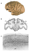
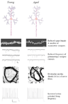
This review is concerned with the effects of normal aging on the structure and function of prefrontal area 46 in the rhesus monkey (Macaca mulatta). Area 46 has complex connections with somatosensory, visual, visuomotor, motor, and limbic systems and a key role in cognition, which frequently declines with age. An important question is what alterations might account for this decline. We are nowhere near having a complete answer, but as will be shown in this review, it is now evident that there is no single underlying cause. There is no significant loss of cortical neurons and although there are a few senile plaques in rhesus monkey cortex, their frequency does not correlate with cognitive decline. However, as discussed in this review, the following do correlate with cognitive decline. Loss of white matter has been proposed to result in some disconnections between parts of the central nervous system and changes in the structure of myelin sheaths reduce conduction velocity and the timing in neuronal circuits. In addition, there are reductions in the inputs to cortical neurons, as shown by regression of dendritic trees, loss of dendritic spines and synapses, and alterations in transmitters and receptors. These factors contribute to alterations in the intrinsic and network physiological properties of cortical neurons. As more details emerge, it is to be hoped that effective interventions to retard cognitive decline can be proposed.
Figures


Similar articles
-
Aging and the myelinated fibers in prefrontal cortex and corpus callosum of the monkey.J Comp Neurol. 2002 Jan 14;442(3):277-91. doi: 10.1002/cne.10099. J Comp Neurol. 2002. PMID: 11774342
-
A review of the structural alterations in the cerebral hemispheres of the aging rhesus monkey.Neurobiol Aging. 2012 Oct;33(10):2357-72. doi: 10.1016/j.neurobiolaging.2011.11.015. Epub 2011 Dec 21. Neurobiol Aging. 2012. PMID: 22192242 Free PMC article. Review.
-
Medial prefrontal cortices are unified by common connections with superior temporal cortices and distinguished by input from memory-related areas in the rhesus monkey.J Comp Neurol. 1999 Aug 2;410(3):343-67. doi: 10.1002/(sici)1096-9861(19990802)410:3<343::aid-cne1>3.0.co;2-1. J Comp Neurol. 1999. PMID: 10404405
-
Age-related reduction in microcolumnar structure correlates with cognitive decline in ventral but not dorsal area 46 of the rhesus monkey.Neuroscience. 2009 Feb 18;158(4):1509-20. doi: 10.1016/j.neuroscience.2008.11.033. Epub 2008 Nov 27. Neuroscience. 2009. PMID: 19105976 Free PMC article.
-
Anatomic organization of the basilar pontine projections from prefrontal cortices in rhesus monkey.J Neurosci. 1997 Jan 1;17(1):438-58. doi: 10.1523/JNEUROSCI.17-01-00438.1997. J Neurosci. 1997. PMID: 8987769 Free PMC article. Review.
Cited by
-
Differential balance of prefrontal synaptic activity in successful versus unsuccessful cognitive aging.J Neurosci. 2013 Jan 23;33(4):1344-56. doi: 10.1523/JNEUROSCI.3258-12.2013. J Neurosci. 2013. PMID: 23345211 Free PMC article.
-
Myelin dystrophy in the aging prefrontal cortex leads to impaired signal transmission and working memory decline: a multiscale computational study.bioRxiv [Preprint]. 2023 Sep 1:2023.08.30.555476. doi: 10.1101/2023.08.30.555476. bioRxiv. 2023. Update in: Elife. 2024 Jul 19;12:RP90964. doi: 10.7554/eLife.90964. PMID: 37693412 Free PMC article. Updated. Preprint.
-
Age-related alterations to working memory and to pyramidal neurons in the prefrontal cortex of rhesus monkeys begin in early middle-age and are partially ameliorated by dietary curcumin.Neurobiol Aging. 2022 Jan;109:113-124. doi: 10.1016/j.neurobiolaging.2021.09.012. Epub 2021 Sep 16. Neurobiol Aging. 2022. PMID: 34715442 Free PMC article.
-
Evidence for neuronal desynchrony in the aged suprachiasmatic nucleus clock.J Neurosci. 2012 Apr 25;32(17):5891-9. doi: 10.1523/JNEUROSCI.0469-12.2012. J Neurosci. 2012. PMID: 22539850 Free PMC article.
-
Promoter methylation and age-related downregulation of Klotho in rhesus monkey.Age (Dordr). 2012 Dec;34(6):1405-19. doi: 10.1007/s11357-011-9315-4. Epub 2011 Sep 16. Age (Dordr). 2012. PMID: 21922250 Free PMC article.
References
-
- Aggleton JP, Burton MJ, Passingham RE. Cortical and subcortical afferents to the amygdala of the rhesus monkey (Macaca mulatta) Brain Res. 1980;190:347–368. - PubMed
-
- Albert M. Neuropsychological and neurophysiological changes in healthy adult humans across the age range. Neurobiol. Aging. 1993;14:623–625. - PubMed
-
- Alexander GE, Fuster JM. Effects of cooling prefrontal cortex on cell firing in the nucleus medialis dorsalis. Brain Res. 1973;61:93–105. - PubMed
-
- Arikuni T, Sako H, Murata A. Ipsilateral connections of the anterior cingulate cortex with the frontal and medial temporal cortices in the macaque monkey. Neurosci. Res. 1994;21:19–39. - PubMed
Publication types
MeSH terms
Grants and funding
LinkOut - more resources
Full Text Sources
Medical
Research Materials

