Distinct signaling pathways after higher or lower doses of radiation in three closely related human lymphoblast cell lines
- PMID: 20005454
- PMCID: PMC7556731
- DOI: 10.1016/j.ijrobp.2009.08.015
Distinct signaling pathways after higher or lower doses of radiation in three closely related human lymphoblast cell lines
Abstract
Purpose: The tumor suppressor p53 plays an essential role in cellular responses to DNA damage caused by ionizing radiation; therefore, this study aims to further explore the role that p53 plays at different doses of radiation.
Materials and methods: The global cellular responses to higher-dose (10 Gy) and lower dose (iso-survival dose, i.e., the respective D0 levels) radiation were analyzed using microarrays in three human lymphoblast cell lines with different p53 status: TK6 (wild-type p53), NH32 (p53-null), and WTK1 (mutant p53). Total RNAs were extracted from cells harvested at 0, 1, 3, 6, 9, and 24 h after higher and lower dose radiation exposures. Template-based clustering, hierarchical clustering, and principle component analysis were applied to examine the transcriptional profiles.
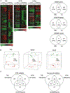
Results: Differential expression profiles between 10 Gy and iso-survival radiation in cells with different p53 status were observed. Moreover, distinct gene expression patterns were exhibited among these three cells after 10 Gy radiation treatment, but similar transcriptional responses were observed in TK6 and NH32 cells treated with iso-survival radiation.
Conclusions: After 10 Gy radiation exposure, the p53 signaling pathway played an important role in TK6, whereas the NFkB signaling pathway appeared to replace the role of p53 in WTK1. In contrast, after iso-survival radiation treatment, E2F4 seemed to play a dominant role independent of p53 status. This study dissected the impacts of p53, NFkB and E2F4 in response to higher or lower doses of gamma-irradiation.
Conflict of interest statement
Conflict of interest: none.
Figures


References
-
- Short SC, Buffa FM, Bourne S, et al. Dose- and time-dependent changes in gene expression in human glioma cells after low radiation doses. Radiat Res 2007;168:199–208. - PubMed
-
- Ding LH, Shingyoji M, Chen F, et al. Gene expression profiles of normal human fibroblasts after exposure to ionizing radiation: A comparative study of low and high doses. Radiat Res 2005;164:17–26. - PubMed
-
- Amundson SA, Bittner M, Meltzer P, et al. Induction of gene expression as a monitor of exposure to ionizing radiation. Radiat Res 2001;156:657–661. - PubMed
-
- Criswell T, Leskov K, Miyamoto S, et al. Transcription factors activated in mammalian cells after clinically relevant doses of ionizing radiation. Oncogene 2003;22:5813–5827. - PubMed
-
- Amundson SA, Lee RA, Koch-Paiz CA, et al. Differential responses of stress genes to low dose-rate gamma irradiation. Mol Cancer Res 2003;1:445–452. - PubMed
Publication types
MeSH terms
Substances
Grants and funding
LinkOut - more resources
Full Text Sources
Research Materials
Miscellaneous

