. 2009 Dec 11;139(6):1119-29.
doi: 10.1016/j.cell.2009.11.002.

A structure-based mechanism for vesicle capture by the multisubunit tethering complex Dsl1

Affiliations

A structure-based mechanism for vesicle capture by the multisubunit tethering complex Dsl1

Yi Ren et al. Cell. .

Abstract

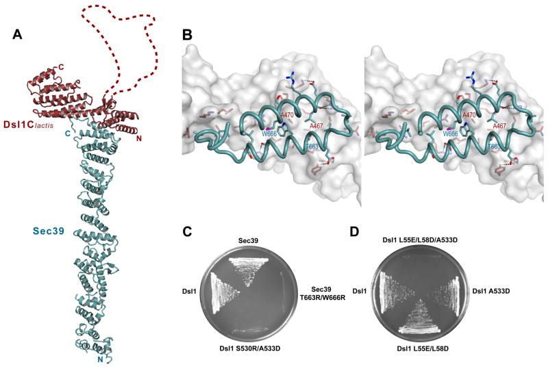
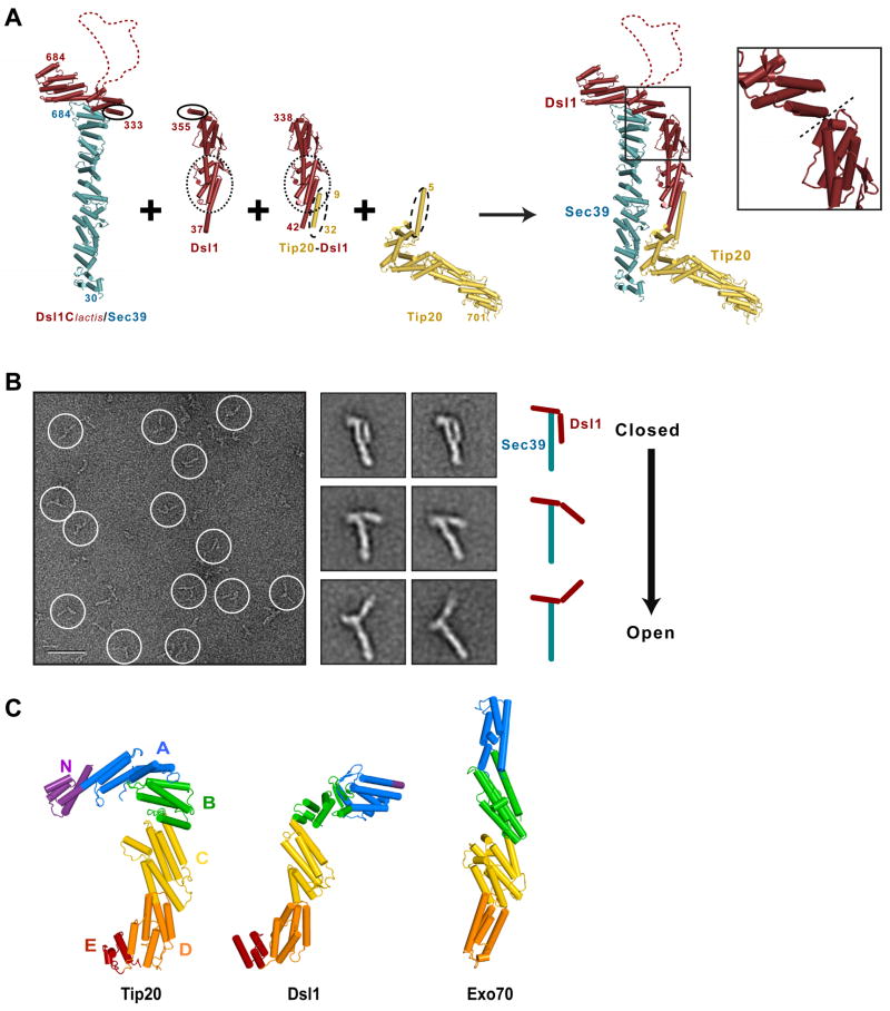
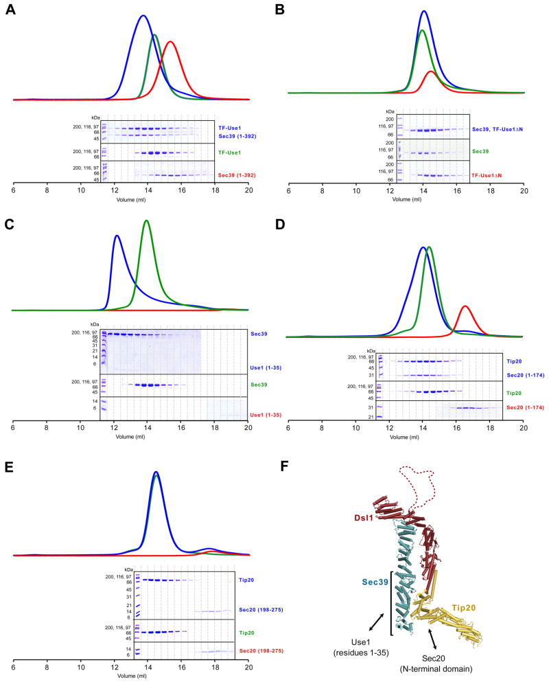
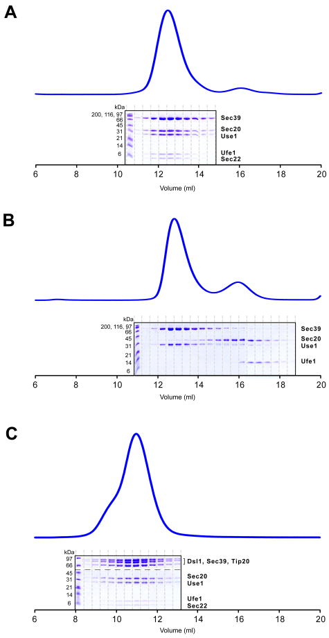
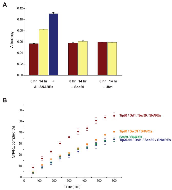
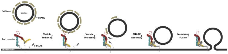
Vesicle trafficking requires membrane fusion, mediated by SNARE proteins, and upstream events that probably include "tethering," an initial long-range attachment between a vesicle and its target organelle. Among the factors proposed to mediate tethering are a set of multisubunit tethering complexes (MTCs). The Dsl1 complex, with only three subunits, is the simplest known MTC and is essential for the retrograde traffic of COPI-coated vesicles from the Golgi to the ER. To elucidate structural principles underlying MTC function, we have determined the structure of the Dsl1 complex, revealing a tower containing at its base the binding sites for two ER SNAREs and at its tip a flexible lasso for capturing vesicles. The Dsl1 complex binds to individual SNAREs via their N-terminal regulatory domains and also to assembled SNARE complexes; moreover, it is capable of accelerating SNARE complex assembly. Our results suggest that even the simplest MTC may be capable of orchestrating vesicle capture, uncoating, and fusion.


Figures

Figure 1
Figure 1. X-ray Crystal Structure of a Dsl1/Sec39 Complex
(A) Ribbon representation of the complex between Dsl1Clactis (residues 332–686; red) and Sec39 (residues 1–709; blue). Dsl1Clactis residues 367–423, which are apparently disordered, are represented by a dashed red line. (B) Stereo view of the Dsl1Clactis/Sec39 binding interface. Dsl1Clactis is shown as a surface representation, while Sec39 is shown as a ribbon representation. Side chains are included for residues involved in binding. (C) Mutations, in either dsl1 or sec39, that disrupted the Dsl1–Sec39 interaction were lethal in yeast. Deletion strains bearing both a CEN-URA plasmid encoding the wild-type protein and a CEN-LEU plasmid encoding the corresponding mutant protein as indicated were grown at 23°C on 5-FOA plates. (D) dsl1 mutations that disrupt the Dsl1–Sec39 interaction (A533D) and the Dsl1–Tip20 interaction (L55E/L58D) are synthetically lethal.
Figure 2
Figure 2. Molecular Architecture of the Yeast Dsl1 Complex
(A) Modeling of the entire Dsl1 complex using crystal structures of Dsl1Clactis–Sec39, Dsl1 (PDB 3ETU), a Tip20-Dsl1 fusion protein (PDB 3ETV), and Tip20 (PDB 3FHN). Sec39, Dsl1, and Tip20 are colored in blue, red, and yellow, respectively. Regions of overlap were overlaid to yield a model representing the intact Dsl1 complex; these include Dsl1 residues 341 to 355 and Dsl1lactis residues 333–347 (indicated by solid ovals), Dsl1 residues 52 to 205 (dotted ovals), and Tip20 residues 9 to 32 (dashed ovals). Inset: Close-up view of the proposed flexible hinge (dashed line) between domains B and C (see panel C) near the center of the Dsl1 subunit. (B) Negative stain EM of the Dsl1–Sec39 complex. Left: typical image of a sample stained with uranyl formate (scale bar = 50 nm). Middle: representative class averages displaying different conformations (side length = 36 nm). Right: cartoon representation of the class averages. (C) Structural alignment of Tip20 (PDB 3FHN), Dsl1 (as modeled in (A)), and Exo70 (PDB 2PFV). Domains C through E were superimposed using DaliLite (Holm and Park, 2000).
Figure 3
Figure 3. Mapping Interacting Regions Between the Dsl1 complex and the ER SNAREs Use1 and Sec20
(A) The N-terminal region of Sec39 (residues 1–392) binds TF-Use1 (residues 1–212 fused to trigger factor). Sec39 alone, Use1 alone, or an equimolar mixture were analyzed by gel filtration. (B) Sec39 does not bind TF-Use1ΔN (residues 36–216 fused to trigger factor). (C) Sec39 binds the N-terminus of Use1 (residues 1–35). Binding of Sec39 to Use1 residues 1–35, but not to Use1 residues 1–212 (data not shown), appears to induce oligomerization as judged by performance on gel filtration. (D) Tip20 binds the N-terminal regulatory domain of Sec20 (residue 1–174). (E) Tip20 does not bind the Sec20 SNARE motif (residues 198–275). (F) Summary of the interactions between the Dsl1 complex and the SNAREs Use1 and Sec20.
Figure 4
Figure 4. SNARE Complexes and Their Interaction with the Dsl1 complex
(A) Three ER SNAREs, Use1 (residues 1–212, stabilized/solubilized by Sec39), Sec20 (residues 1–275), and Ufe1 (residues 240–320), together with the vesicle SNARE Sec22 (residues 125–188), assemble to form complexes. (B) The three ER SNAREs do not interact in the absence of the vesicle SNARE, Sec22. (C) SNARE complexes assemble in the presence of the intact Dsl1 complex. Individual gel filtration fractions were analyzed both by 10% SDS-PAGE gel (to visualize the high molecular weight polypeptides composing the Dsl1 complex) and by tricine SDS-PAGE gel (to visualize SNAREs); in the figure, the gels are separated by a dashed line.
Figure 5
Figure 5. The Dsl1 Complex Accelerates SNARE Assembly
(A) Fluorescence polarization experiments were performed using fluorescein-labeled Sec22 as described in the Experimental Procedures. An increase in anisotropy, signifying SNARE complex assembly, was observed after a 14-hr incubation of Use1 (stabilized/solubilized by Sec39), Sec20, Ufe1, and Sec22. The anisotropy of purified SNARE complexes is also shown. Little change in anisotropy, as compared to t=0, was observed if either Sec20 or Ufe1 was omitted from the mixture. Error bars represent SEM for two independent experiments. (B) SNARE assembly was monitored using fluorescence anisotropy as a function of time. Details are provided in the Experimental Procedures. Error bars represent SEM for three independent experiments.
Figure 6
Figure 6. Working Model for Vesicle Tethering via the Dsl1 Complex
The Dsl1 complex is depicted in a space-filling representation, color-coded as in Figure 2A. Evidence for lasso-COPI interactions has been presented by Schmitt and co-workers (Andag et al., 2001; Andag and Schmitt, 2003; Zink et al., 2009). Although two different conformations of the Dsl1 complex are illustrated, it is not known whether, or in what manner, the conformation and/or flexibility of the complex is affected by its interactions with SNAREs and COPI.

Comment in

Similar articles

Cited by

References

    1. Adams PD, Grosse-Kunstleve RW, Hung LW, Ioerger TR, McCoy AJ, Moriarty NW, Read RJ, Sacchettini JC, Sauter NK, Terwilliger TC. PHENIX: building new software for automated crystallographic structure determination. Acta Crystallogr D. 2002;58:1948–1954. - PubMed
    1. Andag U, Neumann T, Schmitt HD. The coatomer-interacting protein Dsl1p is required for Golgi-to-endoplasmic reticulum retrieval in yeast. J Biol Chem. 2001;276:39150–39160. - PubMed
    1. Andag U, Schmitt HD. Dsl1p, an essential component of the Golgi-endoplasmic reticulum retrieval system in yeast, uses the same sequence motif to interact with different subunits of the COPI vesicle coat. J Biol Chem. 2003;278:51722–51734. - PubMed
    1. Aoki T, Kojima M, Tani K, Tagaya M. Sec22b-dependent assembly of endoplasmic reticulum Q-SNARE proteins. Biochem J. 2008;410:93–100. - PubMed
    1. Bricogne G, Vonrhein C, Flensburg C, Schiltz M, Paciorek W. Generation, representation and flow of phase information in structure determination: recent developments in and around SHARP 2.0. Acta Crystallogr D. 2003;59:2023–2030. - PubMed

Publication types

MeSH terms

Associated data