. 2009 Dec 10;64(5):645-62.
doi: 10.1016/j.neuron.2009.10.017.

A cluster of cholinergic premotor interneurons modulates mouse locomotor activity

Affiliations

A cluster of cholinergic premotor interneurons modulates mouse locomotor activity

Laskaro Zagoraiou et al. Neuron. .

Abstract

Mammalian motor programs are controlled by networks of spinal interneurons that set the rhythm and intensity of motor neuron firing. Motor neurons have long been known to receive prominent "C bouton" cholinergic inputs from spinal interneurons, but the source and function of these synaptic inputs have remained obscure. We show here that the transcription factor Pitx2 marks a small cluster of spinal cholinergic interneurons, V0(C) neurons, that represents the sole source of C bouton inputs to motor neurons. The activity of these cholinergic interneurons is tightly phase locked with motor neuron bursting during fictive locomotor activity, suggesting a role in the modulation of motor neuron firing frequency. Genetic inactivation of the output of these neurons impairs a locomotor task-dependent increase in motor neuron firing and muscle activation. Thus, V0(C) interneurons represent a defined class of spinal cholinergic interneurons with an intrinsic neuromodulatory role in the control of locomotor behavior.


Figures

Figure 1
Figure 1. Origin and neurotransmitter phenotype of spinal Pitx2+ neurons
(A-D) Pitx2 expression in e17.5 (A) and p8 (B-D) spinal cord. Spinal Pitx2 (munc-30) expression has been noted (Nicholson and Goulding, 2001). (E-J) Neurotransmitter phenotype of Pitx2+ neurons in p8 spinal cord. Pitx2 expression in rostral (E) and caudal (F) lumbar levels. ChAT and Pitx2 expression in rostral (G) and caudal (H) lumbar levels. (I-I″) vGluT2 expression in a subset of lumbar Pitx2+ neurons. Arrows in I indicate cells shown in higher magnification in I′ and I″. (J) Cholinergic Pitx2+ neurons predominate at upper lumbar levels whereas at lower lumbar levels most Pitx2+ neurons are glutamatergic. Data from 3 p4 and 2 p8 mice (mean ± S.E.) per 100 μm. >90% of all Pitx2+ neurons can be accounted for by ChAT or vGluT2 expression. (K) Origin and diversity of p0 domain-derived V0 neurons. For details, see text and Lanuza et al. (2004). (L-Q) Pitx2+ neurons derive from p0 domain progenitors. Most Pitx2+ neurons express nuclear LacZ in a Dbx1∷LacZ transgene in e12.5 spinal cord (L, M, M′). Arrows indicate double labeled cells. Pitx2 is expressed in e16.5 Dbx1 heterozygous mice (N) but expression is lost in Dbx1 mutant mice (O). (P) Evx1 expression at lumbar (lu) levels and (Q) thoracic (th) levels at e12.5. Virtually all newly-generated Pitx2+ neurons co-express Evx1, but by e14, Evx1 expression has been extinguished from most neurons. In addition to the Pitx2+ V0 population, rare Pitx2+ neurons are detected in the dorsal spinal cord (1.6 neurons/100 micron; n = 6 mice) (Figure S7). These non-cholinergic neurons likely correspond to neurons described by Polgar et al. (2007). Scale bars = 100μm (A-D), 20μm (G).
Figure 2
Figure 2. Genetic marking of Pitx2+ neurons
(A-D′) Genetic marking of Pitx2+ neurons in Pitx2∷Cre; Thy1.lsl.YFP mice. (A, B) Fluorescent protein (FP) is expressed in a small subset of peri-central canal neurons (cc: central canal). B shows high power view of the peri-central canal region. (C, D) In this Thy1 line, FP is expressed in 66% of Pitx2+ neurons. (C′, D′) FP and vAChT expression at rostral lumbar levels. (E, F) Genetic marking of V0C neurons in Pitx2∷Cre; vAChT.lsl.eGFP mice reveals FP expression in Pitx2+ and vAChT+ neurons.
Figure 3
Figure 3. Genetic tracing of V0C neuronal connections with motor neurons
(A) Spatial distribution of V0C and V0G axons and terminals in lumbar spinal cord of Pitx2∷Cre; Tau.lsl.mGFP reporter mice. (B, C) Co-expression of FP and vAChT in C-boutons. >95% of vAChT+ terminals on motor neurons express FP. The density of FP-labeled boutons on motor neurons that innervate proximal hindlimb muscles was ∼3-fold greater than that on motor neurons innervating distal footpad (plantar) muscles, consistent with the known pattern of C-bouton innervation (Hellstrom, 2004). (D-D‴) FP+ terminals motor neurons express vAChT but not vGluT2. (E-E″) FP+ terminals in the intermediate spinal cord express vGluT2. (F, G) In Pitx2∷Cre; Tau.lsl.mGFP mice, m2 muscarinic receptor (m2) and Kv2.1 channel (Kv) clusters are aligned with FP+ C-boutons. (H, I) FP+ C-boutons are concentrated on motor neuron somata and proximal dendrites. Cholera toxin B (CTB) subunit-labeled tibialis anterior motor neurons in lumbar spinal cord of a p24 Pitx2∷Cre; Tau.lsl.mGFP mouse. FP-labeled terminals are detected on the soma and proximal dendrites (15.7 C-boutons/50 micron length of proximal dendrite, n = 3), but more distal dendritic domains are devoid of FP+ terminals. Scale bars = 20μm (C), 2μm (F, G).
Figure 4
Figure 4. The synaptic circuitry of V0C interneurons
(A-H″) Lack of connectivity of V0C neurons with identified interneurons. (A) Motor neuron (MN), Sox14+ V2a interneuron (V2a) and calbindin+ Renshaw cell (R) position in lumbar spinal cord. (B) GFP+ V2a interneurons in lumbar spinal cord of p15 Sox14∷eGFP mice. (C) GFP labeled Sox14+ neurons are contacted by few vAChT+ terminals (<4 boutons per neuron, 37 neurons). (D-D″) vGluT1+ terminals on dorsally located Sox14+ interneurons. (E, F) In p24 Pitx2cre; Tau/Thy1.lsl.FP mice, calbindin+ Renshaw cells are contacted by vAChT terminals that do not express FP (n = 0/132 boutons, 8 neurons). (G) GFP+ motor neurons in Hb9∷eGFP mice lack FP+ vAChT input. (H-H″) In p21 Hb9∷eGFP mice, most vAChT+ terminals on calbindin+ Renshaw cells express FP. (I-Q‴) Synaptic inputs to p24 V0CG interneurons. (I-J) ChAT+, GFP+ V0C neurons contacted by vGluT2+ boutons. Panel J shows a different neuron. (K-L) ChAToff, GFP+ V0G neurons also contacted by vGluT2+ boutons. Panel L shows a different neuron. (M-O) ChAT+, GFP+ V0C neurons contacted by 5HT+ boutons. Panels N and O show different neurons. (P-P‴) ChAT+, GFP+ V0C neurons contacted by vGluT1+ boutons. (Q-Q‴) ChAT+, GFP+ V0C neurons contacted by GAD67+ boutons. (R) Connectivity of V0C neurons. Scale bar = 2μm (I″, K″, M″, P″, Q″).
Figure 5
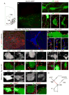
Figure 5. Intrinsic properties of Pitx2+ V0CG interneurons
(A) Hemisected spinal cord preparation used for physiology. (B-D) IR-DIC and fluorescence images of an identified V0CG neuron from a Pitx2∷Cre; Thy1.lsl.YFP mouse. The cell was patch-clamped under IR-DIC optics and filled with Alexa 594 during recording. (E) Slow frequency, tonic activity recorded from a FP-labeled V0CG neuron. Prominent after-hyperpolarization shown in grey inset. (F) Recordings from a FP-labeled V0CG neuron after injection of depolarizing and hyperpolarizing current pulses (1s duration). Bottom trace shows injected current. (G) Instantaneous firing frequency of a FP-labeled V0CG neuron in response to 1s injection of depolarizing current. (H) Steady-state firing frequency - current plot (f-I) for a FP-labeled V0CG neuron upon injection of incremental (1s) depolarizing current steps. (I) Spinal cord slice preparation. (J) Instantaneous firing frequency (moving average of 5 consecutive spikes; top) and corresponding current-clamp recordings (bottom) from a tonically active Thy1 FP-labeled V0CG neuron in a slice preparation. Control (left) and with NMDA (5μM), 5-HT (10μM) and dopamine (50μM) (right).
Figure 6
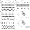
Figure 6. Activity of Pitx2+ V0CG neurons during locomotor episodes
(A) Moving average (5 consecutive spikes) of the instantaneous firing frequency of a tonically active Thy1 FP-labeled V0CG neuron (top trace) along with the corresponding current-clamp recording (2nd trace) and rectified/integrated ventral root recordings (bottom traces) during drug-induced (NMDA 5μM, 5-HT 10μM, dopamine 50μM) locomotor activity in a hemisected spinal cord preparation. (B) Phasic activity recorded from neuron in A during the injection of hyperpolarizing current (top trace) along with rectified/integrated recordings of locomotor activity from ventral roots (bottom traces). (C) Recording from a bursting Thy1 FP-labeled V0CG neuron (top trace) and rectified/integrated recordings of locomotor activity from ventral roots (bottom traces). The coupling between the spiking of this FP-labeled V0CG neuron and ventral root activity is indicated by arrows. (D) Moving average (5 consecutive spikes) of the instantaneous firing frequency of a tonically active vAChT FP-labeled V0C neuron (top trace) along with the corresponding current-clamp recording (2nd trace) and rectified/integrated recordings of locomotor activity from ventral roots (bottom traces). (E) Circular plot for the FP-labeled V0CG neuron shown in C depicting its preferred firing phase (mean vector; arrow) in relation to the locomotor cycle. The start of the locomotor cycle (0.0) is taken as the onset of the burst in the rostral lumbar ventral root. Shaded area highlights the average duration of rostral lumbar root activity. Each point on the circle corresponds to a single action potential. The direction of the mean vector indicates the preferred firing phase of the neuron, and the length of the vector indicates the tuning of action potentials around their mean. (F) Circular plot showing the preferred firing phases (position of mean vectors) for all FP-labeled V0CG neurons, revealing a significant correlation with ventral root bursting (Rayleigh test, p < 0.05). Data include neurons from Pitx2∷Cre; Thy1.lsl.YFP (L1-L3 levels, closed circles; L4-L5 levels, open circles) and Pitx2∷Cre; vAChT mice (L1-L3 levels, open triangles).
Figure 7
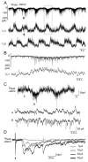
Figure 7. Synaptic inputs to Pitx2+ V0CG interneurons
(A) Voltage-clamp recording of a Thy1 FP-labeled V0CG neuron held at -60mV (top trace) and rectified/integrated ventral root recordings (bottom traces) during drug-induced (NMDA 5μM, 5-HT 10μM, dopamine 50μM) locomotor activity in a hemisected spinal cord preparation. The coupling between EPSCs recorded in this FP-labeled V0CG neuron and L1 ventral root activity is indicated by arrows. (B) A volley of EPSCs recorded from the FP-labeled V0CG neuron in A (top trace) and a simultaneous ventral root burst (bottom trace). These data are outlined by the dotted box in A. (C) Voltage-clamp recordings of a Thy1 FP-labeled V0CG neuron held at -40mV reveal IPSCs throughout the locomotor cycle. Bottom two traces show data from the time points marked ‘a’ and ‘b’ in the top trace. (D) Voltage-clamp recording of EPSCs evoked in a Thy1 FP-labeled V0CG neuron by dorsal root stimulation (5-30 μA, 0.5 ms). Each trace is an average of 5 sweeps, arrowhead points to stimulus artefact).
Figure 8
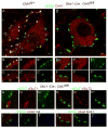
Figure 8. Genetically programmed elimination of ChAT from V0C interneurons
(A-C″) Lumbar motor neurons in p24 ChAT fl/+ mice express ChAT. C-bouton terminals on motor neurons express both vAChT and ChAT. (D-F″) Lumbar motor neurons in p24 Dbx1∷cre; ChAT fl/fl mice express ChAT. Their C-bouton inputs express vAChT, but not ChAT (n = 503 boutons from 2 p24 and 3 p60 mice). (G-H″) In Dbx1∷cre; ChAT fl/fl mice (p24 and p60), ChAT depleted C-boutons do not express vGluT1 (G) or vGluT2 (H). (I-J″) In Dbx1∷cre; ChAT fl/fl mice (p60), m2 muscarinic receptors (I) and Kv2.1 channels (J) are clustered in alignment with vAChT+, ChAT-deficient C-boutons. Scale bar = 2μm (A, D).
Figure 9
Figure 9. Preservation of basic locomotor pattern in mice with ChAT-depleted V0C neurons
(A) EMG recordings from the contralateral tibialis anterior (TAcont, ankle flexor), ipsilateral TA (TAipsi.) and the ipsilateral gastrocnemius (Gsipsi., ankle extensor) in control (Chatfl/fl, left recordings; n = 7) and ChAT-depleted V0C neuron mice (Dbx1∷Cre; Chatfl/fl) (right) recordings; n = 8 mice), during walking. (B) In-phase activation of flexor muscles acting on different joints of the same leg (iliopsoas, Ipipsi, hip flexor and TAipsi, ankle flexor) is preserved in control (Chatfl/fl) (left recordings; n = 5) and ChAT-depleted V0C neuron mice (Dbx1∷Cre; Chatfl/fl) (right recordings; n = 4), during walking.
Figure 10
Figure 10. Task-specific impairment in gastrocnemius muscle activation in mice with ChAT-depleted V0C neurons
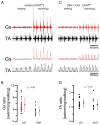
(A) EMG recordings from Gs (red) and TA (black) muscles during walking (left) and swimming (right), in control (Chatfl/fl) mice. Raw and integrated EMG traces are shown. (B) Ratio of Gs EMG amplitudes during walking and swimming in control wt (n = 8) and Chatfl/fl (n = 14 mice) and from V0C ChAT-deficient Dbx1∷Cre; Chatfl/fl mice (n = 12 mice). The Gs data obtained from wt and ChATfl/fl mice were pooled, since they were not statistically different (p=0.13). The ratio of average peak values of Gs EMG activities during swimming and walking was significantly lower for Dbx1∷Cre; Chatfl/fl mice compared to controls. (C) EMG recordings from Gs (red) and TA (black) muscles during walking (left) and swimming (right), in Dbx1∷Cre; Chatfl/fl mice. (D) Ratio of TA EMG amplitudes during walking and swimming in control Chatfl/fl (n = 12) and V0C ChAT-deficient Dbx1∷Cre; Chatfl/fl mice (n = 14 mice).
Figure 11
Figure 11. Intrinsic Neuromodulatory Role of V0C interneurons
A model of the intraspinal circuitry and function of Pitx2+ V0C interneurons. V0C neurons form numerous cholinergic C-bouton synaptic contacts with motor neurons. They receive direct synaptic input from excitatory interneurons involved in the rhythmogenic central pattern generator (CPG) system, inputs from descending pathways, and indirect input from sensory afferents. During walking, the combined influence of these synaptic inputs results in a moderate level activation of the set of V0C neurons that innervate Gs motor neurons, which together with direct CPG input to motor neurons, results in an intermediate rate of Gs motor neuron firing and a modest contraction of the Gs muscle. During swimming, a task-dependent change in the activity of sensory and descending pathways increases the level of activation of V0C neurons, activating muscarinic m2 receptors on motor neurons, enhancing Gs motor neuron firing frequency (Miles et al., 2007), and increasing the amplitude of Gs muscle contraction. For simplicity, we have not depicted direct descending modulatory inputs to motor neurons, which could contribute to the task-dependent modulation of Gs motor neuron activity. The question mark indicates the uncertain nature of the descending and/or sensory inputs that mediate the task-dependent regulation of V0C neuronal activity.

Comment in

Similar articles

Cited by

References

    1. Akay T, Acharya HJ, Fouad K, Pearson KG. Behavioral and electromyographic characterization of mice lacking EphA4 receptors. J Neurophysiol. 2006;96:642–651. - PubMed
    1. Al-Mosawie A, Wilson JM, Brownstone RM. Heterogeneity of V2-derived interneurons in the adult mouse spinal cord. Eur J Neurosci. 2007;26:3003–3015. - PubMed
    1. Alvarez FJ, Fyffe RE. The continuing case for the Renshaw cell. J Physiol. 2007;584:31–45. - PMC - PubMed
    1. Alvarez FJ, Jonas PC, Sapir T, Hartley R, Berrocal MC, Geiman EJ, Todd AJ, Goulding M. Postnatal phenotype and localization of spinal cord V1 derived interneurons. J Comp Neurol. 2005;493:177–192. - PMC - PubMed
    1. Banfield BW, Kaufman JD, Randall JA, Pickard GE. Development of pseudorabies virus strains expressing red fluorescent proteins: new tools for multisynaptic labeling applications. J Virol. 2003;77:10106–10112. - PMC - PubMed

Publication types

MeSH terms

LinkOut - more resources