. 2009 Dec 11;36(5):743-53.
doi: 10.1016/j.molcel.2009.10.014.

The unstructured C-terminal tail of the 9-1-1 clamp subunit Ddc1 activates Mec1/ATR via two distinct mechanisms

Affiliations

The unstructured C-terminal tail of the 9-1-1 clamp subunit Ddc1 activates Mec1/ATR via two distinct mechanisms

Vasundhara M Navadgi-Patil et al. Mol Cell. .

Abstract

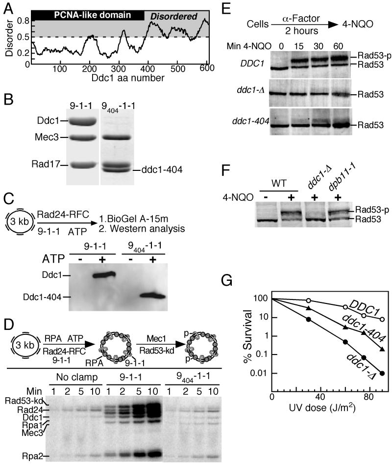
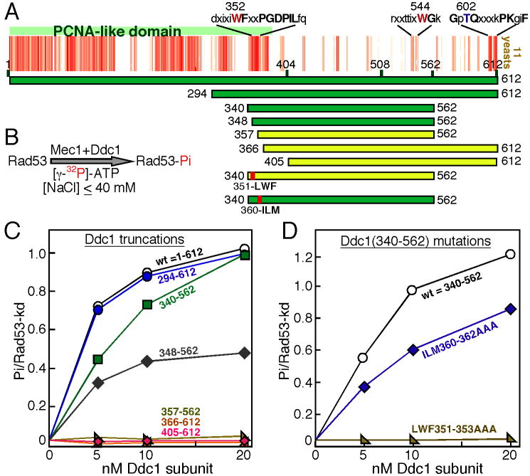
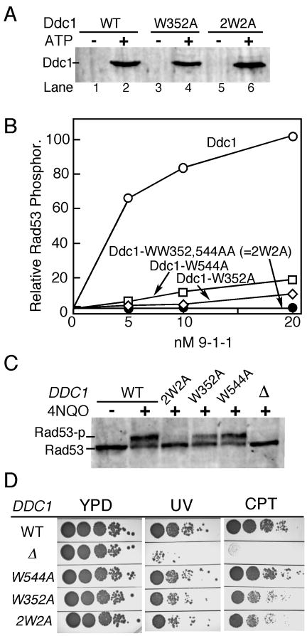
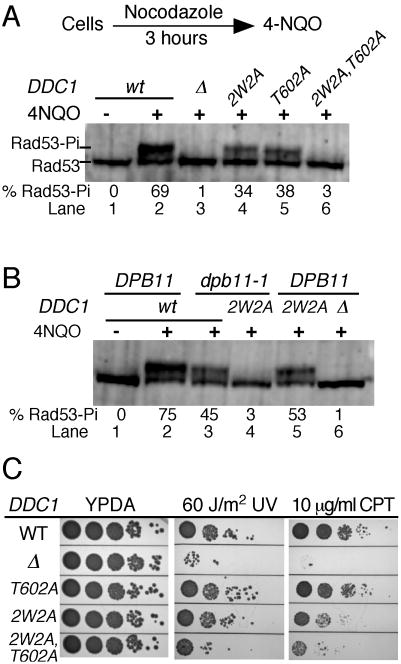
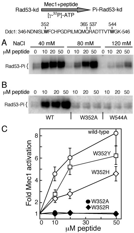
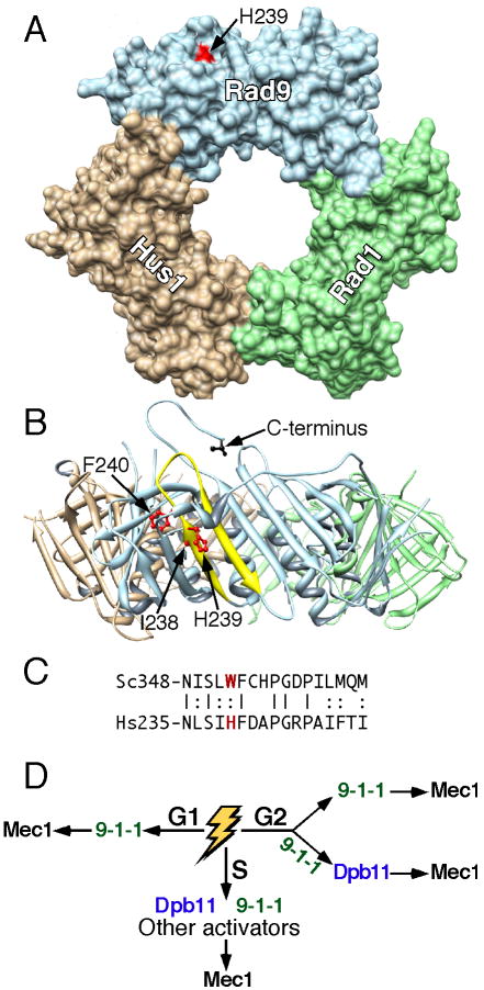
DNA damage checkpoint pathways operate to prevent cell-cycle progression in response to DNA damage and replication stress. In S. cerevisiae, Mec1-Ddc2 (human ATR-ATRIP) is the principal checkpoint protein kinase. Biochemical studies have identified two factors, the 9-1-1 checkpoint clamp and the Dpb11/TopBP1 replication protein, as potential activators of Mec1/ATR. Here, we show that G1 phase checkpoint activation of Mec1 is achieved by the Ddc1 subunit of 9-1-1, while Dpb11 is dispensable. However, in G2, 9-1-1 activates Mec1 by two distinct mechanisms. One mechanism involves direct activation of Mec1 by Ddc1, while the second proceeds by Dpb11 recruitment mediated through Ddc1 T602 phosphorylation. Two aromatic residues, W352 and W544, localized to two widely separated, conserved motifs of Ddc1, are essential for Mec1 activation in vitro and checkpoint function in G1. Remarkably, small peptides that fuse the two tryptophan-containing motifs together are proficient in activating Mec1.


Figures

Figure 1
Figure 1. The Ddc1 C-terminal tail is involved in the activation of Mec1 kinase in the G1 phase
(A) Domain map of Ddc1. The PCNA-like domain (1-385 aa) is indicated in gray. Disordered regions were obtained by submitting the Ddc1 sequence to disorder-prediction programs (IUPRED (iupred.enzim.hu), PONDR (www.pondr.com), and PrDOS (prdos.hgc.jp)), and averaging the output. A disorder value >0.5 is considered to indicate an unstructured protein region. (B) Coomassie stained 10 % SDS-PAGE gel showing the co-purification of Rad17 and Mec3 with wild type Ddc1, or with Ddc1-404 after overexpression in yeast. The migration position of clamp subunits is indicated. (C) The PCNA like domain of Ddc1 is sufficient to form a 9-1-1 clamp that can be loaded onto DNA by Rad24-RFC in an ATP dependent manner. The loaded clamp is detected by Western analysis with anti-Ddc1 antibodies. A flow diagram of the loading assay is shown. See Methods for details. (D) A flow diagram of the complete in vitro phosphorylation assay as described in Methods is shown. Aliquots were taken at the indicated times and analyzed by SDS-PAGE. (E) Western analysis of Rad53 phosphorylation in G1 cells. Wild type, ddc1Δ and ddc1-404 cells were arrested in G1 phase and treated with 4NQO for the indicated times, as described in Methods. The hyperphosphorylated form of Rad53 is indicated as Rad53-p. (F) Strains were grown at 25 °C, arrested in G1, and exposed to 4NQO for 60 min where indicated. A Western analysis with Rad53 antibodies was performed. (G) Dose-response survival curves of the indicated DDC1 mutants (see Methods for details).
Figure 2
Figure 2. Mapping of a Mec1 activation determinant in the PCNA-like domain of Ddc1
(A) Multiple sequence alignment of eleven Ddc1 species from the Saccharomycetales order (budding yeast, see also Supplem Fig. 4) using Kalign with a gap penalty of 5 and a gap extension penalty of 0.5 (default is 11, 0.7). Each red bar indicates strong identity with the consensus and each pink bar indicates conservation with the consensus. Note that the occurrence of multiple gaps has spread out the 404-612 region. The conserved motifs and aa of interest are indicated. Ddc1 domains tested for Mec1 activation in the low-NaCl bypass assay are shown with bars (green is active, yellow is inactive). (B) Flow diagram of the bypass Mec1 activation assay by Ddc1 at 40 mM NaCl, used in (C) and (D). The bypass assay does not require a heterotrimeric 9-1-1 clamp, Rad24-RFC clamp loader, nor DNA (see methods). (C) Quantification of Rad53-kd phosphorylation by Mec1, activated by increasing levels of the Ddc1 domains shown in (A). Background phosphorylation of Rad53-kd by Mec1, obtained in the absence of Ddc1 was substracted. (D) Quantification of Rad53-kd phosphorylation by Mec1, activated by increasing levels of the Ddc1(340-562) protein with triple-point mutants as shown in (A).
Figure 3
Figure 3. Mapping of bi-partite Mec1 activation motifs in Ddc1
(A) Ddc1-W352 and Ddc1-2W2A (WW352,544AA) form functional 9-1-1 clamps that can be loaded onto DNA by Rad24-RFC in an ATP dependent manner. See legend to Fig. 1C and Methods for details. (B) The complete in vitro Mec1 phosphorylation assay was carried out at 125 mM NaCl with indicated levels of (mutant) 9-1-1 clamps (see Fig 1D and Methods for details). Phosphorylation of Rad53-kd is quantified. Background phosphorylation of Rad53-kd by Mec1, obtained in the absence of Ddc1 was substracted. (C) Western analysis of G1-arrested cells exposed to 4NQO for 30 min. (D) Ten-fold serial dilutions of wild type cells and the indicated DDC1 mutants were tested for sensitivity to UV (60 J/m2) or camptothecin (10 μg/ml). Plates were incubated at 30 °C for two days and photographed.
Figure 4
Figure 4. The 9-1-1 clamp activates Mec1 in G2 cells via two distinct mechanisms
(A) Log phase cells were arrested in G2 phase with nocodazole (20 μg/ml) for 3 hours at 30 °C, then treated with 4NQO for 20 min. A western analysis of Rad53 phosphorylation was carried out as described in Methods. The % of hyperphosphorylated Rad53 is shown beneath the lanes. (B) as in (A), except that the entire experiment was carried out at 23 °C (dpb11-1 is temperature-sensitive for growth), nocodazole treatment was for 4 hours, and 4NQO treatment for 20 min. (C) Ten-fold serial dilutions of wild type cells and the indicated DDC1 mutants were tested for sensitivity to UV (60 J/m2) or camptothecin (10 μg/ml). Plates were incubated at 30 °C for two days and photographed.
Figure 5
Figure 5. A Ddc1 peptide containing the bipartite Mec1 activation sequence activates Mec1
A 30 mer peptide containing the bipartite Ddc1 sequence from 346 to 365 aa and 537 to 546 aa is shown. Residues Trp352 and Trp544 required for Mec1 activation are indicated. (A) Kinase activity of Mec1 was detected using Rad53-kd as a substrate in buffer containing the indicated levels of NaCl. Increasing levels of Ddc1 peptide were added and phosphorylated Rad53-kd analyzed after 10 min. (B) Ddc1 peptides with W352A or W544A mutations are inactive for Mec1 stimulation. The assay as in (A) was carried out in the presence of 60 mM NaCl. (C) Quantification of Mec1 activation by Ddc1 peptides in (B), and those with W352Y, W352H, W352A, and W352R mutations. Average of three experiments with error bars are shown.
Figure 6
Figure 6. Role of 9-1-1 in two distinct checkpoint pathways
(A) Surface representation of the 9-1-1 crystal structure (Dore et al., 2009). Head-on view with I238, H239, and F240 in red. Note that only H239 is surface-exposed. The Rad9 C-terminus is in back of the donut. The Chimera molecular viewer was used (Pettersen et al., 2004). (B) Ribbon representation after a 90° x-axis rotation. The Rad9 (aa235-252) β-strand-loop-β-strand is in yellow with I238, H239, and F240 as stick models in red. The C-terminal aa273 of truncated Rad9 is shown in black. (C) Alignment of the β-strand-loop-β-strand of human Rad9 with yeast Ddc1. (D) Model for the participation of 9-1-1 in two pathways depending on the phase of the cell cycle.

Comment in

References

    1. Araki H, Leem SH, Phongdara A, Sugino A. Dpb11, which interacts with DNA polymerase II(epsilon) in Saccharomyces cerevisiae, has a dual role in S-phase progression and at a cell cycle checkpoint. Proc Natl Acad Sci USA. 1995;92:11791–11795. - PMC - PubMed
    1. Bakkenist CJ, Kastan MB. Initiating cellular stress responses. Cell. 2004;118:9–17. - PubMed
    1. Bonilla CY, Melo JA, Toczyski DP. Colocalization of sensors is sufficient to activate the DNA damage checkpoint in the absence of damage. Mol Cell. 2008;30:267–276. - PMC - PubMed
    1. Choi JH, Lindsey-Boltz LA, Sancar A. Reconstitution of a human ATR-mediated checkpoint response to damaged DNA. Proc Natl Acad Sci U S A. 2007;104:13301–13306. - PMC - PubMed
    1. Delacroix S, Wagner JM, Kobayashi M, Yamamoto K, Karnitz LM. The Rad9-Hus1-Rad1 (9-1-1) clamp activates checkpoint signaling via TopBP1. Genes Dev. 2007;21:1472–1477. - PMC - PubMed

Publication types

MeSH terms

Substances