. 2009 Dec 11;36(5):794-804.
doi: 10.1016/j.molcel.2009.11.015.

Ubiquitinated proteins activate the proteasome by binding to Usp14/Ubp6, which causes 20S gate opening

Affiliations

Ubiquitinated proteins activate the proteasome by binding to Usp14/Ubp6, which causes 20S gate opening

Andreas Peth et al. Mol Cell. .

Abstract

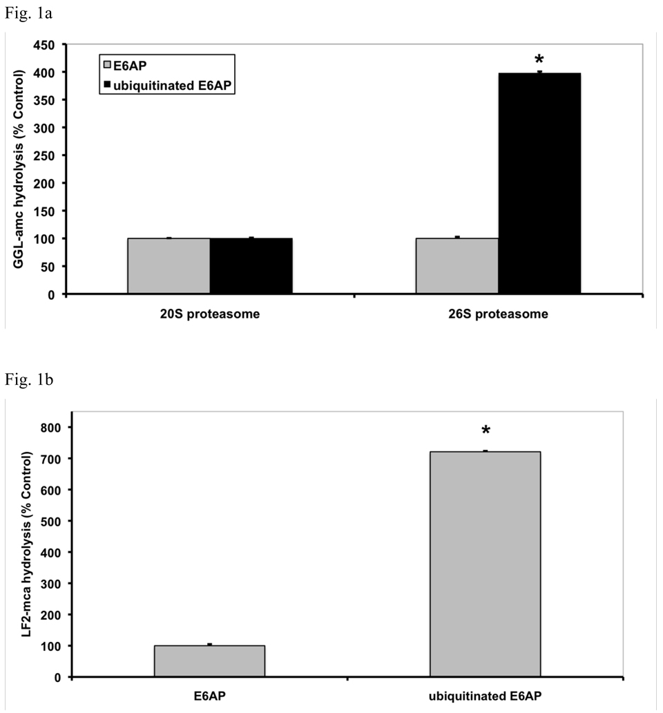
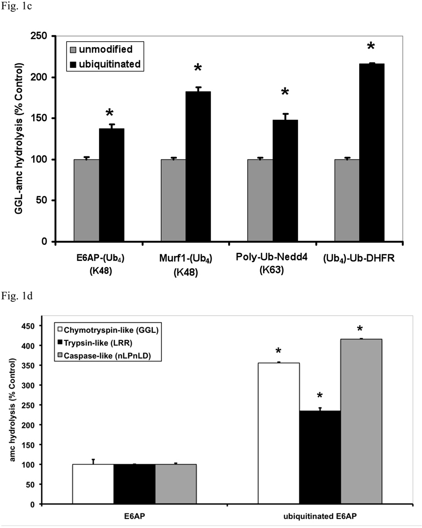
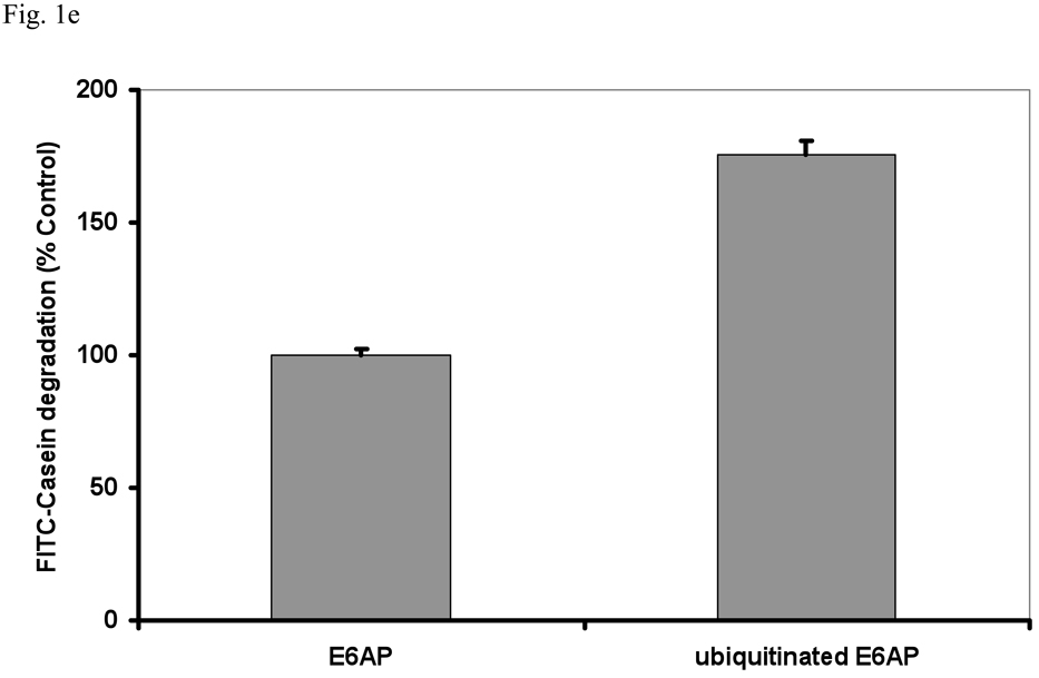
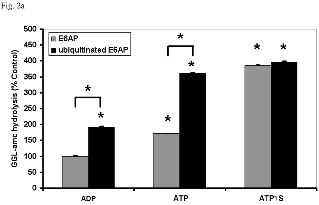
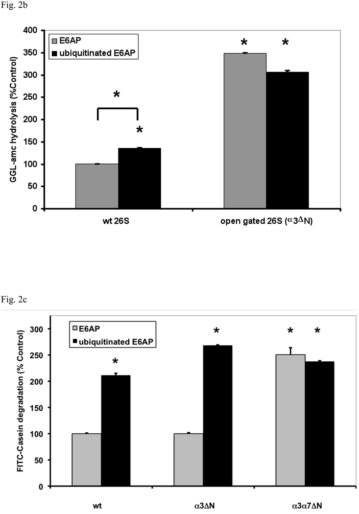
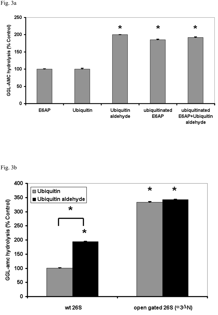
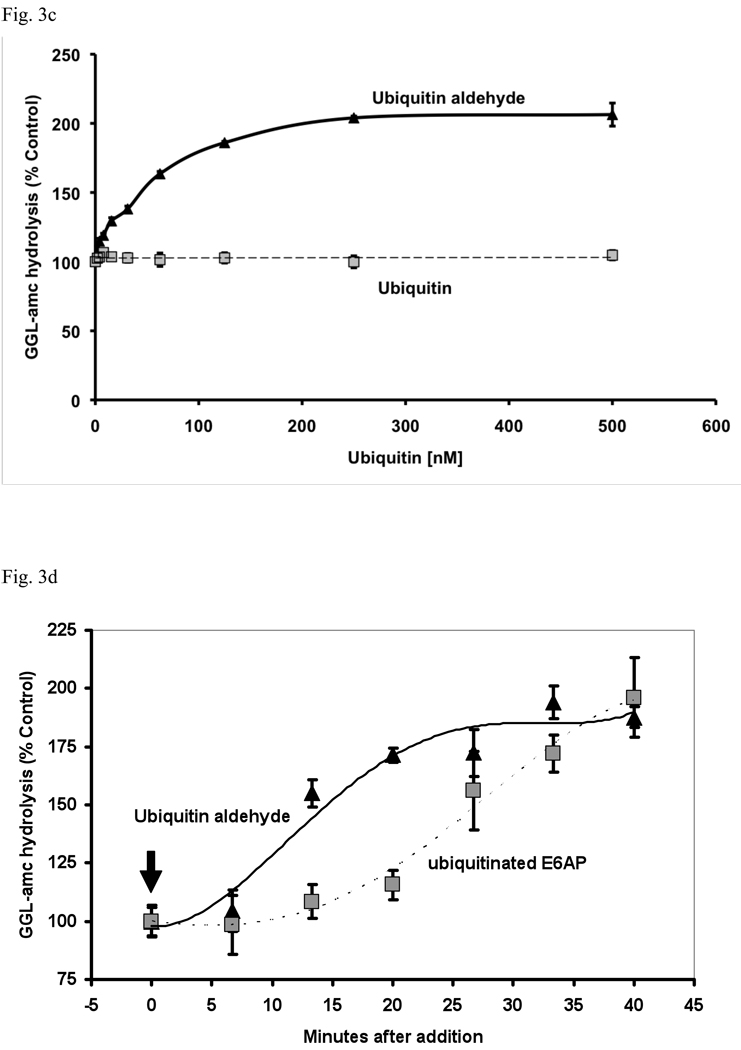
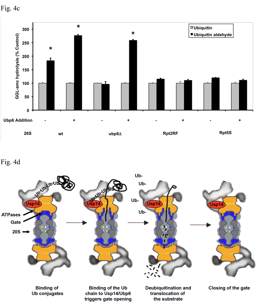
In eukaryotic cells, ubiquitination of proteins leads to their degradation by the 26S proteasome. We tested if the ubiquitin (Ub) chain also regulates the proteasome's capacity for proteolysis. After incubation with polyubiquitinated proteins, 26S proteasomes hydrolyzed peptides and proteins 2- to 7-fold faster. Ub conjugates enhanced peptide hydrolysis by stimulating gate opening in the 20S proteasome. This stimulation was seen when this gate was closed or transiently open, but not maximally open. Gate opening requires conjugate association with Usp14/Ubp6 and also occurs if Ub aldehyde occupies this isopeptidase's active site. No stimulation was observed with 26S from Ubp6Delta mutants, but this effect was restored upon addition of Usp14/Ubp6 (even an inactive Ubp6). The stimulation of gate opening by Ub conjugates through Usp14/Ubp6 requires nucleotide binding to the gate-regulatory ATPases. This activation enhances the selectivity of the 26S proteasome for ubiquitinated proteins and links their deubiquitination to their degradation.


Figures

Fig. 1
Fig. 1. Ub conjugates stimulate peptide hydrolysis by 26S, but not 20S proteasomes
(a) Equal concentrations of purified rabbit 20S and 26S proteasomes (5 nM) were incubated with an excess (500 nM) of E6AP or ubiquitinated E6AP. The chymotrypsin-like activity was measured using the fluorogenic substrate, GGL-amc, (10 µM) at 30°C. Proteasomal activities were expressed relative to that seen with unmodified E6AP, a 2–4 fold increase in activity was observed, depending on the 26S purification. The asterisk indicates P< 0.05. (b) The hydrolysis of the tripeptide GGL-amc and the nonapeptide LF2-mca by 26S proteasomes were measured in the presence of E6AP or ubiquitinated E6AP as described in (a). (c) A variety of autoubiquitinated Ub ligases (E6AP, Nedd4 or MuRF1) and the proteasome substrate, Ub4-Ub-DHFR, stimulate 26S activity. E6AP forms a K48 chain during pre-incubation with E1, E2 and ATP, Nedd4 forms a K63 chain and MuRF1, which was allowed to autoubiquitinate in the presence of K48-linked tetra-Ub, forms predominantly (~66%) a tetra-Ub derivative. Proteasomal activities were measured as described in (a). (d) The activity of the three different peptidase sites were stimulated by ubiquitinated E6AP, but not by E6AP. The trypsin-like activity was measured with LRR-amc, the chymotrypsin-like with GGL-amc and the caspase-like activity with nLPnLD-amc in the presence of ATP. The asterisk indicates P< 0.05. (e) FITC-casein was degraded faster by the 26S proteasome in the presence of ubiquitinated E6AP than E6AP. FITC-casein degradation by the 26S alone was taken as 100%.
Fig. 2
Fig. 2. Ub conjugates stimulate peptide hydrolysis by inducing gate opening in the 20S particle
(a) Stimulation by Ub conjugates occurs with ADP, which maintains the closed conformation of the gate, and ATP which supports gate opening, but not ATPγS where the open gate conformation is maintained continuously. 26S proteasomes were incubated with E6AP or ubiquitinated E6AP in the presence of ADP, ATP or ATPγS (1 mM). Peptide hydrolysis was measured using GGL-amc. 26S activity in the presence of ADP and E6AP was taken as 100%. The asterisk indicates P< 0.05 difference from the control. (b) Ubiquitinated E6AP increased peptide hydrolysis by 26S proteasomes from wt yeast, but not the open gated mutant (α3ΔN). The proteasomes were purified and equal amounts incubated with E6AP or ubiquitinated E6AP as in Fig. 1. The stimulation observed with wt yeast 26S proteasomes is consistently smaller than with mammalian for Ub conjugates but not for Ub aldehyde (see below). The wt incubated with E6AP was taken as 100%. The asterisk indicates P< 0.05 from wt and E6AP. (c) Ub conjugates increase casein hydrolysis in the wt and α3ΔN open-gated 26S particles but not in the α3α7ΔN 26S proteasomes. FITC-casein degradation was measured as described in Fig. 1. The asterisk indicates P< 0.05 from wt and E6AP treated controls.
Fig. 3
Fig. 3. The transition-state inhibitor of 26S-associated deubiquitinating enzymes, Ub aldehyde, causes gate opening
(a) 26S proteasomes purified from rabbit muscle were incubated with E6AP, ubiquitinated E6AP, Ub (500 nM), Ub aldehyde or Ub aldehyde together with ubiquitinated E6AP. Proteasomal activity measured in the presence of E6AP was taken as 100% as in Fig. 1. The asterisk indicates P< 0.05. (b) Unlike Ub, Ub aldehyde stimulates peptide hydrolysis of wt 26S, but not the open gated α3ΔN mutant. The activity of wild type 26S proteasomes incubated with Ub was taken as 100%. The asterisk indicates P< 0.05 from wt. (c) Effect of increasing concentrations of Ub or Ub aldehyde (0 – 500nM) on the activity of 26S proteasomes (5 nM). The estimated K1/2 was 50 nM under these conditions. Proteasomal activities were measured as described in Fig. 1a. (d) Ub aldehyde stimulates peptide hydrolysis more rapidly than Ub conjugates. The time course of 26S peptide hydrolysis was monitored after addition of ubiquitinated E6AP or Ub aldehyde, which were added after the start of the assay as indicated by the arrow. GGL-amc hydrolysis before the addition was taken as 100%.
Fig. 4
Fig. 4. Gate opening by Ub conjugates or Ub aldehyde requires Ubp6 protein in the proteasome but not its deubiquitinating activity
(a) Ubiquitinated E6AP and Ub aldehyde stimulate activity of 26S proteasomes purified from wt yeast and strains expressing the UbpC118A active site mutant, but not from the ubp6 deletion mutant. The activity of untreated wild type 26S proteasomes was taken as 100%. The asterisk indicates P< 0.05. The stimulation by Ub aldehyde in wt yeast 26S proteasomes was consistently greater than by ubiquitinated E6AP. A large stimulation by Ub conjugates and free Ub was seen only in the enzymatically inactive Ubp6C188A mutant. (b) Reconstitution of stimulation (seen in Fig. 4a) was observed in Ubp6-deleted proteasomes after addition of purified wt or C188A mutant Ubp6. 26S proteasomes (5 nM) purified form ubp6 deletion strains were supplemented with wt Ubp6 or Ubp6C118A (20 nM). Peptide hydrolysis was monitored after the addition of Ub or Ub aldehyde. Wt and ubp6Δ proteasomes were used as positive and negative controls for stimulation by Ub aldehyde. The asterisk indicates P< 0.05. (c) Nucleotide binding to the ATPase subunits Rpt2 or Rpt5 is required for stimulation of 26S proteasomes by Ub aldehyde. Peptide hydrolysis was monitored after the addition of Ub or Ub aldehyde, no increase was observed in the Rpt2RF and Rpt5S proteasomes carrying mutations in their ATP-binding motif. 26S particles purified from wt and ubp6Δ strains were used as positive and negative controls for stimulation with Ub aldehyde. Addition of recombinant Ubp6 restores stimulation by Ub aldehyde in ubp6Δ, but not in Rpt2RF and Rpt5S proteasomes. The asterisk indicates P< 0.05. (d) Summary: The binding of Ub conjugates to Usp14 induces gate opening in the 26S proteasome. A cross section of the 26S proteasome shows binding of a ubiquitinated substrate to the 19S regulatory particle. Subsequently, the Ub chain interacts with Usp14, and during its cleavage, it induces maximal gate opening in the 20S particle. The Ub chain is disassembled, and the substrate is translocated into the 20S and hydrolyzed.

Similar articles

Cited by

References

    1. Bajorek M, Finley D, Glickman MH. Proteasome disassembly and downregulation is correlated with viability during stationary phase. Curr Biol. 2003;13:1140–1144. - PubMed
    1. Bech-Otschir D, Helfrich A, Enenkel C, Consiglieri G, Seeger M, Holzhutter HG, Dahlmann B, Kloetzel PM. Polyubiquitin substrates allosterically activate their own degradation by the 26S proteasome. Nat Struct Mol Biol. 2009;16:219–225. - PubMed
    1. Benaroudj N, Zwickl P, Seemuller E, Baumeister W, Goldberg AL. ATP hydrolysis by the proteasome regulatory complex PAN serves multiple functions in protein degradation. Mol Cell. 2003;11:69–78. - PubMed
    1. Besche H, Haas W, Gygi S, Goldberg A. Isolation of mammalian 26S proteasomes and p97/VCP complexes using the ubiquitin-like domain from HHR23B reveals novel proteasome-associated proteins. Biochemistry. 2009 - PMC - PubMed
    1. Borodovsky A, Kessler BM, Casagrande R, Overkleeft HS, Wilkinson KD, Ploegh HL. A novel active site-directed probe specific for deubiquitylating enzymes reveals proteasome association of USP14. Embo J. 2001;20:5187–5196. - PMC - PubMed

Publication types

MeSH terms

LinkOut - more resources