. 2009 Dec 11;36(5):861-71.
doi: 10.1016/j.molcel.2009.11.022.

Optimizing protein stability in vivo

Affiliations

Optimizing protein stability in vivo

Linda Foit et al. Mol Cell. .

Abstract

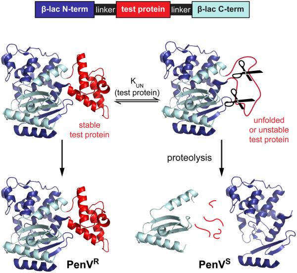
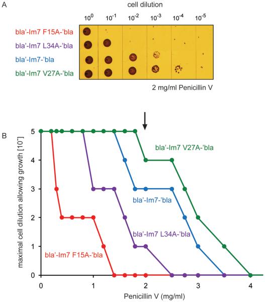
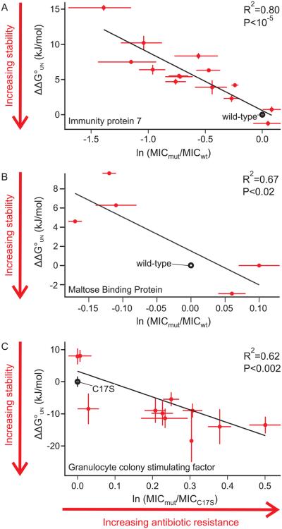
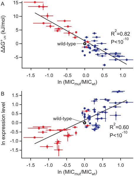
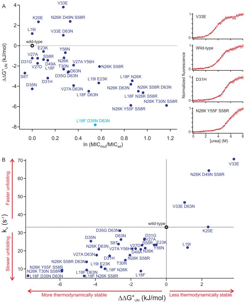
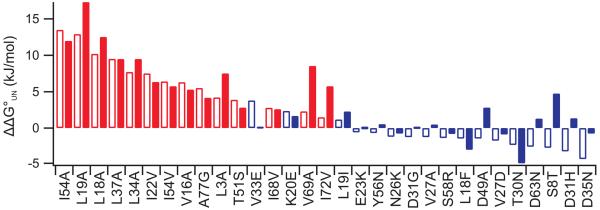
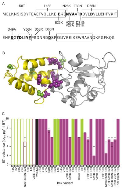
Identifying mutations that stabilize proteins is challenging because most substitutions are destabilizing. In addition to being of immense practical utility, the ability to evolve protein stability in vivo may indicate how evolution has formed today's protein sequences. Here we describe a genetic selection that directly links the in vivo stability of proteins to antibiotic resistance. It allows the identification of stabilizing mutations within proteins. The large majority of mutants selected for improved antibiotic resistance are stabilized both thermodynamically and kinetically, indicating that similar principles govern stability in vivo and in vitro. The approach requires no prior structural or functional knowledge and allows selection for stability without a need to maintain function. Mutations that enhance thermodynamic stability of the protein Im7 map overwhelmingly to surface residues involved in binding to colicin E7, showing how the evolutionary pressures that drive Im7-E7 complex formation have compromised the stability of the isolated Im7 protein.


Figures

Figure 1
Figure 1. Schematic Diagram of the Tripartite System for the Evolution of Improved Protein Stability
The test protein (red) was inserted into TEM1-β-lactamase as part of a tripartite fusion. Poor folding and/or low stability of the test protein should result in an increased sensitivity of the tripartite fusion to cellular proteases and in turn to low levels of resistance to β-lactam antibiotics such as penicillin V. In the case of proper folding and/or increased stability of the test protein, the fusion protein should remain intact and confer high levels of antibiotic resistance. The level of in vivo penicillin resistance may thus be related to the folding/unfolding equilibrium constant of the inserted protein, designated KUN. PenVS = penicillin V sensitive; PenVR = penicillin V resistant; linker = glycine-serine rich linkers of the following lengths: 27 residues for G-CSF, 33 for Im7 and cyt b562, and 68 residues for MBP.
Figure 2
Figure 2. Protein Stability Correlates with Antibiotic Resistance
Mid-log phase cells of E. coli NEB10-β expressing TEM1-β-lactamase fused with wild-type Im7, the destabilized variants Im7 L34A or F15A, or the stabilized variant Im7 V27A were normalized to A600 nm = 1. Serial dilutions of cultures from 100 to 10−5 were spotted on LB plates containing different concentrations of penicillin V. After 18 hr incubation at 37°C, growth or no growth for each dilution at each penicillin V concentration was scored and used to calculate MIC values as described in Figure S1. (A) Spot titer from a single LB plate supplemented with 2 mg/ml penicillin V. (B) Maximal cell dilution that allowed growth plotted against penicillin V concentration. The arrow indicates the penicillin V concentration shown in Figure 2A (2 mg/ml). The published ΔG°UN values for wild-type Im7, F15A, V27A, and L34A are −24.9, −9.7, −27.0, and −17.2 kJ/mol, respectively (Capaldi et al., 2002), which correspond to KUN (the ratio of folded to unfolded protein) values of 40,000, 62, 96,000, and 1,500, respectively.
Figure 3
Figure 3. Antibiotic Resistance Correlates with Stability for a Variety of Proteins
Thermodynamically destabilized or stabilized mutants of Im7 (A), MBP (B), or G-CSF (C) were inserted into β-lactamase via flexible linkers. The level of antibiotic resistance for cells expressing the corresponding fusion constructs was determined as described in Figure S1. The average MIC for penicillin V, relative to the wild-type protein, is plotted against ΔΔG°UN, where ΔΔG°UN = ΔG°UN (mutant) − ΔG°UN (wild-type). In the case of G-CSF, the pseudo wild-type protein C17S was used because wild-type G-CSF has a high tendency to aggregate (Bishop et al., 2001). Error bars represent the standard deviation of the mean of the MIC measurements and the standard errors of the ΔΔG°UN measurements when available.
Figure 4
Figure 4. Antibiotic Resistance Correlates with Stability and Expression Levels of the Variant Proteins
Data points include published destabilized mutants (red) (Capaldi et al., 2002) and mutants selected on the basis of their increased level of antibiotic resistance (blue), see also Figure S2 and Table S1. (A) Thermodynamic stabilities of Im7 variants selected on the basis of an increased MIC. Stabilities were measured by denaturant titration (see Experimental Procedures and Supplemental Data). (B) Expression levels of tripartite fusion proteins that contained thermodynamically destabilized or stabilized mutants of Im7. Fusion proteins were detected by Western blot using a β-lactamase antibody. Band intensities for tripartite fusions containing Im7 mutants were normalized to the expression level of β-lactamase containing wild-type Im7.
Figure 5
Figure 5. Im7 Mutants Selected for Increased Antibiotic Resistance Exhibit Enhanced Thermodynamic and Kinetic Stability
(A) Relative antibiotic resistance, expressed as ln(MICmut/MICwt) is plotted against relative thermodynamic stability, ΔΔG°UN, where ΔΔG°UN = ΔG°UN (mutant) − ΔG°UN wild-type). The designed variant L18F D35N D63N is highlighted in cyan. Equilibrium denaturant titrations for wild-type Im7 and different selected variants are shown on the right. Solid lines show the fit to a two-state equilibrium unfolding equation (Ferguson et al., 1999). (B) Relative contribution of thermodynamic stability (ΔΔG°UN measured by equilibrium urea titration) and kinetic stability (measured as the unfolding rate of the native protein) of selected (blue) and designed (cyan) Im7 variants, see also Figure S3 and Table S2.
Figure 6
Figure 6. Stabilizing Mutations Are Poorly Predicted by Computational Methods
The effect of mutations on Im7 stability was determined experimentally (open bars) and calculated (solid bars) using the program FoldX, see also Figure S4. Only single mutations are considered here. Red bars indicate the data for hydrophobic truncation mutations; blue bars indicate mutations selected for antibiotic resistance. The variants are ordered in each plot according to the experimentally measured thermodynamic stability (least stable on the left and most stable on the right). FoldX predicted the stabilities of the published destabilized Im7 variants with some success (R2 value was 0.62), it was unable to efficiently predict the effects of selected mutants (R2 value was 0.02).
Figure 7
Figure 7. Stabilizing Mutants in bla′-Im7-′bla Are Concentrated in Functional Regions of the Inserted Protein
(A) Im7 sequence showing the 14 single mutations that show a thermodynamically stabilizing effect (Table S2). Binding interface residues identified in the Im7:colicin E7 co-crystal structure ((Ko et al., 1999), PDB code 7CEI) are indicated by bold type. Binding residues that were found in mutants that are thermodynamically stabilized are underlined. The native α-helices are shown by boxes, see also Figure S5 and Table S3. (B) Co-crystal structure of Im7 (yellow) bound to the nuclease domain of colicin E7 (grey). Mutated residues selected for enhanced antibiotic resistance in variants containing single point mutations that increase thermodynamic stability and are also involved in the binding interface are highlighted in purple; selected residues that increase thermodynamic stability but are not directly involved in the binding interface are colored green. (C) In vivo activity of all selected variants, measured by the resistance of E. coli cells expressing each Im7 variant to grow in the presence of colicin E7. The variants are ordered by thermodynamic stability with the most stable on the right. Thermodynamically stabilized variants are shown in solid bars; thermodynamically destabilized variants are shown in open bars; colors are as in (B). Wild-type Im7 is shown in black. Three mutants created by deletion of hydrophobic core residues I22V, L34A, and F41A (Capaldi et al., 2002) (far left) were assayed for comparison with the selected variants. These show no measurable reduction in activity.

Comment in

Similar articles

Cited by

References

    1. Bahar I, Chennubhotla C, Tobi D. Intrinsic dynamics of enzymes in the unbound state and relation to allosteric regulation. Curr. Opin. Struct. Biol. 2007;17:633–640. - PMC - PubMed
    1. Bishop B, Koay DC, Sartorelli AC, Regan L. Reengineering granulocyte colony-stimulating factor for enhanced stability. J. Biol. Chem. 2001;276:33465–33470. - PubMed
    1. Bloom JD, Labthavikul ST, Otey CR, Arnold FH. Protein stability promotes evolvability. Proc. Natl. Acad. Sci. USA. 2006;103:5869–5874. - PMC - PubMed
    1. Bloom JD, Raval A, Wilke CO. Thermodynamics of neutral protein evolution. Genetics. 2007;175:255–266. - PMC - PubMed
    1. Capaldi AP, Kleanthous C, Radford SE. Im7 folding mechanism: misfolding on a path to the native state. Nat. Struct. Biol. 2002;9:209–216. - PubMed

Publication types

LinkOut - more resources