. 2010 Feb 15;494(2):216-25.
doi: 10.1016/j.abb.2009.12.011. Epub 2009 Dec 22.

Assembly of the Sos1-Grb2-Gab1 ternary signaling complex is under allosteric control

Affiliations

Assembly of the Sos1-Grb2-Gab1 ternary signaling complex is under allosteric control

Caleb B McDonald et al. Arch Biochem Biophys. .

Abstract

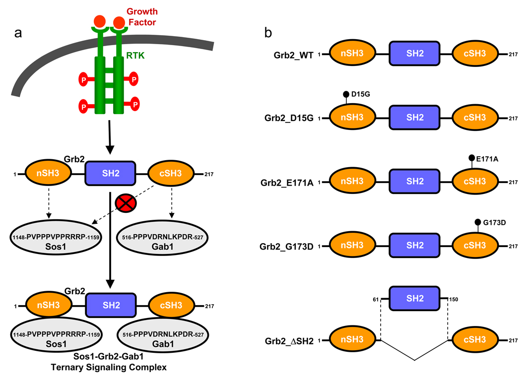
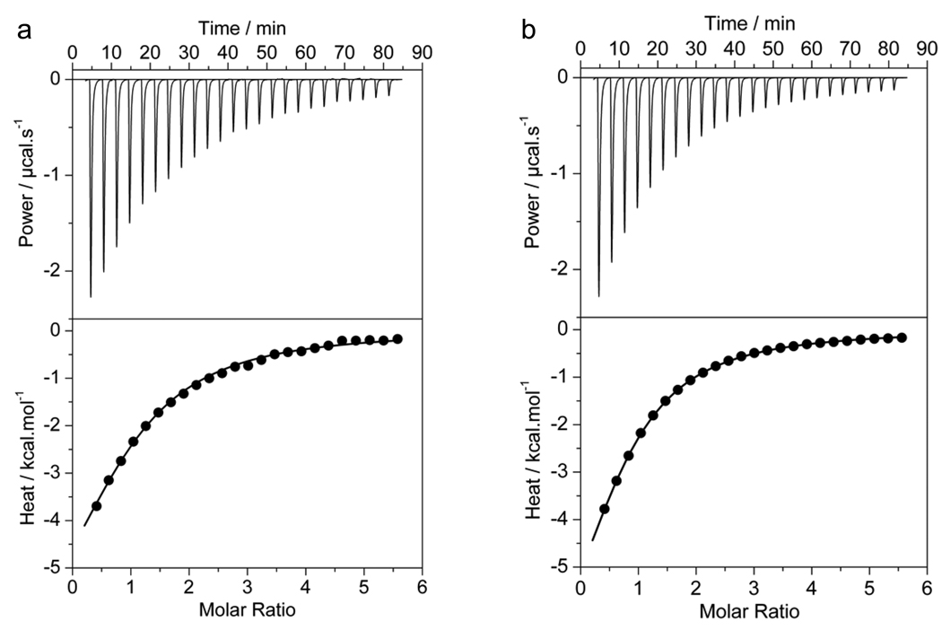
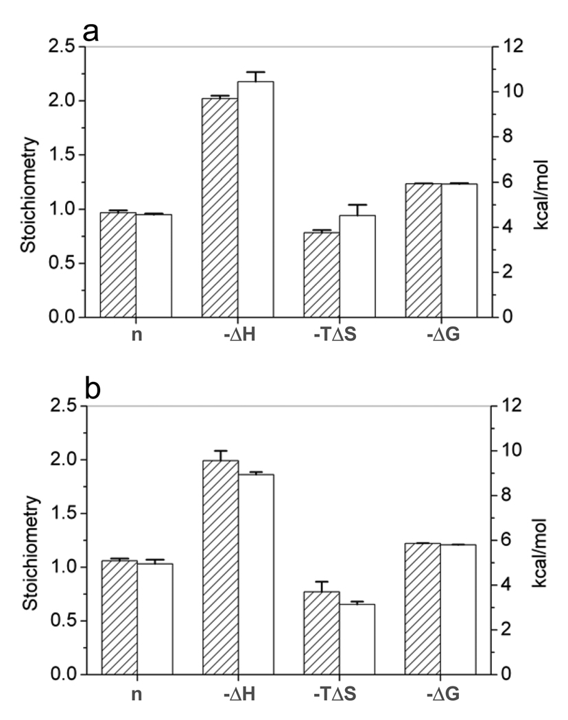
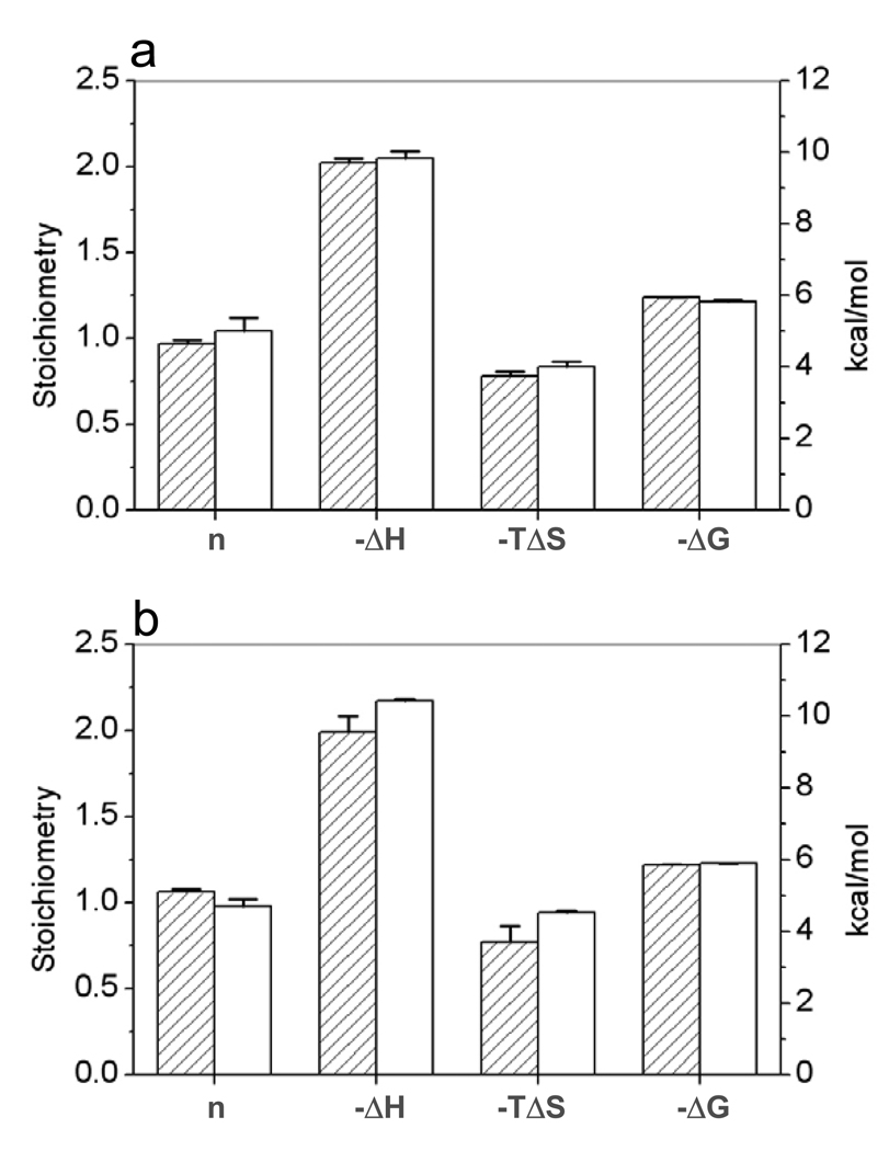
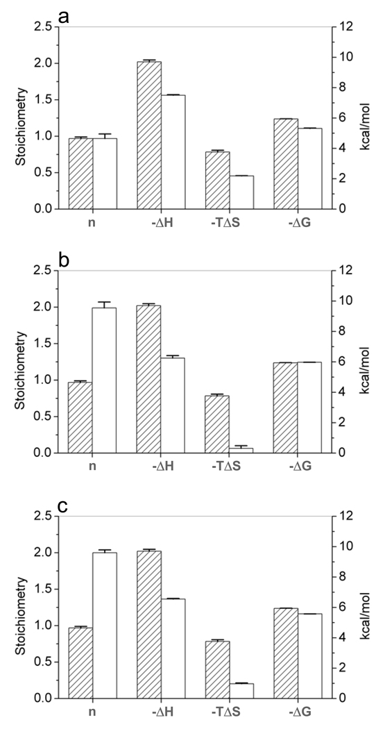
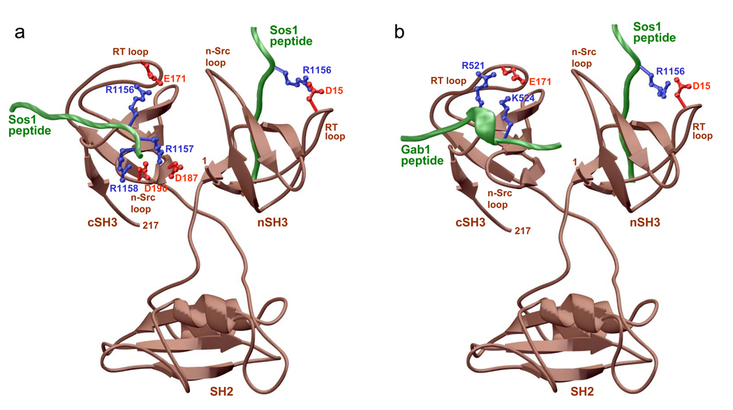
Allostery has evolved as a form of local communication between interacting protein partners allowing them to quickly sense changes in their immediate vicinity in response to external cues. Herein, using isothermal titration calorimetry (ITC) in conjunction with circular dichroism (CD) and macromolecular modeling (MM), we show that the binding of Grb2 adaptor--a key signaling molecule involved in the activation of Ras GTPase--to its downstream partners Sos1 guanine nucleotide exchange factor and Gab1 docker is under tight allosteric regulation. Specifically, our findings reveal that the binding of one molecule of Sos1 to the nSH3 domain allosterically induces a conformational change within Grb2 such that the loading of a second molecule of Sos1 onto the cSH3 domain is blocked and, in so doing, allows Gab1 access to the cSH3 domain in an exclusively non-competitive manner to generate the Sos1-Grb2-Gab1 ternary signaling complex.


Figures

Figure 1
Figure 1
Role of Grb2 in cellular signaling. (a) A schematic showing the assembly of Sos1-Grb2-Gab1 ternary signaling complex. Upon receptor stimulation, Grb2 is recruited to the inner membrane surface via the binding of its SH2 domain to tyrosine-phosphorylated (pY) sequences in the context of pYXN motif located within the cytoplasmic tails. The SH3 domains in turn recruit Sos1 and Gab1 as indicated in a mutually inclusive manner to generate the Sos1-Grb2-Gab1 ternary complex. Although the isolated cSH3 domain can bind to both Sos1 and Gab1, allosteric communication between the SH3 domains of Grb2 ensures that Sos1 solely binds to the nSH3 domain and thereby handing the cSH3 domain exclusive right to bind to Gab1. Note that the amino acid sequences of Sos1 and Gab1 shown correspond to respective peptides employed in this study. (b) Illustration of various Grb2 constructs used in this study. These include the wildtype Grb2 (Grb2_WT), D15G mutant of Grb2 (Grb2_D15G), E171A mutant of Grb2 (Grb2_E171A), G173D mutant of Grb2 (Grb2_G173D) and the truncated construct of Grb2 in which the central SH2 domain was excised out and the terminal SH3 domains were stitched together (Grb2_ΔSH2). Note that the D15G mutation abolishes the binding of nSH3 domain to Sos1, E171A mutation abolishes the binding of cSH3 domain to Sos1, and the G173D mutation renders the cSH3 domain into nSH3-mimetic in that it binds to Sos1 with comparable eneregtics to that observed for the binding of nSH3 domain [28].
Figure 2
Figure 2
Representative ITC isotherms obtained for the binding of Sos1 and Gab1 peptides to full-length wildtype Grb2 (Grb2_WT) in Tris buffer. (a) Binding of Sos1 peptide to Grb2_WT. (b) Binding of Gab1 peptide to Grb2_WT. The upper panels show the raw ITC data expressed as change in thermal power with respect to time over the period of titration. In the lower panels, change in molar heat is expressed as a function of peptide-to-protein molar ratio. The solid lines in the lower panels show the fit of data to a one-site model, based on the binding of a ligand to a macromolecule [33], as incorporated in the Microcal Origin software.
Figure 3
Figure 3
Comparison of thermodynamic parameters n, ΔH, TΔS and ΔG for the binding of Sos1 and Gab1 peptides to full-length wildtype Grb2 (Grb2_WT) under competitive and non-competitive settings in Tris buffer. (a) Binding of Sos1 peptide to Grb2_WT alone (shaded columns) and Grb2_WT pre-equilibrated with Gab1 peptide (unshaded columns). (b) Binding of Gab1 peptide to Grb2_WT alone (shaded columns) and Grb2_WT pre-equilibrated with Sos1 peptide (unshaded columns). All parameters were directly determined from ITC analysis.
Figure 4
Figure 4
Comparison of thermodynamic parameters n, ΔH, TΔS and ΔG for the binding of Sos1 and Gab1 peptides to full-length wildtype Grb2 (Grb2_WT) under different buffer conditions. (a) Binding of Sos1 peptide to Grb2_WT in Tris buffer (shaded columns) and Phosphate buffer (unshaded columns). (b) Binding of Gab1 peptide to Grb2_WT in Tris buffer (shaded columns) and Phosphate buffer (unshaded columns). All parameters were directly determined from ITC analysis.
Figure 5
Figure 5
Effect of various point and deletion mutations on the thermodynamic parameters n, ΔH, TΔS and ΔG for the binding of Sos1 peptide to various constructs of Grb2 in Tris buffer. (a) Binding of Sos1 peptide to Grb2_WT (shaded columns) and Grb2_D15G (unshaded columns). (b) Binding of Sos1 peptide to Grb2_WT (shaded columns) and Grb2_G173D (unshaded columns). (c) Binding of Sos1 peptide to Grb2_WT (shaded columns) and Grb2_ΔSH2 (unshaded columns). All parameters were directly determined from ITC analysis.
Figure 6
Figure 6
Representative CD spectra of full-length wildtype Grb2 alone and in complex with Sos1 and Gab1 peptides (with the contributions from the Trx-tag and peptides removed). (a) Far-UV spectra of Grb2 alone (black), Grb2 bound to Sos1 peptide (red), Grb2 bound to Gab1 peptide (green) and, Grb2 bound to both Sos1 and Gab1 peptides (blue). (b) Near-UV spectra of Grb2 alone (black), Grb2 bound to Sos1 peptide (red), Grb2 bound to Gab1 peptide (green) and, Grb2 bound to both Sos1 and Gab1 peptides (blue). Note that all spectra are expressed in terms of molar ellipticity ([θ]) as a function of wavelength (λ) of electromagnetic radiation.
Figure 7
Figure 7
3D structural models of full-length Grb2 in complex with Sos1 and Gab1 peptides obtained using the MODELLER software based on homology modeling [35]. (a) Sos1 peptides bound to both nSH3 and cSH3 domains within Grb2. (b) Sos1 peptide bound to the nSH3 domain and Gab1 peptide bound to the cSH3 domain within Grb2. The backbone of Grb2 is depicted in brown, while the backbones of Sos1 and Gab1 peptides are shown in green. The sidechain atoms of residues within Grb2 and the peptides involved in intermolecular interaction are colored red and blue, respectively.

Similar articles

Cited by

References

    1. Kendrew JC, Bodo G, Dintzis HM, Parrish RG, Wyckoff H, Phillips DC. Nature. 1958;181:662–666. - PubMed
    1. Monod J, Changeux JP, Jacob F. J Mol Biol. 1963;6:306–329. - PubMed
    1. Monod J, Wyman J, Changeux JP. J Mol Biol. 1965;12:88–118. - PubMed
    1. Kuriyan J, Eisenberg D. Nature. 2007;450:983–990. - PubMed
    1. Chardin P, Cussac D, Maignan S, Ducruix A. FEBS Lett. 1995;369:47–51. - PubMed

Publication types