

Modulating cortical connectivity in stroke patients by rTMS assessed with fMRI and dynamic causal modeling
- PMID: 20005962
- PMCID: PMC8020334
- DOI: 10.1016/j.neuroimage.2009.12.029
Modulating cortical connectivity in stroke patients by rTMS assessed with fMRI and dynamic causal modeling
Abstract
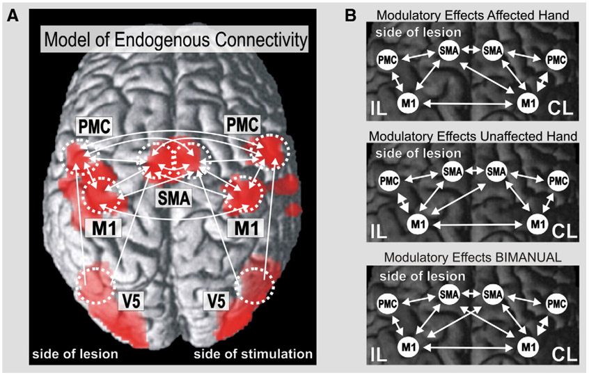
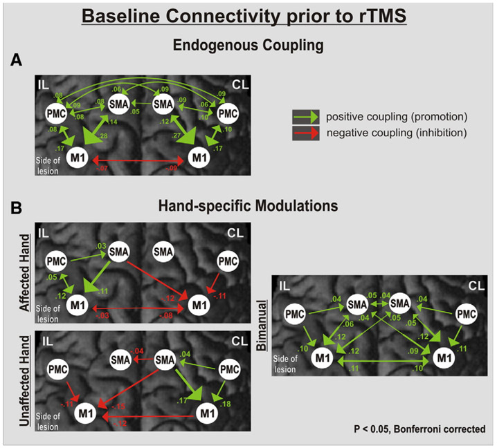
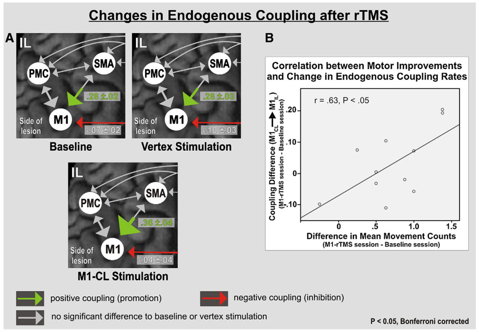
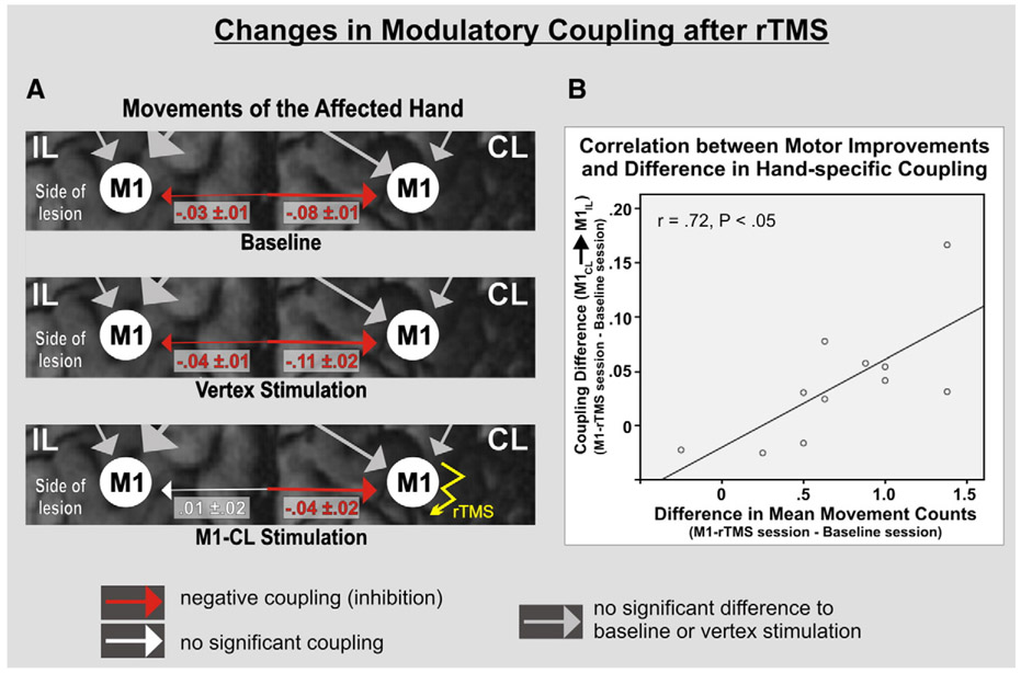
Data derived from transcranial magnetic stimulation (TMS) studies suggest that transcallosal inhibition mechanisms between the primary motor cortex of both hemispheres may contribute to the reduced motor performance of stroke patients. We here investigated the potential of modulating pathological interactions between cortical motor areas by means of repetitive TMS using functional magnetic resonance imaging (fMRI) and dynamic causal modeling (DCM). Eleven subacute stroke patients were scanned 1-3 months after symptom onset while performing whole hand fist closure movements. After a baseline scan, patients were stimulated with inhibitory 1-Hz rTMS applied over two different locations: (i) vertex (control stimulation) and (ii) primary motor cortex (M1) of the unaffected (contralesional) hemisphere. Changes in the endogenous and task-dependent effective connectivity were assessed by DCM of a bilateral network comprising M1, lateral premotor cortex, and the supplementary motor area (SMA). The results showed that rTMS applied over contralesional M1 significantly improved the motor performance of the paretic hand. The connectivity analysis revealed that the behavioral improvements were significantly correlated with a reduction of the negative influences originating from contralesional M1 during paretic hand movements. Concurrently, endogenous coupling between ipsilesional SMA and M1 was significantly enhanced only after rTMS applied over contralesional M1. Therefore, rTMS applied over contralesional M1 may be used to transiently remodel the disturbed functional network architecture of the motor system. The connectivity analyses suggest that both a reduction of pathological transcallosal influences (originating from contralesional M1) and a restitution of ipsilesional effective connectivity between SMA and M1 underlie improved motor performance.
Copyright (c) 2009 Elsevier Inc. All rights reserved.
Figures
References
-
- Ameli M, Grefkes C, Kemper F, Riegg F, Rehme AK, Karbe H, Fink GR, Nowak DA, 2009. Differential effects of high-frequency rTMS over ipsilesional primary motor cortex in cortical and subcortical MCA stroke. Ann. Neurol 66 (3), 298–309. - PubMed
-
- Ashburner J, Friston K, 2005. Unified segmentation. Neuroimage 26, 839–851. - PubMed
-
- Biernaskie J, Szymanska A, Windle V, Corbett D, 2005. Bi-hemispheric contribution to functional motor recovery of the affected forelimb following focal ischemic brain injury in rats. J. Neurosci 21, 989–999. - PubMed
-
- Bonita R, Beaglehole R, 1988. Modification of Rankin Scale: recovery of motor function after stroke. Stroke 19, 1497–1500. - PubMed
-
- Bestmann S, Baudewig J, Siebner HR, Rothwell JC, Frahm J, 2005. BOLD MRI responses to repetitive TMS over human dorsal premotor cortex. Neuroimage 28, 22–29. - PubMed
Publication types
MeSH terms
Grants and funding
LinkOut - more resources
Full Text Sources
Medical
