

Overproduction and localization of Mycobacterium tuberculosis ParA and ParB proteins
- PMID: 20006309
- PMCID: PMC3226068
- DOI: 10.1016/S1472-9792(09)70015-0
Overproduction and localization of Mycobacterium tuberculosis ParA and ParB proteins
Abstract
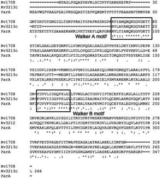
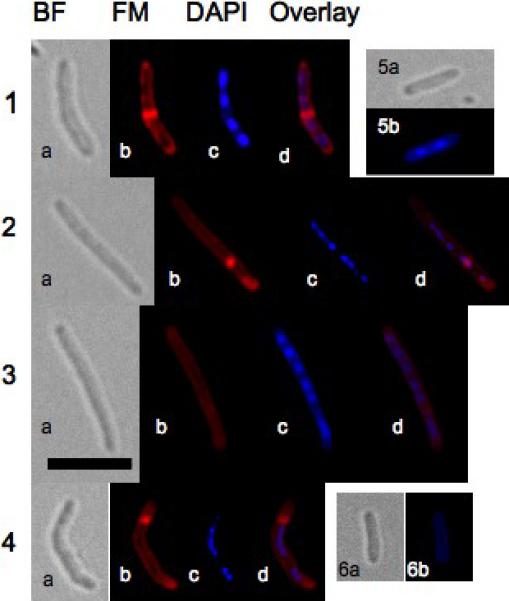
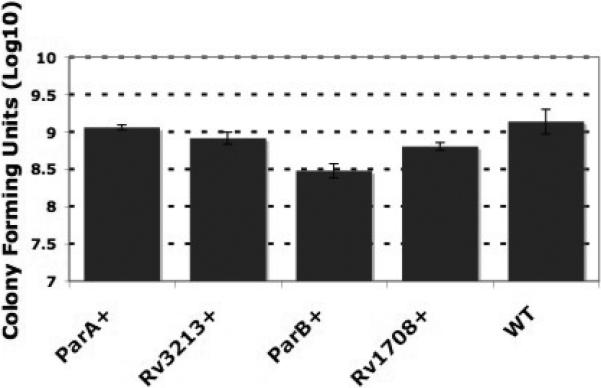
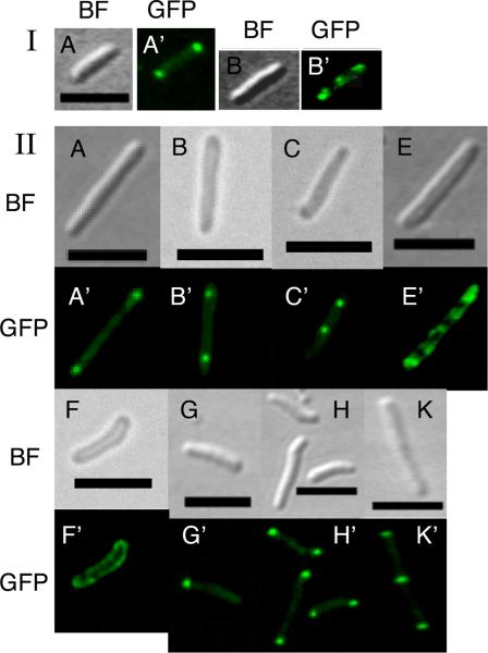
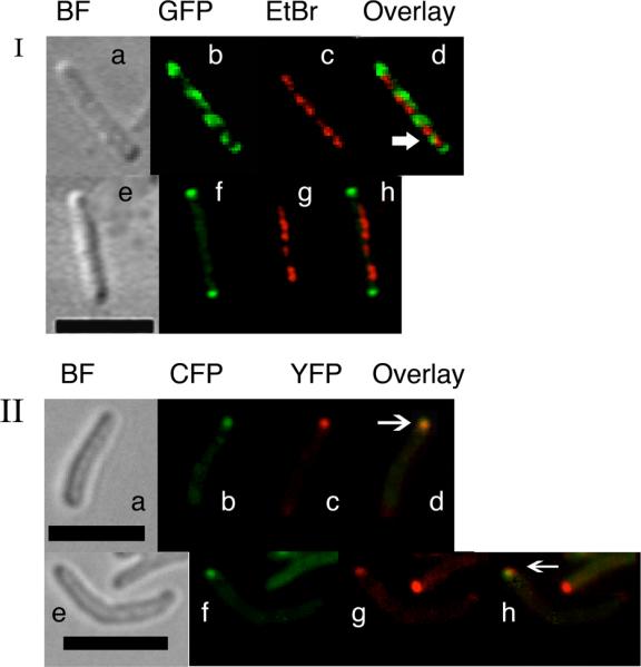
The ParA and ParB family proteins are required for accurate partitioning of replicated chromosomes. The Mycobacterium tuberculosis genome contains parB, parA and two parA homologs, Rv1708 and Rv3213c. It is unknown if parA and its homologs are functionally related. To understand the roles of ParA and ParB proteins in M. tuberculosis cell cycle, we have evaluated the consequences of their overproduction and visualized their localization patterns in M. smegmatis. We show that cells overproducing ParA, Rv1708 and Rv3213c and ParB are filamentous and multinucleoidal indicating defects in cell-cycle progression. Visualization of green-fluorescent protein fusions of ParA and its homologues showed similar localization patterns with foci at poles, quarter-cell, midcell positions and spiral-like structures indicating that they are functionally related. On the other hand, the ParB- GFP fusion protein localized only to the cell poles. The cyan- and yellow-fluorescent fusion proteins of ParA and ParB, respectively, colocalized at the cell poles indicating that these proteins interact and possibly associate with the chromosomal origin of replication. Collectively our results suggest that the M. tuberculosis Par proteins play important roles in cell-cycle progression.
Figures
Similar articles
-
The studies of ParA and ParB dynamics reveal asymmetry of chromosome segregation in mycobacteria.Mol Microbiol. 2017 Aug;105(3):453-468. doi: 10.1111/mmi.13712. Epub 2017 Jun 22. Mol Microbiol. 2017. PMID: 28517109
-
Characterization of the mycobacterial chromosome segregation protein ParB and identification of its target in Mycobacterium smegmatis.Microbiology (Reading). 2007 Dec;153(Pt 12):4050-4060. doi: 10.1099/mic.0.2007/011619-0. Microbiology (Reading). 2007. PMID: 18048919
-
par genes in Mycobacterium bovis and Mycobacterium smegmatis are arranged in an operon transcribed from "SigGC" promoters.BMC Microbiol. 2008 Mar 27;8:51. doi: 10.1186/1471-2180-8-51. BMC Microbiol. 2008. PMID: 18371202 Free PMC article.
-
Prokaryotic ParA-ParB-parS system links bacterial chromosome segregation with the cell cycle.Plasmid. 2012 Jan;67(1):1-14. doi: 10.1016/j.plasmid.2011.08.003. Epub 2011 Sep 6. Plasmid. 2012. PMID: 21924286 Review.
-
The bacterial ParA-ParB partitioning proteins.J Biotechnol. 2001 Sep 13;91(1):1-34. doi: 10.1016/s0168-1656(01)00293-0. J Biotechnol. 2001. PMID: 11522360 Review.
Cited by
-
Identification of Mycobacterial Genes Involved in Antibiotic Sensitivity: Implications for the Treatment of Tuberculosis with β-Lactam-Containing Regimens.Antimicrob Agents Chemother. 2017 Jun 27;61(7):e00425-17. doi: 10.1128/AAC.00425-17. Print 2017 Jul. Antimicrob Agents Chemother. 2017. PMID: 28438925 Free PMC article.
-
Tracking of chromosome and replisome dynamics in Myxococcus xanthus reveals a novel chromosome arrangement.PLoS Genet. 2013;9(9):e1003802. doi: 10.1371/journal.pgen.1003802. Epub 2013 Sep 19. PLoS Genet. 2013. PMID: 24068967 Free PMC article.
-
Mild Nutrient Starvation Triggers the Development of a Small-Cell Survival Morphotype in Mycobacteria.Front Microbiol. 2016 Jun 16;7:947. doi: 10.3389/fmicb.2016.00947. eCollection 2016. Front Microbiol. 2016. PMID: 27379076 Free PMC article.
-
Catching a Walker in the Act-DNA Partitioning by ParA Family of Proteins.Front Microbiol. 2022 May 26;13:856547. doi: 10.3389/fmicb.2022.856547. eCollection 2022. Front Microbiol. 2022. PMID: 35694299 Free PMC article. Review.
-
Mycobacterial Growth.Cold Spring Harb Perspect Med. 2015 May 8;5(10):a021097. doi: 10.1101/cshperspect.a021097. Cold Spring Harb Perspect Med. 2015. PMID: 25957314 Free PMC article. Review.
References
-
- Bignell C, Thomas CM. The bacterial ParA-ParB partitioning proteins. J Biotechnol. 2001;91:1–34. - PubMed
Publication types
MeSH terms
Substances
Grants and funding
LinkOut - more resources
Full Text Sources
Molecular Biology Databases
