. 2010 Feb 4;9(2):109-19.
doi: 10.1016/j.dnarep.2009.11.002. Epub 2009 Dec 16.

DNA polymerase beta-dependent long patch base excision repair in living cells

Affiliations

DNA polymerase beta-dependent long patch base excision repair in living cells

Kenjiro Asagoshi et al. DNA Repair (Amst). .

Abstract

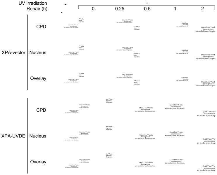
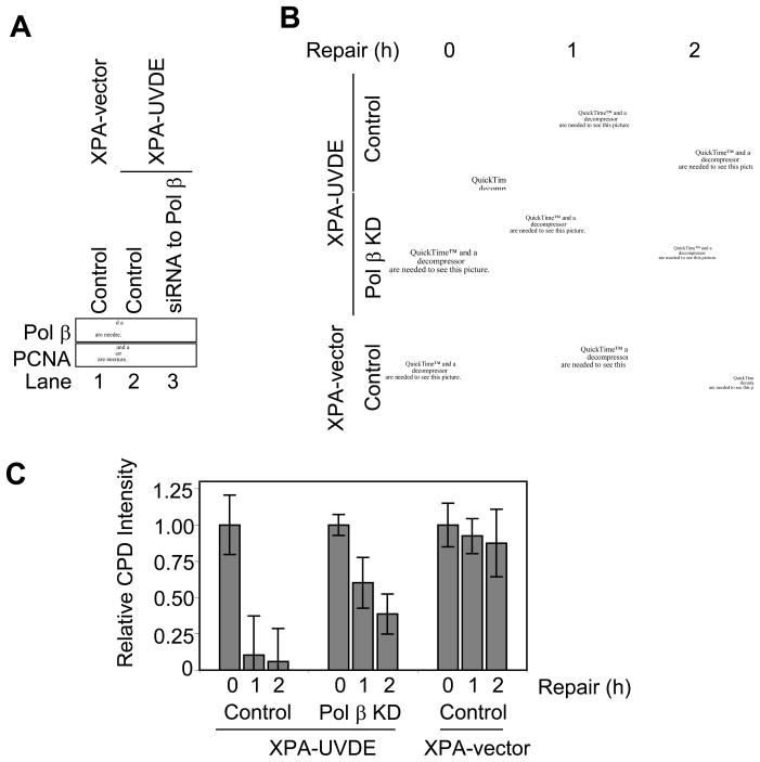
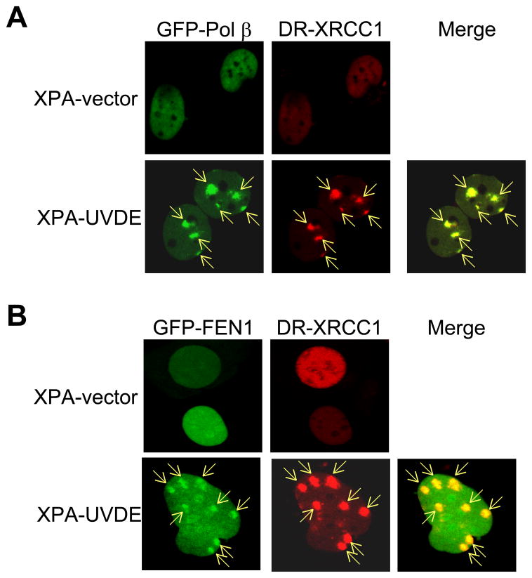
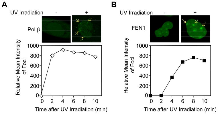
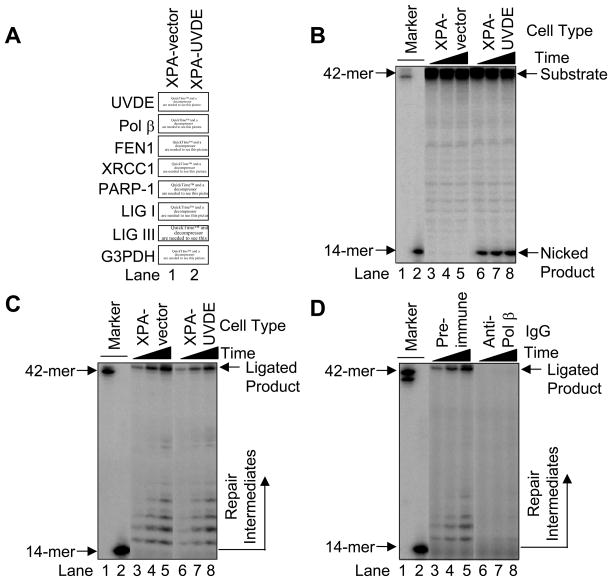
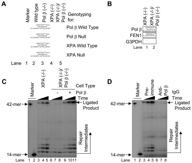
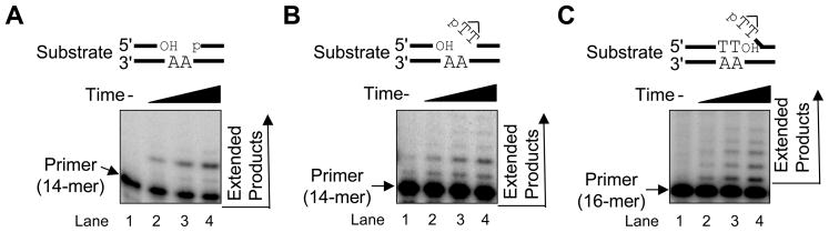
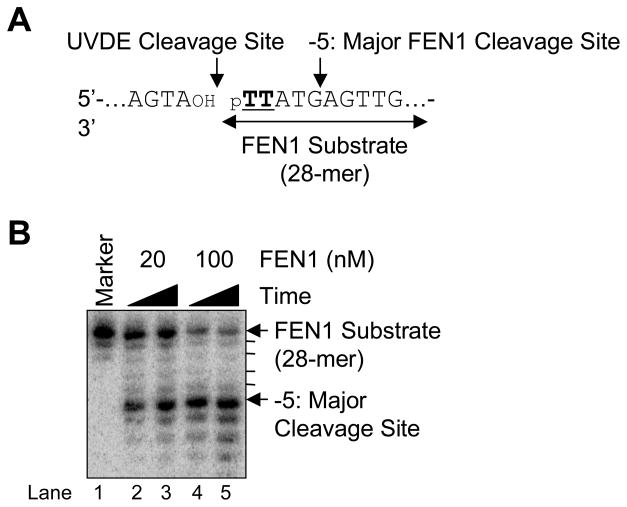
We examined a role for DNA polymerase beta (Pol beta) in mammalian long patch base excision repair (LP BER). Although a role for Pol beta is well known in single-nucleotide BER, information on this enzyme in the context of LP BER has been limited. To examine the question of Pol beta involvement in LP BER, we made use of nucleotide excision repair-deficient human XPA cells expressing UVDE (XPA-UVDE), which introduces a nick directly 5' to the cyclobutane pyrimidine dimer or 6-4 photoproduct, leaving ends with 3'-OH and 5'-phosphorylated UV lesion. We observed recruitment of GFP-fused Pol beta to focal sites of nuclear UV irradiation, consistent with a role of Pol beta in repair of UV-induced photoproducts adjacent to a strand break. This was the first evidence of Pol beta recruitment in LP BER in vivo. In cell extract, a 5'-blocked oligodeoxynucleotide substrate containing a nicked 5'-cyclobutane pyrimidine dimer was repaired by Pol beta-dependent LP BER. We also demonstrated Pol beta involvement in LP BER by making use of mouse cells that are double null for XPA and Pol beta. These results were extended by experiments with oligodeoxynucleotide substrates and purified human Pol beta.


Conflict of interest statement

Conflict of interest statement

None declared.

Figures

Fig. 1
Fig. 1
(A) In vivo repair of SSB-bearing CPDs in human XPA cells. The distribution of CPDs was detected by anti-CPD monoclonal antibody as described in Section 2. Disappearance of CPD signal was measured in human XPA-vector and XPA-UVDE cells for the indicated repair incubation times (0–2 h) after 254 nm UV exposure of 30 J/m2. Nuclei were visualized after staining with DAPI. The DAPI and CPD staining was superimposed in the panels marked “overlay.”
Fig. 2
Fig. 2
(A) Immunoblotting for Pol β in siRNA knockdown (KD) XPA-vector and XPA-UVDE cells. Cell extracts were prepared from human XPA-vector cells with control siRNA (lane 1) and human XPA-UVDE cells with control siRNA (lane 2) or siRNA to Pol β (lane 3). They were immunoblotted with antibody for Pol β and PCNA as a loading control. (B) In vivo repair of SSB-bearing CPDs in siRNA knockdown XPA-UVDE and XPA-vector cells. The experiment was conducted as described in Fig. 1. (C) Relative intensities of CPD signal during the indicated repair period after 254 nm local UV exposure of 30 J/m2 in control and Pol β KD XPA-UVDE cells and XPA-vector cells.
Fig. 3
Fig. 3
Recruitment of Pol β, FEN1, and XRCC1 in human XPA-vector and XPA-UVDE cells after 254 nm local UV exposure using a polycarbonate isopore membrane filter as described in Section 2. (A) GFP-fused Pol β and DR-fused XRCC1 and (B) GFP-fused FEN1 and DR-fused XRCC1 were measured 5 min after localized UV irradiation with 100 J/m2 in XPA-vector (top) and XPA-UVDE (bottom) cells. The sites of localized UV irradiation are indicated by arrows. All sites of XRCC1 accumulation were positive for Pol β accumulation and FEN1 accumulation.
Fig. 4
Fig. 4
Recruitment of Pol β and FEN1 in human XPA-UVDE cells before and after local UV irradiation at 254 nm. Accumulation of (A) GFP-fused Pol β and (B) GFP-fused FEN1 was measured before (left) and 5 min after local UV irradiation with 100 J/m2 (right). The sites of UV irradiation are indicated by arrows. Quantification of the time course of accumulation of Pol β (open diamonds) and FEN1 (solid squares) is shown below each image.
Fig. 5
Fig. 5
(A) Immunoblotting for UVDE and known LP BER proteins. Cell extracts were prepared from human XPA-vector (lane 1) and XPA-UVDE (lane 2) cells and immunoblotted with antibody for N. crassa UVDE, Pol β, FEN1, XRCC1, PARP-1, LIG I, LIG III, and G3PDH as a loading control as described in Section 2. (B) Nicking activity of human XPA-vector and XPA-UVDE cell extract on CPD-containing ODN substrate. The in vitro cell extract-based nicking analysis was performed as described in Section 2. The incubation was conducted for 15 (lanes 3 and 6), 30 (lanes 4 and 7), and 60 min (lanes 5 and 8). The substrate (42-mer) and nicked product (14-mer) are shown. (C) Repair activity of human XPA-vector and XPA-UVDE cell extracts on the nicked CPD ODN substrate. The incubation was conducted for 5 (lanes 3 and 6), 15 (lanes 4 and 7), and 30 min (lanes 5 and 8). The position of repair intermediates and ligated product were shown. The mobility of the 42-mer marker was slightly faster than the ligated product due to the presence of 5′-phosphate. (D) Neutralization of Pol β in the in vitro cell extract-based LP BER. The in vitro repair reaction was conducted with pre-immune IgG (lanes 3–5) and anti-Pol β IgG (lanes 6–8) as described in Section 2. The incubation was conducted for 5 (lanes 3 and 6), 15 (lanes 4 and 7), and 30 min (lanes 5 and 8). The position of repair intermediates and ligated products are shown.
Fig. 6
Fig. 6
(A) PCR genotyping for Pol β and XPA in mouse XPA/Pol β cell lines. PCR was conducted as described in Section 2 and the resulting product was analyzed by 1.5% agarose gel electrophoresis. (B) Immunoblotting of Pol β and FEN1 in mouse XPA null and XPA/Pol β double null cells. WCEs were prepared and immunoblotted as described in Section 2. The blots were probed with anti-G3PDH as a loading control. Repair activity of mouse XPA null (lanes 3–5) and XPA/Pol β double null (lanes 6–11) cell extracts on the nicked CPD ODN substrate. The in vitro cell extract-based LP BER assay was carried out as described in Section 2 using a nicked CPD substrate. The incubation was conducted for 5 (lanes 3, 6 and 9), 15 (lanes 4, 7 and 10), and 30 min (lanes 5, 8 and 11). Purified Pol β was added to the XPA/Pol β double null cell extract reaction mixture for complementation analysis (lanes 9–11). The positions of repair intermediate and ligated product are shown. The mobility of the 42-mer marker was slightly faster than the ligated product due to the presence of 5′-phosphate. (D) Repair activity of mouse XPA null cell extracts in the presence of pre-immune IgG (lanes 3–5) or anti-Pol β IgG (lanes 6–8) and incubated with [α-32P]dTTP. The incubation was conducted for 5 (lanes 3 and 6), 15 (lanes 4 and 7), and 30 min (lanes 5 and 8). The positions of repair intermediate and ligated product are shown.
Fig. 7
Fig. 7
In vitro Pol β synthesis with LP BER DNA intermediates. The Pol β synthesis reaction was carried out as described in Section 2 using (A) the 2-nucleotide gapped substrate, (B) the nicked CPD substrate, and (C) the nicked CPD-flap substrate for 0 (lane 1), 0.5 (lane 2), 1 (lane 3), and 1.5 (lane 4) min at 37°C. The positions of the primer band (14-mer for A and B; 16-mer for C) and extended product are shown.
Fig. 8
Fig. 8
FEN1 cleavage on the nicked CPD ODN substrate. (A) Schematic diagram of the FEN1 cleavage reaction where TT indicates a CPD. The UVDE cleavage site and the major FEN1 cleavage site are indicated along with FEN1 substrate. (B) The in vitro FEN1 cleavage reaction was carried out as described in Section 2 using human FEN1 (20 nM and 100 nM) for 5 min (lanes 2 and 4) and 15 min (lanes 3 and 5). The positions of DNA substrate and product are shown.
Fig. 9
Fig. 9
Coordination of Pol β and FEN1 on the nicked CPD ODN substrate. (A) Schematic diagram of the reaction. The Pol β substrate was the 5′-end labeled 14-mer upstream strand and the product was expected to be observed beyond 14-mer (left side scheme). The FEN1 substrate was the 3′-end labeled 28-mer downstream strand containing CPD lesion (TT) at the 5′-end. The observed FEN1 cleavage product was expected to less than 28-mer (right side scheme). (B) Typical result of Pol β and FEN1 coordination reaction is shown. The in vitro Pol β synthesis and FEN1 cleavage reaction was simultaneously conducted using Pol β (1 nM) and FEN1 [20 nM (lane 1) and 100 nM (lane 2)]. The positions of Pol β synthesis product and FEN1 cleavage product are shown.

Similar articles

Cited by

References

    1. Friedberg EC, Walker GC, Siede W, Wood RD, Schultz RA, Ellenberger T. DNA Repair and Mutagenesis. ASM Press; Washington, DC: 2006.
    1. Biade S, Sobol RW, Wilson SH, Matsumoto Y. Impairment of proliferating cell nuclear antigen-dependent apurinic/apyrimidinic site repair on linear DNA. Journal of Biological Chemistry. 1998;273:898–902. - PubMed
    1. Blank A, Kim B, Loeb LA. DNA polymerase δ is required for base excision repair of DNA methylation damage in Saccharomyces cerevisiae. Proceedings of the National Academy of Sciences, USA. 1994;91:9047–9051. - PMC - PubMed
    1. Fortini P, Pascucci B, Parlanti E, Sobol RW, Wilson SH, Dogliotti E. Different DNA polymerases are involved in the short- and long-patch base excision repair in mammalian cells. Biochemistry. 1998;37:3575–3580. - PubMed
    1. Frosina G, Fortini P, Rossi O, Carrozzino F, Raspaglio G, Cox LS, Lane DP, Abbondandolo A, Dogliotti E. Two pathways for base excision repair in mammalian cells. Journal of Biological Chemistry. 1996;271:9573–9578. - PubMed

Publication types

MeSH terms

LinkOut - more resources