. 2010 Apr 1;316(6):1050-60.
doi: 10.1016/j.yexcr.2009.12.005. Epub 2009 Dec 16.

Non-cell-autonomous stimulation of stem cell proliferation following ablation of Tcf3

Affiliations

Non-cell-autonomous stimulation of stem cell proliferation following ablation of Tcf3

Fei Yi et al. Exp Cell Res. .

Abstract

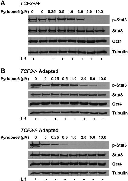
A combination of cell intrinsic factors and extracellular signals determine whether mouse embryonic stem cells (ESC) divide, self-renew, and differentiate. Here, we report a new interaction between cell intrinsic aspects of the canonical Wnt/Tcf/beta-catenin signaling pathway and extracellular Lif/Jak/Stat3 stimulation that combines to promote self-renewal and proliferation of ESC. Mutant ESC lacking the Tcf3 transcriptional repressor continue to self-renew in the absence of exogenous Lif and through pharmacological inhibition of Lif/Jak/Stat3 signaling; however, proliferation rates of TCF3-/- ESC were significantly decreased by inhibiting Jak/Stat3 activity. Cell mixing experiments showed that stimulation of Stat3 phosphorylation in TCF3-/- ESC was mediated through secretion of paracrine acting factors, but did not involve elevated Lif or LifR transcription. The new interaction between Wnt and Lif/Jak/Stat3 signaling pathways has potential for new insights into the growth of tumors caused by aberrant activity of Wnt/Tcf/beta-catenin signaling.


Figures

Figure 1
Figure 1. Long-term phosphorylation of Stat3 at tyrosine 705 in TCF3−/− ESC in the absence of exogenous Lif
A) Western blot analysis for the phosphorylation of Stat3 in short-term Lif withdrawal assay. TCF3+/+ and TCF3−/− were cultured in media containing 1000 U/ml Lif (Lif+) for 24 hours. Cells were washed twice by PBS and fed with media without supplemented Lif (Lif−). Protein samples were isolated at indicated time points. Phosphorylation of Stat3 was diminished in both ESC lines 15 and 30 minutes after Lif withdrawal, and returned to normal levels within 30 minutes when switch back to the Lif+ media. B) Stat3 was phosphorylated at a lower but substantial level comparing to the Lif+ cultures in all three independently-derived adapted TCF3−/− ESC. C) Western blot analysis for the phosphorylation of Stat3 in long-term Lif withdrawal assay. Stat3 was not significantly phosphorylated until the third passage (P3) and maintained throughout the assay (P10). D) Flow cytometry analysis for cell cycle using propidium iodide staining for total DNA content. Similarly to the TCF3+/+ and naïve TCF3−/− ESC, adapted TCF3−/− ESC exhibited a typical self renewing ESC cell cycle structure.
Figure 2
Figure 2. Jak Kinase activity is necessary for Stat3 phosphorylation in adapted TCF3−/− ESC
Western blot analysis for the indicated antigens (right) was performed using protein lysates after six hours of pyridone 6 treatment. A) 5.0-10.0 μM Jak kinase specific inhibitor pyridine 6 inhibited Stat3 phosphorylation of TCF3+/+ ESC in Lif+ culture. B) Upper panel: 5.0-10.0 μM pyridone 6 similar inhibited the Stat3 phosphorylation in adapted TCF3−/− ESC in Lif+ culture comparing to the TCF3+/+ ESC, but a higher concentration was needed to achieve a complete inhibition; Lower panel: 0.5-1.0 μM pyridone 6 inhibited the phosphorylation of Stat3 in adapted TCF3−/− ESC in Lif− culture.
Figure 3
Figure 3. Adapted TCF3−/− ESC self renew under blockade of Jak phosphorylation of Stat3
Adapted TCF3−/− ESC were treated with 0-1.0 μM of pyridone 6 in Lif− culture (A-D). A) Total numbers of cells were counted at the end of each passage(P#). Each column represents a mean number of total cell counts in duplicated assays with standard deviation. B) Western blot analysis of protein samples isolated at the end of the fifth passage (P5). Adapted TCF3−/− ESC lost the Stat3 phosphorylation with 0.5-1.0 μM pyridone 6 while the expression of Nanog and Oct4 were not affected by pyridone 6. C) Percentages of alkaline phosphatase-positive colonies were determined at the end of the fifth passage (P5). Inhibition of Stat3 phosphorylation by pyridone 6 affected the morphology of colonies without substantially increasing the percentage of differentiated colonies. D) Key for different categories of colonies counted for graphs in panel C.
Figure 4
Figure 4. Adaptation of TCF3−/− ESC in long-term Lif− culture occurs without Jak activity
TCF3+/+ and naïve TCF3−/− ESC were passaged in Lif− culture with or without 1.0 μM pyridone 6. The numbers of cells were counted at the end of each passage. Each column represents a mean number of total cell counts in duplicated assays with standard deviation. TCF3+/+ ESC failed to proliferate and underwent apoptosis during the first three passages. Treatment with 1.0μM pyridone 6 reduced the proliferation of naïve TCF3−/− ESC throughout the assay but did not prevent the naïve TCF3−/− ESC from adapting to Lif− culture.
Figure 5
Figure 5. Lif and LifR were not upregulated in the adapted TCF3−/− ESC
Quantitative real-time RT PCR (qPCR) was used to measure the levels of Lif and LifR mRNA. A) Comparing to the mouse embryonic fibroblasts (MEFs) and TCF3+/+ ESC, both naïve and adapted TCF3−/− ESC did not express sufficient levels of Lif to support Stat3 phosphorylation and self renewal. B) The LifR level was also not significantly higher in adapted TCF3−/− ESC comparing to TCF3+/+ ESC.
Figure 6
Figure 6. Stat3 phosphorylation in adapted TCF3−/− ESC is partially LifR-independent
Western blot analysis of TCF3+/+ and TCF3−/− ESC in Lif+ culture and adapted TCF3−/− ESC in Lif− culture treated with 0-10 nM Lif antagonist (LA) (A-C). A) 1-10 nM LA blocked the Stat3 phosphorylation in TCF3+/+ ESC in Lif+ culture. B) 1-10 nM LA blocked the Stat3 phosphorylation in TCF3−/− ESC in Lif+ culture. C) 1-10 nM LA diminished but did not completely block the Stat3 phosphorylation in adapted TCF3−/− ESC in Lif− culture. D) Western blot analysis of TCF3+/+ ESC in Lif+ culture and adapted TCF3−/− ESC in Lif− culture under either pyridone 6 or LA treatment. 1 μM pyridone 6 completely blocked Stat3 phosphorylation in adapted TCF3−/− ESC but only caused a minor decrease in TCF3+/+ ESC; while 1 nM and 10 nM LA decreased the Stat3 phosphorylation in both TCF3+/+ and adapted TCF3−/− ESC to comparable levels.
Figure 7
Figure 7. Lif-independent Stat3 phosphorylation occurred non-cell-autonomously
A) Similar to Figure 1A, Western blot analysis for Stat3 phosphorylation in short-term Lif withdrawal assay. TCF3+/+, TCF3−/− were cultured in media containing 1000 U/ml Lif (Lif+) while adapted TCF3−/− ESC were cultured without supplemented Lif (Lif−) for 24 hours. All cells were washed twice by PBS and fed with media without supplemented Lif (Lif−). Protein samples were isolated at indicated time points. Phosphorylation of Stat3 was diminished in all three ESC lines 15 and 30 minutes after Lif withdrawal, and returned to normal levels within 30 minutes when switch back to the Lif+ media. B-D) Indirect immunofluorescent staining for Tcf3 (Red; B) or phosphoStat3 (Red; C-D). B) Individual colony from TCF3+/+ ESC and EGFP-labeled adapted TCF3−/− ESC mixed-culture exhibited mosaic patterns of expression of Tcf3 (Red) and EGFP (Green). Multiple colonies were shown for staining (C-D). C) Stat3 phosphorylation (Red) in TCF3+/+ and TCF3−/− ESC was detected in Lif+ culture (upper two panels) and absent from Lif− culture (middle two panels). In Lif− culture, Stat3 was phosphorylated in adapted TCF3−/− ESC and the EGFP (Green) was ubiquitously expressed (bottom panel). D) Stat3 was uniformly phosphorylated in all cells in individual colonies in mixed-culture of TCF3+/+ (upper panel) or TCF3−/− (lower panel) ESC with adapted TCF3−/− ESC in the absence of Lif. EGFP was detected in a mosaic pattern in the culture confirming the mixture of cell types in individual colonies.

Similar articles

Cited by

References

    1. Nusse R. Wnt signaling and stem cell control. Cell Res. 2008;18:523–527. - PubMed
    1. Brannon M, Gomperts M, Sumoy L, Moon RT, Kimelman D. A beta-catenin/XTcf-3 complex binds to the siamois promoter to regulate dorsal axis specification in Xenopus. Genes Dev. 1997;11:2359–2370. - PMC - PubMed
    1. Molenaar M, van de Wetering M, Oosterwegel M, Peterson-Maduro J, Godsave S, Korinek V, Roose J, Destree O, Clevers H. XTcf-3 transcription factor mediates beta-catenin-induced axis formation in Xenopus embryos. Cell. 1996;86:391–399. - PubMed
    1. van de Wetering M, Cavallo R, Dooijes D, van Beest M, van Es J, Loureiro J, Ypma A, Hursh D, Jones T, Bejsovec A, Peifer M, Mortin M, Clevers H. Armadillo coactivates transcription driven by the product of the Drosophila segment polarity gene dTCF. Cell. 1997;88:789–799. - PubMed
    1. Tutter AV, Fryer CJ, Jones KA. Chromatin-specific regulation of LEF-1-beta-catenin transcription activation and inhibition in vitro. Genes Dev. 2001;15:3342–3354. - PMC - PubMed

Publication types

MeSH terms