Review
. 2010 Feb 25;160(1-3):153-9.
doi: 10.1016/j.regpep.2009.12.007. Epub 2009 Dec 16.

Chromogranin A: a new proposal for trafficking, processing and induction of granule biogenesis

Affiliations
Review

Chromogranin A: a new proposal for trafficking, processing and induction of granule biogenesis

Hisatsugu Koshimizu et al. Regul Pept. .

Abstract

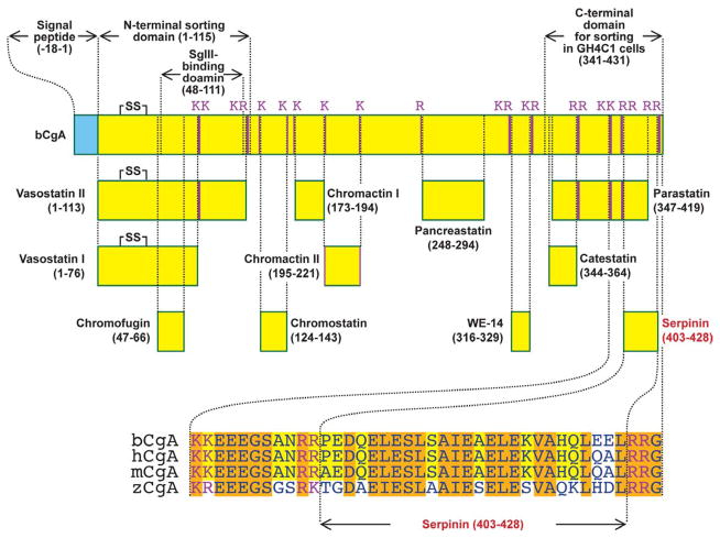
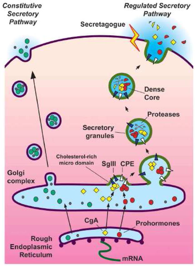
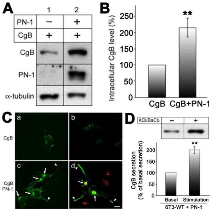
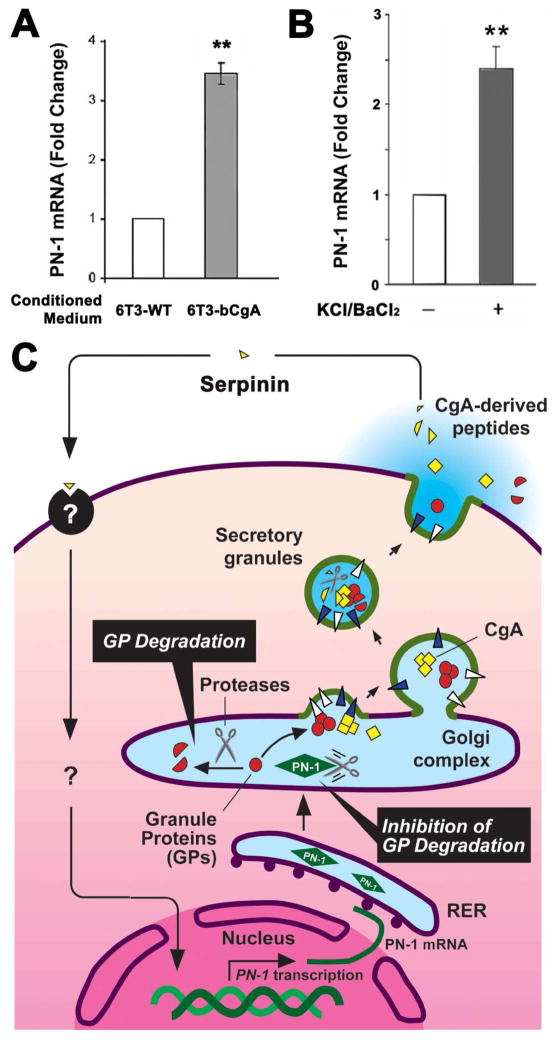
Chromogranin A (CgA), a member of the granin family serves several important cell biological roles in (neuro)endocrine cells which are summarized in this review. CgA is a "prohormone" that is synthesized at the rough endoplasmic reticulum and transported into the cisternae of this organelle via its signal peptide. It is then trafficked to the Golgi complex and then to the trans-Golgi network (TGN) where CgA aggregates at low pH in the presence of calcium. The CgA aggregates provide the physical driving force to induce budding of the TGN membrane resulting in dense core granule (DCG) formation. Within the granule, a small amount of the CgA is processed to bioactive peptides, including a predicted C-terminal peptide, serpinin. Upon stimulation, DCGs undergo exocytosis and CgA and its derived peptides are released. Serpinin, acting extracellularly is able to signal the increase in transcription of a serine protease inhibitor, protease nexin-1 (PN-1) that protects DCG proteins against degradation in the Golgi complex, which then enhances DCG biogenesis to replenish those that were released. Thus CgA and its derived peptide, serpinin, plays a significant role in granule formation and regulation of granule biogenesis, respectively, in (neuro) endocrine cells.


Figures

Fig. 1
Fig. 1
Schematic representation of the structure of CgA. Bovine CgA (bCgA; NCBI accession number P05059) showing functional domains and paired and single basic residue cleavage sites. N-terminal domain (1–115) and SgIII binding site (48–111) were identified in rat CgA but the location of these regions is identical with that of bCgA. The C-terminal domain which is required for sorting in GH4C1 cells (341–431) was identified in bCgA. Known CgA-derived peptides and a novel CgA-derived peptide, Serpinin (403–428) are displayed. Serpinin residues which are conserved in three mammalian species: bovine CgA (bCgA), human CgA (hCgA; accession number AAH09384) and mouse CgA (mCgA; NP031719), are highlighted in yellow. Residues conserved in the three mammalian species and zebra fish (zCgA; accession number NP001006059) are highlighted in orange. C-terminal domain and Serpinin have highly conserved sequences.
Fig. 2
Fig. 2
Secretory pathways and dense core granule biogenesis in (neuro)endocrine cells. CgA and prohormones which are synthesized in the rough endoplasmic reticulum (RER), are transported to the Golgi complex, aggregated and sorted into granules at cholesterol-rich membrane microdomains in the trans-Golgi network via sorting receptors, such as SgIII and CPE, respectively. Aggregated CgA and prohormones induce the budding of the TGN membrane to form the dense core granule (DCG) in the regulated secretory pathway. Prohormones and CgA (partially) are cleaved by proteases in DCGs to yield biologically active peptides. Stimulation of DCGs by secretagogues triggers exocytosis and secretion of hormones. In contrast, vesicles of the constitutive secretory pathway which is present in all cell types release their contents without stimulation.
Fig. 3
Fig. 3
A. Effect of CgA antisense knock-out on granule biogenesis. Electron micrographs (EM) of wild-type PC12 cells (WT, left panel) and clone CGAAS-5 cells which stably express antisense constructs against CgA sense sequences (right panel). Dense-core secretory granules (arrows) are abundant in the wild-type PC12 cells but scarce in clone CGAAS-5 cells. B–E. Effect of CgA knock-in on granule biogenesis: B. 6T3-WT cells, a variant AtT-20 pituitary endocrine cell line lacking DCGs were stably transfected with bCgA. Bar graph shows EM morphometric analysis of the number of DCGs in 6T3-WT cells and 6T3-bCgA cells. DCGs were counted from 20 (6T3-WT) or 10 (6T3-bCgA) individual EM micrographs, and the number of DCGs per square micrometer was calculated by dividing the number of DCGs with the cytoplasmic area measured from each micrograph. 6T3-WT cells had 0.35 ± 0.06 DCG/μm2 (total of 112 μm2 of the cytoplasmic area measured), while 6T3-bCgA cells had 1.82 ± 0.29 DCG/μm2 (total of 48 μm2 of the cytoplasmic area measured) (***P < 0.0001). C. Western blot of release medium in the presence of 50 mM KCl/2 mM BaCl2 in 6T3 cells stably transfected with CgA (6T3-bCgA) shows that CgA secretion was significantly stimulated indicating that CgA expression in these mutant endocrine cells was able to restore the regulated secretory pathway. D. Western blots of release media from wild-type (6T3-WT) and CgA transfected 6T3 cells (6T3-bCgA). In 6T3-WT cells, transfected bovine POMC was secreted at a high basal level, and no stimulation was detected with 50 mM KCl/2 mM BaCl2. In 6T3-bCgA cells, stimulated secretion of exogenous POMC was restored. Thus, CgA alone was sufficient to rescue regulated secretion in a CgA-deficient mutant corticotroph endocrine cell line lacking the regulated secretory pathway. E. Degradation of transiently transfected CgB (6T3-CgB) was rescued by treatment with NH4Cl. 6T3 cells after transfection with bovine CgB construct did not show any detectable CgB immunoreactivity without NH4Cl treatment. Treatment with 10 mM NH4Cl (90 min) restored the level of expressed CgB. (Data taken from Figs. in Kim et al., 2001, Cell. [27], with permission and from Fig. in Kim et al., 2006, Mol. Biol. Cell. [36])
Fig. 4
Fig. 4
A. To identify the compartment responsible for degradation of DCG proteins in 6T3-WT cells, the degradation of exogenously expressed rat CgB as a representative DCG protein was tracked in CgB expressing 6T3-WT cells. When vesicular trafficking from the ER to the Golgi apparatus was blocked in these cells by BFA (5 μg/ml; 2 h) [37], the cellular level of CgB was significantly increased. When 6T3-WT cells expressing CgB were incubated at 20°C for 2 h, the level of CgB was not recovered, indicating that degradation of CgB was initiated in the Golgi complex. Monensin treatment (10 μg/ml; 2 h) [38, 42], did not recover the intracellular CgB level in 6T3-WT cells, further indicating that degradation is initiated in the early compartments of the Golgi apparatus. B-D. Up-regulation of PN-1 expression by CgA: B. Real-time RT-PCR analyses revealed that PN-1 mRNA levels in 6T3-bCgA cells were 4.01 ± 0.33-fold (± SEM; n = 3; *P < 0.05) higher than 6T3-WT cells. C. Western blot analysis showing the expression of PN-1 in mouse astrocytes (positive control), 6T3-WT, and three different clones of 6T3-bCgA cells (1~3). CgA expression in 6T3-bCgA clones is higher than that in 6T3-WT cells. D. Immunofluorescence microscopy for endogenous PN-1 [7] in 6T3-bCgA cells consistently showed a perinuclear, Golgi-like distribution (a and c, arrows), overlapping with CgA (b and c, arrows). Similarly, when coexpressed with CFP-tagged GalT, a Golgi marker, PN-1 (d and f) colocalized with GalT-CFP (GalT) in 6T3-WT cells (e and f, arrows), indicating that exogenously expressed PN-1 is also localized to the Golgi apparatus. However, PN-1 did not colocalize with CgA along and at the tips of cell processes, where DCGs reside (a–c, arrowheads). (Data taken from Figs. in Kim et al., 2006, Mol. Biol. Cell. [36])
Fig. 5
Fig. 5
A. Western blotting analysis of CgB and PN-1 from 6T3-WT cells infected with CgB expressing Tet-On adenovirus (lane 1), or with CgB and PN-1 adenovirus (lane 2). B. Bar graph shows CgB level in the cells expressing PN-1 (217 ± 29%, ± SEM; n = 7; **P < 0.01) compared with cells without PN-1 expression (100% as a control). α-tubulin was used as a loading control in A and used for normalization of CgB level for the graph in B. C. Immunofluorescence microscopy on CgB in 6T3-WT cells was performed after transfection of CgB alone (a and b) or both CgB and PN-1 adenoviruses (c and d). Arrows indicate the Golgi apparatus positive for a Golgi marker, GRASP65 (d; red). Arrowheads indicate CgB immunoreactivity in the processes. Bar, 10 μm. Immunoreactive CgB was distributed in a punctate manner along the processes and tips of these cells, characteristic of localization in DCGs. D. Western blotting analysis of CgB in conditioned medium from CgB- and PN-1-coexpressing 6T3-WT cells treated with (+) or without (−) 50mM KCl/2 mM BaCl2 (upper panel). CgB secretion was significantly increased to 201 ± 16% (± SEM; n = 4; **P < 0.01) with stimulation compared with basal secretion (100% as control) (lower panel). Taken together, these data indicate that CgB recovered by PN-1 expression was packaged into regulated secretion-competent DCGs in 6T3-WT cells. (Data taken from Figs. in Kim et al., 2006, Mol. Biol. Cell. [36])
Fig. 6
Fig. 6
A. CgA-dependent up-regulation of PN-1 mRNA expression in pituitary cell lines. Bar graphs show the effect of 20h treatment of 6T3-WT cells with conditioned medium from 6T3-WT cells, which lack CgA expression, or 6T3-bCgA cells, which express CgA, on PN-1 mRNA expression. Cells treated with 6T3-bCgA cell-conditioned medium showed a significant increase in PN-1 mRNA expression (3.30 ± 0.17 fold, ± SEM, **P < 0.01, N = 3) relative to cells treated with 6T3-WT cell-conditioned medium (1.00 fold as control, N =3). B. AtT-20 cells were stimulated with 50 mM KCl/2mM BaCl2. The bar graph shows that the fold change in PN-1 mRNA of stimulated cells was 2.40 ± 0.24 (± SEM, N = 3, *P < 0.05) relative to unstimulated cells (1.00 fold as control, N = 3). The PN-1 mRNA quantification was performed as previously described [36]. (Koshimizu et al., in preparation) C. Model for serpinin-inducing PN-1-dependent granule biogenesis in (neuro)endocrine cells. CgA is proteolytically cleaved to form serpinin which is secreted in an activity-dependent manner. Secreted serpinin binds to a cognate receptor and up-regulates PN-1 transcription. The increase in PN-1 protein stabilizes the secretory granule proteins at the Golgi apparatus to increase their levels which then promotes biogenesis of dense core granules.

Republished in

Similar articles

Cited by

References

    1. O’Connor DT, Frigon RP. Chromogranin A, the major catecholamine storage vesicle soluble protein. Multiple size forms, subcellular storage, and regional distribution in chromaffin and nervous tissue elucidated by radioimmunoassay. J Biol Chem. 1984;259:3237–3247. - PubMed
    1. Helle KB, Corti A, Metz-Boutigue MH, Tota B. The endocrine role for chromogranin A: a prohormone for peptides with regulatory properties. Cell Mol Life Sci. 2007;64:2863–2886. - PMC - PubMed
    1. Lee JC, Hook V. Proteolytic fragments of chromogranins A and B represent major soluble components of chromaffin granules, illustrated by two-dimensional proteomics with NH(2)-terminal Edman peptide sequencing and MALDI-TOF MS. Biochemistry. 2009;48:5254–5262. - PMC - PubMed
    1. Zhou A, Webb G, Zhu X, Steiner DF. Proteolytic processing in the secretory pathway. J Biol Chem. 1999;274:20745–20748. - PubMed
    1. Aardal S, Helle KB. The vasoinhibitory activity of bovine chromogranin A fragment (vasostatin) and its independence of extracellular calcium in isolated segments of human blood vessels. Regul Pept. 1992;41:9–18. - PubMed

Publication types