

Development and use of the lens epithelial explant system to study lens differentiation and cataractogenesis
- PMID: 20006728
- PMCID: PMC2964862
- DOI: 10.1016/j.preteyeres.2009.12.001
Development and use of the lens epithelial explant system to study lens differentiation and cataractogenesis
Abstract
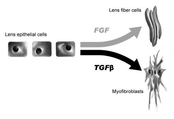
Over the last two decades much progress has been made in identifying and characterizing many of the molecules involved in understanding normal lens biology and its pathology. Much of this has been made possible through the establishment and use of the lens epithelial explant system. This simplistic tissue culture model, comprised of a sheet of lens epithelium on its native substratum, has been used effectively to study many cellular processes, including lens epithelial cell proliferation, fiber cell differentiation, cell apoptosis as well as epithelial-to-mesenchymal transformation of cells. In doing so, a number of key growth factors and cytokines, including members of the FGF, Wnt and TGFbeta family have been shown to play essential roles in many of these cellular events. This has led to further studies exploring the signaling pathways downstream of these molecules in the lens, paving the way for the development of a number of in situ models (primarily transgenic mouse lines) to further explore in more detail the nature of these molecular and cellular interactions. To reciprocate, the lens epithelial explant system is increasingly being used to further characterize the nature of many complex phenotypes and pathologies observed in these in situ models, allowing us to selectively isolate and examine the direct impact of an individual molecule on a specific cellular response in lens cells. There is no question that the lens epithelial explant system has served as a powerful tool to further our understanding of lens biology and pathology, and there is no doubt that it will continue to serve in such a capacity, as new developments are realized and putative treatments for aberrant lens cell behavior are to be trialed.
Copyright 2009 Elsevier Ltd. All rights reserved.
Figures


References
-
- Awasthi N, Wagner BJ. Suppression of human lens epithelial cell proliferation by proteasome inhibition, a potential defense against posterior capsular opacification. Invest Ophthalmol Vis Sci. 2006;47(10):4482–9. - PubMed
-
- Banh A, Deschamps PA, et al. The role of Hsp70 and Hsp90 in TGF-beta-induced epithelial-to-mesenchymal transition in rat lens epithelial explants. Mol Vis. 2007;13:2248–62. - PubMed
-
- Campbell MT, McAvoy JW. Onset of fibre differentiation in cultured rat lens epithelium under the influence of neural retina-conditioned medium. Exp Eye Res. 1984;39(1):83–94. - PubMed
-
- Campbell MT, McAvoy JW. A lens fibre differentiation factor from calf neural retina. Exp Cell Res. 1986;163(2):453–66. - PubMed
Publication types
MeSH terms
Grants and funding
LinkOut - more resources
Full Text Sources
Medical
