

Spectral domain optical coherence tomography for detection of foveal morphology in patients with nystagmus
- PMID: 20006817
- PMCID: PMC2917909
- DOI: 10.1016/j.jaapos.2009.09.019
Spectral domain optical coherence tomography for detection of foveal morphology in patients with nystagmus
Abstract
Purpose: To evaluate the feasibility of spectral domain optical coherence tomography (SD-OCT) macular scanning as a means of studying the afferent visual system in nystagmus patients.
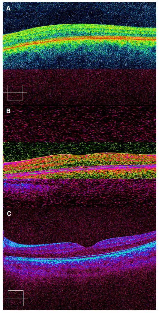
Methods: Nystagmus patients who underwent SD-OCT, clinical evaluation, and eye movement recordings were recruited for this prospective, single-center, noncomparative study. Three SD-OCT macular three-dimensional cube scans per eye (200 x 200 x 1024 samplings in a 6 x 6 mm region) were obtained for qualitative retinal morphology analysis.
Results: Nineteen patients (6-68 years; average, 19 years) were analyzed. Of these, 17 patients had infantile nystagmus syndrome, and 2 had fusion maldevelopment nystagmus; 17 patients (89%) had associated sensory system abnormalities, including 9 (47%) with albinism. Macular images were successfully obtained in all but 1 patient (95%). Of the 8 successfully imaged oculocutaneous patients, 7 patients demonstrated "fovea plana," and all demonstrated abnormal morphology.
Conclusion: SD-OCT reliably provides detailed structural imaging of the fovea in nystagmus patients.
Figures
References
-
- Wojtkowski M, Bajraszewski T, Gorczynska I, et al. Ophthalmic imaging by spectral optical coherence tomography. Am J Ophthalmol. 2004;138:412–19. - PubMed
-
- Jiao S, Knighton RW, Huang X, Gregori G, Puliafito CA. Simultaneous acquisition of sectional and fundus ophthalmic images with spectral-domain optical coherence tomography. Optics Express. 2005;13:444–52. - PubMed
MeSH terms
Grants and funding
LinkOut - more resources
Full Text Sources
