. 2009 Dec 17;6(6):513-22.
doi: 10.1016/j.chom.2009.11.004.

Hepatitis C virus blocks interferon effector function by inducing protein kinase R phosphorylation

Affiliations

Hepatitis C virus blocks interferon effector function by inducing protein kinase R phosphorylation

Urtzi Garaigorta et al. Cell Host Microbe. .

Abstract

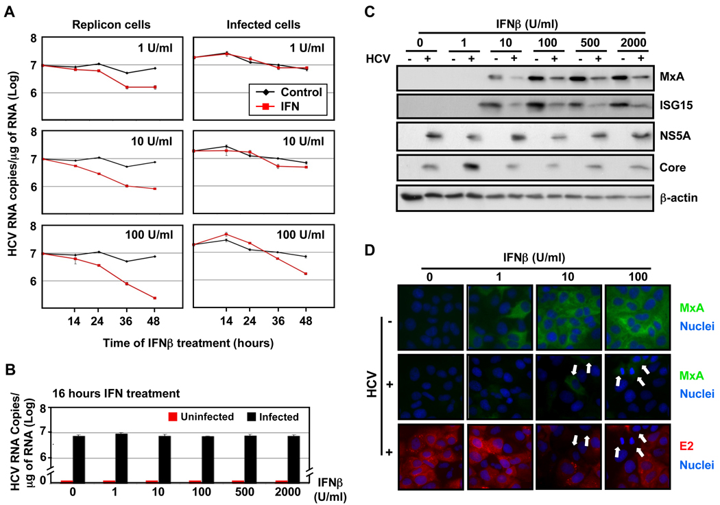
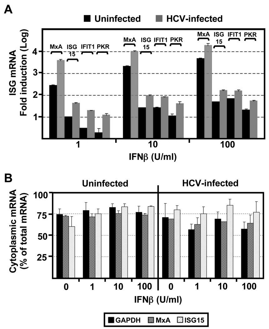
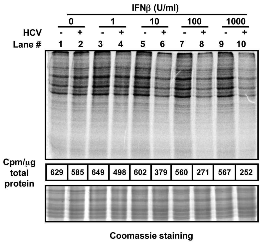
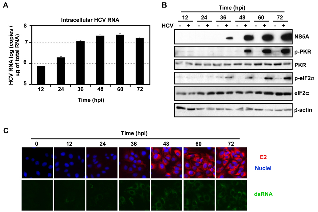
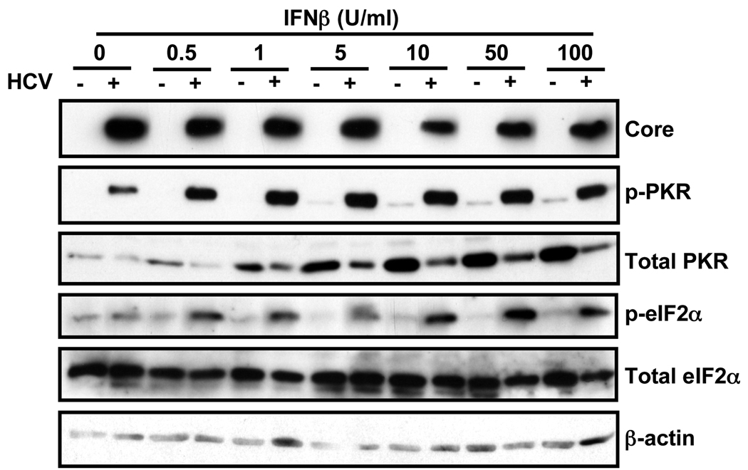
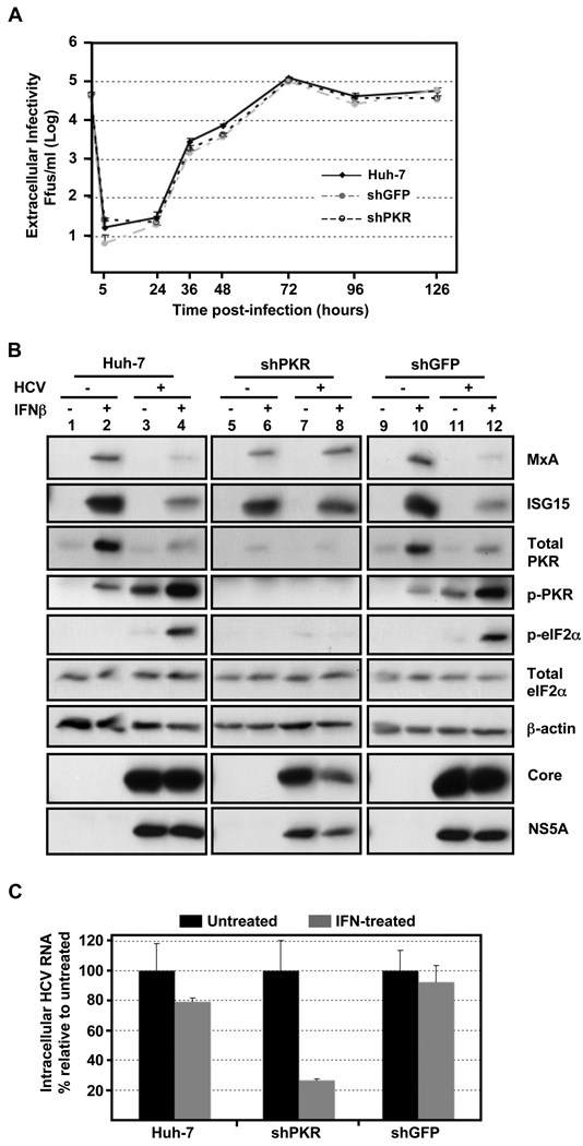
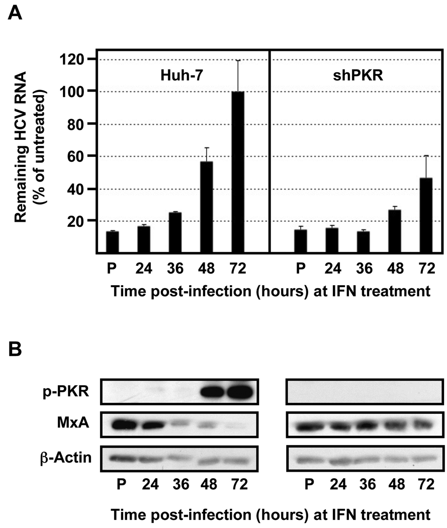
Hepatitis C virus (HCV) is a single-stranded RNA virus encoding a single polyprotein whose translation is driven by an internal ribosome entry site (IRES). HCV infection strongly induces antiviral interferon-stimulated gene (ISG) expression in the liver, yet it persists, suggesting that HCV can block ISG effector function. We now show that HCV infection triggers phosphorylation and activation of the RNA-dependent protein kinase PKR, which inhibits eukaryotic translation initiation factor eIF2 alpha and attenuates ISG protein expression despite normal ISG mRNA induction. ISG protein induction is restored and the antiviral effects of interferon are enhanced when PKR expression is suppressed in interferon-treated infected cells. Whereas host protein translation, including antiviral ISGs, is suppressed by activated PKR, HCV IRES-dependent translation is not. These results suggest that the ability of HCV to activate PKR may, paradoxically, be advantageous for the virus during an IFN response by preferentially suppressing the translation of ISGs.


Figures

Fig. 1
Fig. 1. Suppression of ISG protein induction in HCV-infected cells
(A) Persistently infected Huh-7 cells and JFH-1 full-length stable replicon cells were treated with different doses of IFNβ as indicated in the figure, and at various times thereafter the intracellular HCV RNA and GAPDH mRNA (for normalization) were quantified by RT-qPCR. Data are represented as Mean ± AVEDEV; n=3. This experiment is representative of 2 independent experiments. (B–D) Huh-7 cells were infected with JFH-1 virus at moi=0.2 and at day 5 post-infection uninfected (−) and infected (+) cells were treated for 16 hours with different doses of IFNβ. (B) Quantification of the intracellular HCV RNA by RT-qPCR in total RNA isolated from HCV-infected or uninfected cells. GAPDH mRNA quantification was used for normalization. Data are represented as Mean ± AVEDEV; n=3. This experiment is representative of 3 independent experiments. (C) Analysis by Western-blotting of ISG (MxA and ISG15) and viral (NS5A and core) protein expression induced by IFNβ in cell extracts of HCV-infected or uninfected cells. β-actin expression was examined as protein loading control. Each sample represented a pool of three replicas. (D) Immunofluorescence analysis of MxA (in green) and viral E2 (in red) protein expression in uninfected and HCV-infected cells after IFNβ treatment. The nuclei were stained by Hoechst solution (blue). White arrows in HCV-infected panels point to cells with high MxA expression levels and with no or very low E2 staining. These images are representative of three different experiments.
Fig. 2
Fig. 2. The accumulation and nucleo-cytoplasmic distribution of ISG mRNAs is normal in HCV-infected cells
Huh-7 cells were infected with JFH-1 virus at moi=0.2. At day 5 post-infection uninfected and HCV-infected cells were treated with different doses of IFNβ for 16 hours. (A) Quantification of ISG (MxA, ISG15, IFIT1 and PKR) mRNA levels by RT-qPCR in total RNA samples isolated from uninfected and HCV-infected cells. GAPDH mRNA quantification from the same samples was used for normalization. Results display the average fold induction for each ISG mRNA relative to its expression level in untreated uninfected Huh-7 cells (Mean ± AVEDEV; n=3). This experiment is representative of 3 independent experiments. (B) Quantification of MxA, ISG15 and GAPDH mRNAs in the nuclear and cytoplasmic fractions of uninfected and HCV-infected cells. Results displayed as percentage of cytoplasmic versus total mRNA content for each sample. Each bar in the graph corresponds to the average and the average deviation (Mean ± AVEDEV; n=3) of three replicas.
Fig. 3
Fig. 3. Global cellular protein synthesis is inhibited in IFNβ-treated HCV-infected cells
Huh-7 cells were infected with JFH-1 virus at moi=0.2. At day 5 post-infection uninfected (−) and HCV-infected (+) cells were treated with different doses of IFNβ for 16 hours, after which the cultures were metabolically labeled for 1 hour with 35S-Met. Protein extracts prepared in RIPA buffer were quantified by BCA. Equivalent amounts from each sample were analyzed by SDS-PAGE, Coomassie staining (bottom panel) and autoradiography (top panel). The figure is representative of 2 independent experiments with 2 replicas per sample. The radioactivity present after methanol-chloroform (4:1) precipitation of each sample was quantified and the results were displayed as counts per minute (cpm) per microgram of total protein (middle panel).
Fig. 4
Fig. 4. HCV infection triggers the phosphorylation of PKR and eIF2α proteins
Huh-7 cells were infected (+) or not (−) with JFH-1 d183 virus at moi=3. (A) At the indicated time points post-inoculation, total RNA was isolated from HCV-infected or uninfected cells and the intracellular HCV RNA levels were quantified by RT-qPCR. GAPDH mRNA quantification was used for normalization. Data are represented as Mean ± AVEDEV; n=3. (B) At the indicated time points cellular extracts were prepared in RIPA buffer, their protein content was quantified by BCA and the accumulation of cellular eIF2α , phospho-eIF2α (p-eIF2α), PKR, phospho-PKR (p-PKR) and viral NS5A proteins was analyzed by Western-blotting.β-actin expression was examined as protein loading control. The results displayed are representative of 3 independent experiments. (C) Cultures infected in parallel were fixed at the indicated time points and processed for immunofluorescence for the detection of viral E2 (in red) or dsRNA (in green). The nuclei were stained by Hoechst solution (blue).
Fig. 5
Fig. 5. HCV infection strongly enhances phosphorylation of PKR and eIF2α proteins in IFNβ-treated cells
Huh-7 cells were infected with JFH-1 virus at moi=0.2. At day 5 post-infection uninfected (−) and infected (+) cells were treated for 16 hours with different doses of IFNβ. Total cellular protein in extracts prepared in RIPA buffer was quantified by BCA. Equivalent amounts from each sample were subjected to Western-blot analysis for the detection of viral core and cellular eIF2α, p-eIF2α, PKR and p-PKR proteins. β-actin expression was examined as protein loading control. The figure is representative of 2 independent experiments with 3 replicas per sample.
Fig. 6
Fig. 6. PKR downregulation does not affect HCV infection kinetics, restores HCV-induced ISG protein suppression and enhances the antiviral effect of IFN
Huh-7 cells were transduced with lentiviral vectors expressing GFP and shRNAs against GFP or PKR. (A) PKR-, GFP-down-regulated and Huh-7 cells, used as controls, were infected with JFH-1 d183 virus at moi=5 and the extracellular infectivity was determined at various times thereafter. Data are represented as Mean ± AVEDEV; n=3. The graph is representative of 3 independent experiments. (B) Transduced and control cells were infected (+) or not (−) with JFH-1 d183 virus at moi=5. At 70 hpi the cultures were treated (+) or not (−) with 100 U/ml of IFNβ for 20 hours, after which cellular extracts were prepared in RIPA buffer. Equivalent amounts of total protein were analyzed by Western-blotting for core, NS5A, MxA, ISG15, PKR, p-PKR, eIF2α and p-eIF2α proteins. β-actin expression was examined as protein loading control. (C) The HCV RNA present in samples generated in parallel was extracted and analyzed by RT-qPCR. GAPDH mRNA quantification of the same samples was used for normalization. IFN-treated samples are represented by grey bars and untreated controls by black bars. The results are displayed as Mean ± AVEDEV; n=3. Panels are representative of 3 independent experiments with 3 replicas per sample.
Fig. 7
Fig. 7. The HCV resistance to IFN correlates with PKR phosphorylation and is inversely related to ISG protein expression
Huh-7 cells were transduced with lentiviral vectors expressing shRNAs against PKR, as indicated in Figure 6. Transduced and control cells were pre-treated with 100 U/ml of IFN (P) or remain untreated (the rest). After 16 hours all the cells were infected with JFH-1 d183 virus at moi=3 and at the indicated times post infection cells were treated with 100 U/ml of IFNβ for 20 hours, after which cellular extracts were prepared for RNA and protein analysis. (A) The HCV RNA present in those samples was analyzed by RT-qPCR. The quantification of GAPDH mRNA was used for normalization. Data are displayed as percentage of remaining HCV RNA in IFN-treated samples relative to that in the untreated ones at each time point. Bars in the graph represent the Mean ± AVEDEV; n=3. The graph is representative of 2 independent experiments with 3 replicas per sample. (B) Equivalent amounts of total protein from samples generated in parallel were analyzed by Western-blotting for MxA and p-PKR protein detection. β-actin expression was examined as protein loading control. Panels are representative of 2 independent experiments.

Comment in

Similar articles

Cited by

References

    1. Alter HJ, Seeff LB. Recovery, persistence, and sequelae in hepatitis C virus infection: a perspective on long-term outcome. Semin Liver Dis. 2000;20:17–35. - PubMed
    1. Cheng G, Zhong J, Chisari FV. Inhibition of dsRNA-induced signaling in hepatitis C virus-infected cells by NS3 protease-dependent and -independent mechanisms. Proc Natl Acad Sci U S A. 2006;103:8499–8504. - PMC - PubMed
    1. Choo QL, Richman KH, Han JH, Berger K, Lee C, Dong C, Gallegos C, Coit D, Medina-Selby R, Barr PJ, et al. Genetic organization and diversity of the hepatitis C virus. Proc Natl Acad Sci U S A. 1991;88:2451–2455. - PMC - PubMed
    1. Deutsch M, Hadziyannis SJ. Old and emerging therapies in chronic hepatitis C: an update. J Viral Hepat. 2008;15:2–11. - PubMed
    1. Dreux M, Boson B, Ricard-Blum S, Molle J, Lavillette D, Bartosch B, Pecheur EI, Cosset FL. The exchangeable apolipoprotein ApoC-I promotes membrane fusion of hepatitis C virus. J Biol Chem. 2007;282:32357–32369. - PubMed

Publication types

MeSH terms