. 2009 Dec 17;6(6):523-35.
doi: 10.1016/j.chom.2009.11.006.

Adenovirus transport via direct interaction of cytoplasmic dynein with the viral capsid hexon subunit

Affiliations

Adenovirus transport via direct interaction of cytoplasmic dynein with the viral capsid hexon subunit

K Helen Bremner et al. Cell Host Microbe. .

Abstract

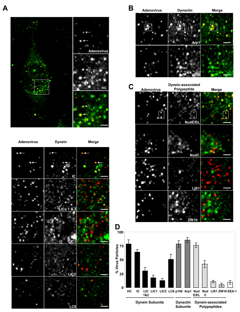
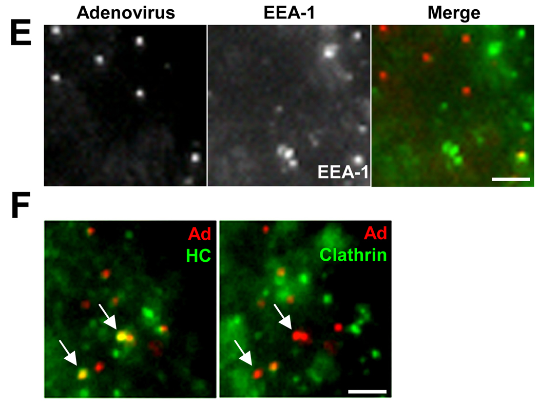
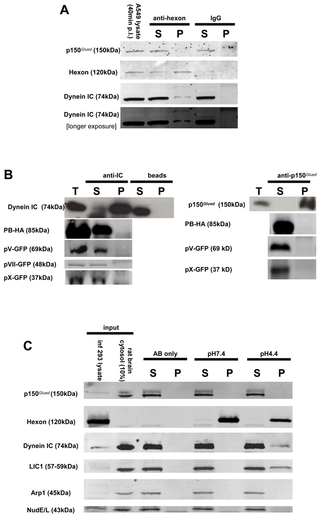
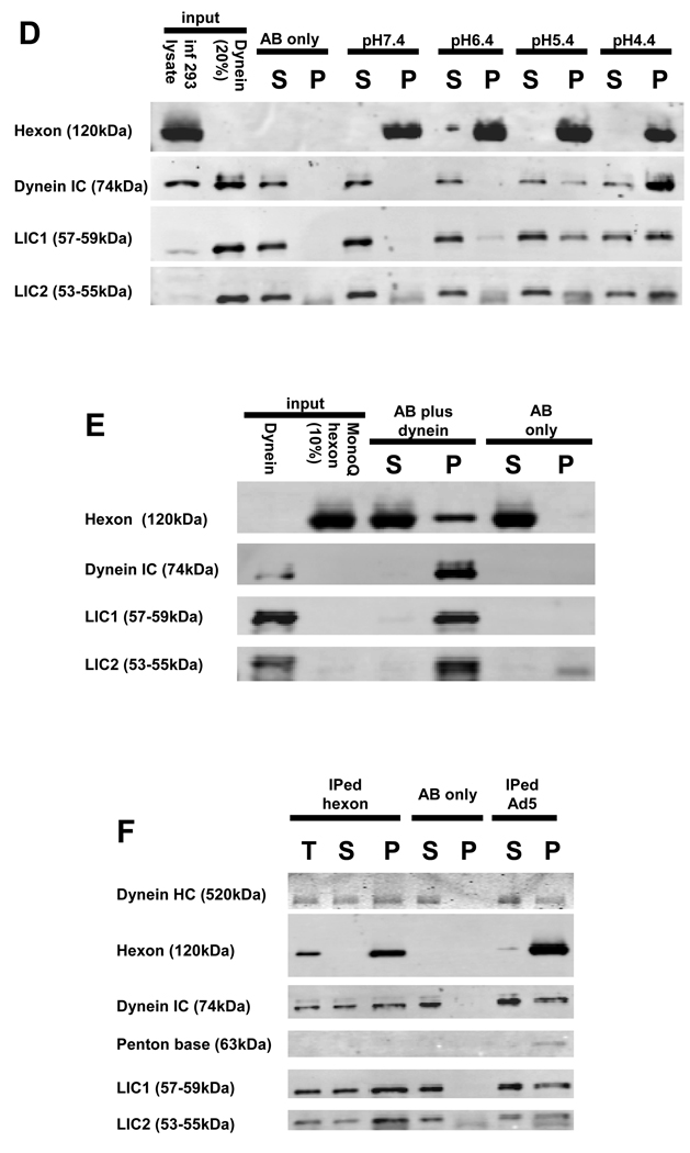
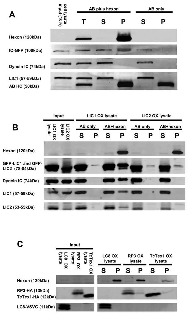
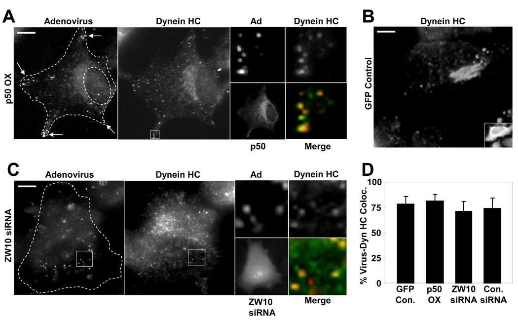
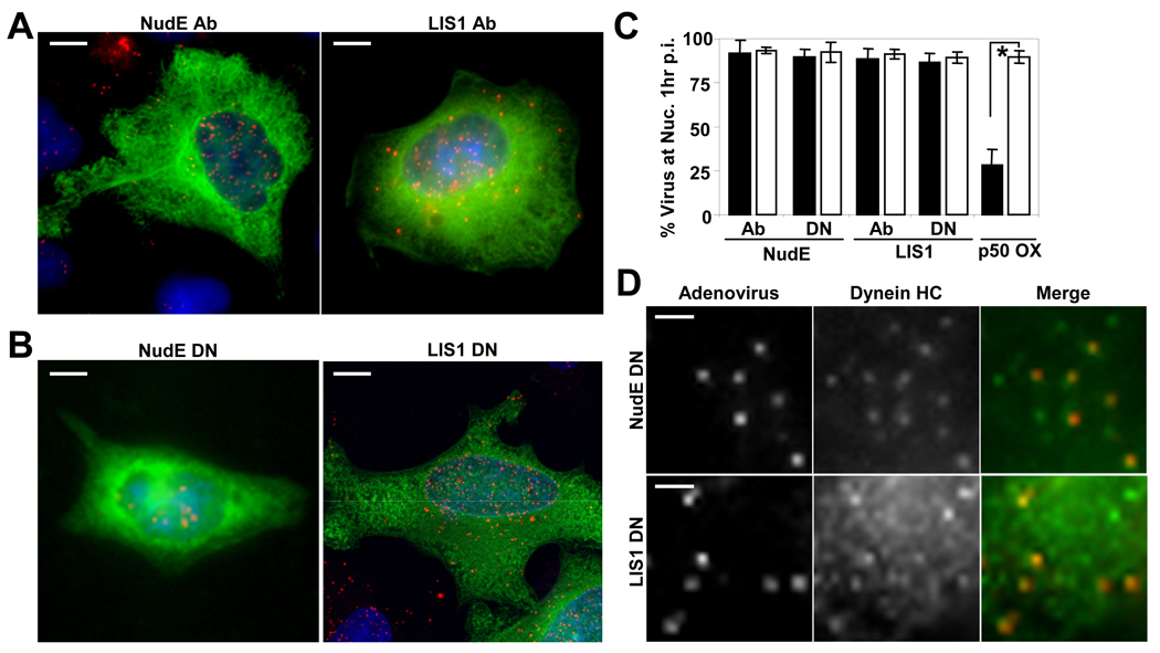
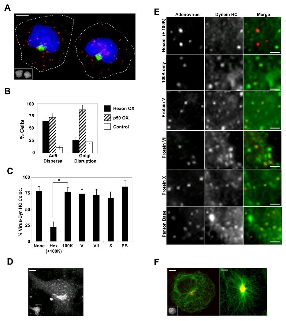
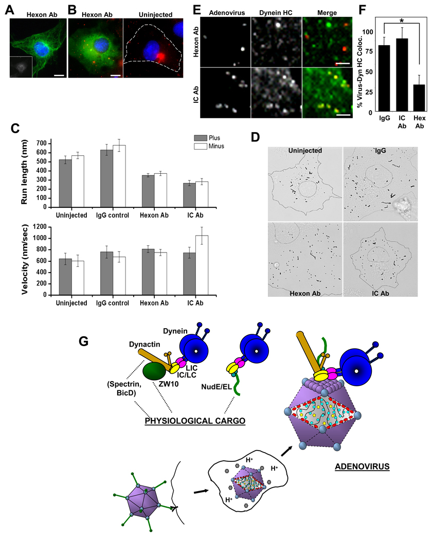
Early in infection, adenovirus travels to the nucleus as a naked capsid using the microtubule motor cytoplasmic dynein. How the dynein complex is recruited to viral cargo remains unclear. We find that cytoplasmic dynein and its associated proteins dynactin and NudE/NudEL, but not LIS1 or ZW10, colocalized with incoming, postendosomal adenovirus particles. However, in contrast to physiological cargos, dynein binding to adenovirus was independent of these dynein-associated proteins. Dynein itself directly interacted through its intermediate and light intermediate chains with the adenovirus capsid subunit hexon in a pH-dependent manner. Expression of hexon or injection of anti-hexon antibody inhibited virus transport but not physiological dynein function. These results identify hexon as a direct receptor for cytoplasmic dynein and demonstrate that hexon recruits dynein for transport to the nucleus by a mechanism distinct from that for physiological dynein cargo.


Figures

Figure 1
Figure 1. Colocalization of cytoplasmic dynein and associated factors with incoming adenovirus capsids in situ
HeLa cells were infected with Ad5 (red) for 60 min and processed for immunofluorescence microscopy using antibodies (green) to (A) dynein, (B) dynactin, or (C) dynein regulatory proteins. A whole cell is shown in A, as well as expanded regions from confocal images of Ad5 chemically labeled with Cy3 (See Fig S1 for additional whole cell images.). Arrows or boxed regions highlight examples of virus colocalization with dynein and other antigens. All images are merged stacks and the scale bars represent 1µm. (D) Percentage of Ad5 particles +/− SD staining positively for costained markers. Virus particles in 10 cells from each of three independent experiments were analyzed for each antigen. (E) Double labeling of adenovirus at 60 min p.i. with the early endosomal marker EEA-1 (green), showing low colocalization. (F) Triple labeling of adenovirus at 15 min p.i with anti-dynein HC and anti-clathrin. Arrows show examples of HC-positive virus particles which are negative for clathrin, consistent with recruitment of dynein after exit of the virus from the early endosome. The panels show the same region of the same cell with dynein HC (green) on the left and clathrin (green) on the right.
Figure 2
Figure 2. Adenovirus interacts with dynein, but not dynactin, via the hexon capsid subunit
(A) Ad5 was immunoprecipitated from infected A549 cell lysate 40min p.i. and the pellets were immunoblotted using anti-dynein IC or anti-dynactin p150Glued antibodies. Dynein but not dynactin was present in the pellets. (B) Dynein (IC) and dynactin (p150Glued) were immunoprecipitated from lysates of COS-7 cells expressing the recombinant viral capsid polypeptides protein V, penton base, and proteins VII and X, none of which were found in the pellets. (C) Analysis of dynein and dynactin binding to hexon. Hexon was immunoprecipitated from infected 293A cell lysates, exposed to pH 7.4 or pH 4.4 buffer for 30 min, and then returned to pH 7.4, mixed with rat brain cytosol, and immunoprecipitated. The dynein complex, detected by blotting for IC and LIC1 subunits, clearly bound to the pH 4.4-treated, but not the control hexon. The dynactin complex, detected by blotting for p150Glued and Arp1 subunits, was absent in both cases, as were NudE and NudEL. (D) Hexon was exposed to a range of pH values, washed at pH 7.4, incubated with purified rat brain cytoplasmic dynein, and immunoprecipitated. The purified cytoplasmic dynein showed a pH-dependent interaction with hexon, which increased markedly with hexon exposure to decreasing pH. (E) Purified rat brain cytoplasmic dynein was bound to anti-dynein IC antibody 74.1 and incubated with pH 4.4 pretreated hexon purified by anion exchange chromatography from adenovirus infected cell lysates. Hexon was clearly present in the dynein pellet. (F) Immunopurified (IPed) hexon or intact adenovirus was acidified at pH 4.4, restored to pH 7.4, incubated with purified rat brain cytoplasmic dynein, and immunoprecipitated. The dynein complex as revealed by staining with anti- HC, IC, LIC1, and LIC2 antibodies was clearly present in both the hexon and adenovirus pellets. T (total), S (supernatant), and P (pellet)
Figure 3
Figure 3. Hexon interacts with dynein IC and LIC1
Hexon was immunoisolated from late stage adenovirus infected 293A cell lysates, exposed to pH 4.4, and combined at pH 7.4 with lysates of COS-7 cells expressing individual dynein subunits. (A) Hexon pull-downs from IC2C-GFP-expressing cell lysates were blotted with antibodies to dynein IC and LIC1. The recombinant IC-GFP at 100kDa clearly associated with hexon. The lack of immunoreactivity at the positions of the endogenous IC and LIC1 indicates that endogenous dynein was present at too low levels to be detected. (B) Hexon pull-downs from LIC1-GFP- or LIC2-GFP-expressing cell lysates were blotted with anti-GFP, plus anti-IC, -LIC1, and LIC2 antibodies. The recombinant LIC1-GFP clearly associated with hexon. In this experiment endogenous dynein complex also bound to hexon as indicated by the presence of all three dynein antigens in the hexon pull-down. Recombinant LIC2 showed little to no hexon binding relative the antibody-only control. (C) Hexon pull-downs from LC8-VSVG-, RP3-HA-, or TcTex1-HA-expressing cell lysates were blotted with antibodies to the respective epitope tags. None of the three recombinant LCs bound to hexon. OX (overexpression), T (total), S (supernatant), and P (pellet)
Figure 4
Figure 4. Dynein recruitment to adenovirus is independent of dynactin and ZW10
(A) HeLa cells were transfected with a cDNA encoding dynamitin-myc, infected at 24 hr with Cy3-Ad5 (red), and fixed and stained at 60 min p.i. for dynamitin and dynein HC (green). Dynein HC is still clearly detectable on virus particles (arrows) in whole cell image (boxed region is expanded at right), which are found largely in the cell periphery as a result of dynamitin overexpression. However, dynein HC localization to the Golgi apparatus was strongly inhibited vs control Golgi staining shown in panel B. (The weak juxtanuclear staining in the dynein HC panel (A) represents diffuse cytoplasmic staining in a thickened cell region, as further indicated by distribution of soluble myc-dynamitin.) (C) HeLa cells were transfected with ZW10 siRNA, infected with Cy3-Ad5 on day 3, fixed, and stained for dynein HC. Ad5 particles localized to the nucleus, consistent with a lack of function for ZW10 in Ad5 transport. Dynein HC staining at the Golgi apparatus was greatly reduced but was still clearly present on Ad5 particles (boxed region expanded at right). (D) Percent Ad5 particles colocalizing with dynein HC in dynamitin-overexpressing and ZW10 RNAi cells vs controls. All values are statistically insignificant compared to the control case (P > 0.2; t-test). Error bars represent SD. (Scale bars = 5 µm)
Figure 5
Figure 5. Ad5 transport and dynein recruitment are unaffected by NudE/NudEL and LIS1 inhibition
(A) A549 cells were microinjected with function-blocking NudE/NudEL or LIS1 antibodies (green), then infected with Cy3-Ad5 (red) before fixing and staining 60 min p.i. Nuclear accumulation of virus was observed in each case. (Scale bars = 5 µm) (B) HeLa cells were transfected with NudE or LIS1 dominant negative (DN) cDNA constructs (green). 24 hours after transfection, cells were infected with Cy3-Ad5 (red) and fixed 60 min p.i.. Again, clear nuclear accumulation of virus was observed. (Scale bars = 5 µm) (C) Percentage of cells exhibiting accumulation of Ad5 at the nucleus was determined for NudE/NudEL- and LIS1-inhibited cells (black bars). The effect of dynamitin (p50) overexpression on Ad5 nuclear accumulation is included for comparison. Open bars indicate results from control injected or transfected cells. Error bars represent SD. * indicates P<0.01; t-test. (D) Cy3-Ad5 (red) in HeLa cells overexpressing NudE or LIS1 DN cDNAs still colocalize with dynein HC (green). Panels show enlarged regions from a single cell. (Scale bars = 1 µm)
Figure 6
Figure 6. Hexon expression displaces dynein from incoming Ad5 and prevents viral accumulation at the nucleus
(A) HeLa cells expressing hexon (stained with anti-hexon monoclonal antibody, inset) show dispersed Ad5 particles (red) at 60 min p.i., but the centrosome-centered organization of the Golgi apparatus stained with anti-giantin (green) is unaffected. (scale bar: 5 µm) (B) Quantitative comparison of hexon and dynamitin effects on Ad5 distribution vs Golgi organization. Ad5 dispersal: Cells with below 70% of virions at nucleus at 60 min p.i.. Error bars represent +/− SD. (C) Percent of Ad5 particles positive for dynein HC +/− SD, determined as described for Fig 1. * indicates P < 0.01; t-test. (D) Dynein HC staining of the Golgi apparatus persists in hexon-expressing cell (inset). (scale bar: 5 µm) (E) HeLa cells transfected with cDNAs encoding viral polypeptides were infected with Cy3-Ad5 (red) 48 hrs post-transfection, fixed at 60 min p.i. and stained for dynein HC (green). Panels show enlarged regions from individual cells. Dynein HC colocalization with adenovirus is disrupted by expression of hexon, but not of the other virus subunits. (scale bars: 1 µm) (F) Partial microtubule disorganization (anti-tubulin antibody, green) in a hexon-expressing cell (left), but with no apparent effect on Golgi organization (red), compared with the control (right). (scale bars: 5 µm)
Figure 7
Figure 7. Anti-hexon antibody interferes with Ad5 transport
(A) COS-7 cell injected with monoclonal anti-hexon antibody (inset, stained with Cy5-conjugated secondary antibody) shows normal microtubule distribution as indicated by anti-tubulin immunostaining (green). (Scale bars: 5 µm) (B) COS-7 cells injected with anti-hexon antibody were infected with Alexa-Ad5, fixed 60 min p.i. and stained with Cy2-conjugated secondary antibody (green) and DAPI (blue). Ad5 particles were dispersed throughout the cytoplasm, whereas in control cells (right panel) the virus accumulated at the centrosome. (Scale bars: 5 µm) (C) Uninjected or antibody-injected cells were infected with Alexa-Ad5, and movies were acquired between 20 – 90 min p.i. for 1 min at 16 frames/sec. Anti-hexon and anti-dynein IC antibodies each significantly inhibited virus run length in both plus and minus directions, but had minimal effect on velocity. Error bars represent +/− SEM. Motility data were extracted from analyzing 13 uninjected, 18 IgG injected, 27 anti-hex antibody injected, and 9 anti-dynein IC antibody injected cells. (D) Virus tracks from the duration of a representative 1 min movie were overlaid onto brightfield images to illustrate the effect of anti-hexon and anti-dynein IC on run length. Overall, shorter runs were observed in both antibody inhibited cases compared to the uninjected or IgG controls. (See also Supplemental Movies 1–4). (E) Antibody-injected, Alexa-Ad5 (red) infected cells were fixed 60 min p.i. and stained for dynein HC (green). Anti-hexon injection decreased colocalization of dynein HC with Ad5, whereas anti-dynein IC did not. (Scale bars: 1 µm) (F) Quantitative analysis of anti-hexon and anti-dynein antibody injection reveals loss of dynein HC from Alexa-Ad5 particles in anti-hexon, but not anti-IC, injected cells. * indicates P < 0.01; t-test. Error bars represent +/− SD. (G) Schematic representation of dynein association with adenovirus vs physiological cargo forms. Initial stages of infection are depicted, with dynein-binding occurring following release of the capsid from the early endosome. Dynein is shown binding directly to the capsid protein hexon (purple), as found in the current study. Diagram also depicts known Golgi and kinetochore dynein recruitment factors dynactin, ZW10, NudE, NudEL, Spectrin , and BicD. Dynactin is involved in both Golgi and kinetochore dynein recruitment, and NudE/NudEL in kinetochore as well as centrosome dynein recruitment (see text). These factors localize to adenovirus particles in the current study, but do not participate in dynein recruitment. Dynactin is, instead, found to regulate virus motility.

Similar articles

Cited by

  • The precise function of alphaherpesvirus tegument proteins and their interactions during the viral life cycle.
    Cui Y, Wang M, Cheng A, Zhang W, Yang Q, Tian B, Ou X, Huang J, Wu Y, Zhang S, Sun D, He Y, Zhao X, Wu Z, Zhu D, Jia R, Chen S, Liu M. Cui Y, et al. Front Microbiol. 2024 Jul 2;15:1431672. doi: 10.3389/fmicb.2024.1431672. eCollection 2024. Front Microbiol. 2024. PMID: 39015737 Free PMC article. Review.
  • Adenovirus flow in host cell networks.
    Flatt JW, Butcher SJ. Flatt JW, et al. Open Biol. 2019 Feb 28;9(2):190012. doi: 10.1098/rsob.190012. Open Biol. 2019. PMID: 30958097 Free PMC article. Review.
  • E3 ubiquitin ligase Mindbomb 1 facilitates nuclear delivery of adenovirus genomes.
    Sarbanes SL, Blomen VA, Lam E, Heissel S, Luna JM, Brummelkamp TR, Falck-Pedersen E, Hoffmann HH, Rice CM. Sarbanes SL, et al. Proc Natl Acad Sci U S A. 2021 Jan 5;118(1):e2015794118. doi: 10.1073/pnas.2015794118. Epub 2020 Dec 21. Proc Natl Acad Sci U S A. 2021. PMID: 33443154 Free PMC article.
  • The expanding clinical and genetic spectrum of DYNC1H1-related disorders.
    Möller B, Becker LL, Saffari A, Afenjar A, Coci EG, Williamson R, Ward-Melver C, Gibaud M, Sedláčková L, Laššuthová P, Libá Z, Vlčková M, William N, Klee EW, Gavrilova RH, Lévy J, Capri Y, Scavina M, Körner RW, Valivullah Z, Weiß C, Möller GM, Frazier Z, Roberts A, Gener B, Scala M, Striano P, Zara F, Thiel M, Sinnema M, Kamsteeg EJ, Donkervoort S, Duboc V, Zaafrane-Khachnaoui K, Elkhateeb N, Selim L, Margot H, Marin V, Beneteau C, Isidor B, Cogne B, Keren B, Küsters B, Beggs AH, Sveden A, Chopra M, Genetti CA, Nicolai J, Dötsch J, Koy A, Bönnemann CG, von der Hagen M, von Kleist-Retzow JC, Voermans NC, Jungbluth H, Dafsari HS. Möller B, et al. Brain. 2025 Feb 3;148(2):597-612. doi: 10.1093/brain/awae183. Brain. 2025. PMID: 38848546 Free PMC article.
  • LIS1 and NudE induce a persistent dynein force-producing state.
    McKenney RJ, Vershinin M, Kunwar A, Vallee RB, Gross SP. McKenney RJ, et al. Cell. 2010 Apr 16;141(2):304-14. doi: 10.1016/j.cell.2010.02.035. Cell. 2010. PMID: 20403325 Free PMC article.

References

    1. Alonso C, Miskin J, Hernaez B, Fernandez-Zapatero P, Soto L, Canto C, Rodriguez-Crespo I, Dixon L, Escribano JM. African swine fever virus protein p54 interacts with the microtubular motor complex through direct binding to light-chain dynein. J Virol. 2001;75:9819–9827. - PMC - PubMed
    1. Burkhardt JK, Echeverri CJ, Nilsson T, Vallee RB. Overexpression of the dynamitin (p50) subunit of the dynactin complex disrupts dynein-dependent maintenance of membrane organelle distribution. J Cell Biol. 1997;139:469–484. - PMC - PubMed
    1. Cotten M, Weber JM. The adenovirus protease is required for virus entry into host cells. Virology. 1995;213:494–502. - PubMed
    1. Dignam JD, Martin PL, Shastry BS, Roeder RG. Eukaryotic gene transcription with purified components. Methods Enzymol. 1983;101:582–598. - PubMed
    1. Dohner K, Nagel CH, Sodeik B. Viral stop-and-go along microtubules: taking a ride with dynein and kinesins. Trends Microbiol. 2005;13:320–327. - PubMed

Publication types

LinkOut - more resources