

Cellular DNA ligase I is recruited to cytoplasmic vaccinia virus factories and masks the role of the vaccinia ligase in viral DNA replication
- PMID: 20006844
- PMCID: PMC2846536
- DOI: 10.1016/j.chom.2009.11.005
Cellular DNA ligase I is recruited to cytoplasmic vaccinia virus factories and masks the role of the vaccinia ligase in viral DNA replication
Erratum in
- Cell Host Microbe. 2010 Jan 21;7(1):82
Abstract
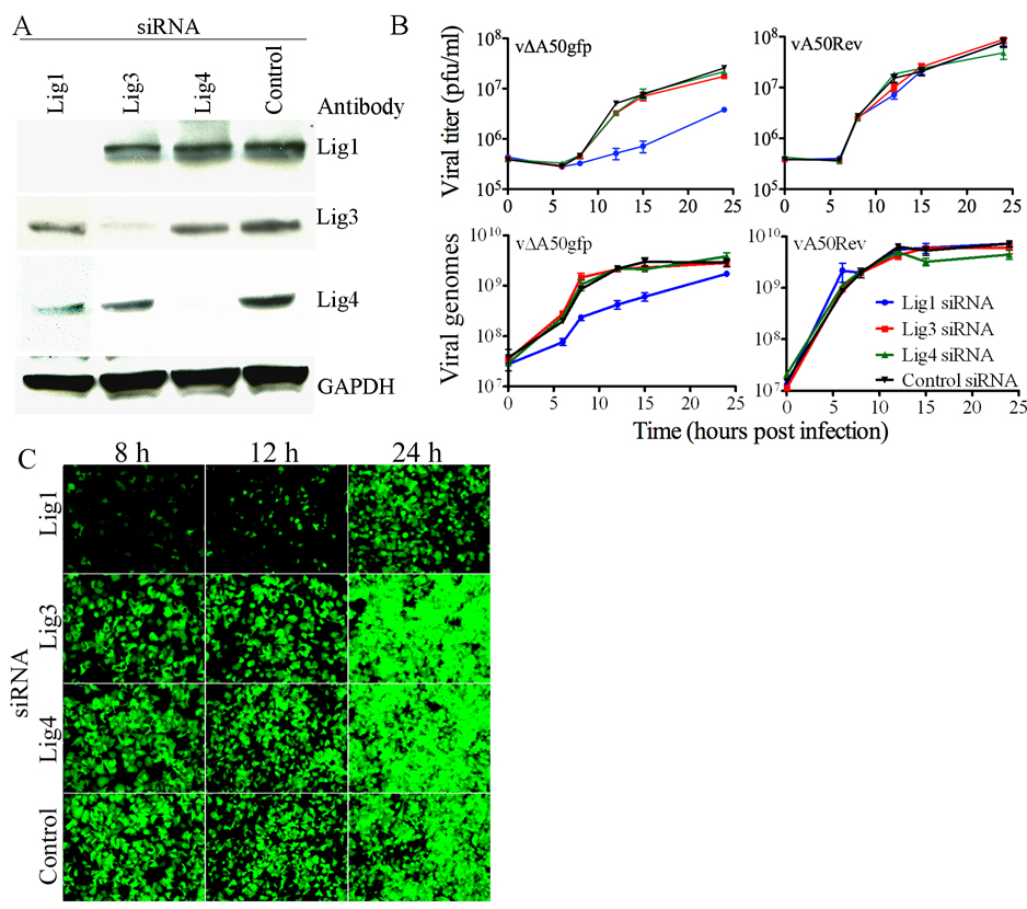
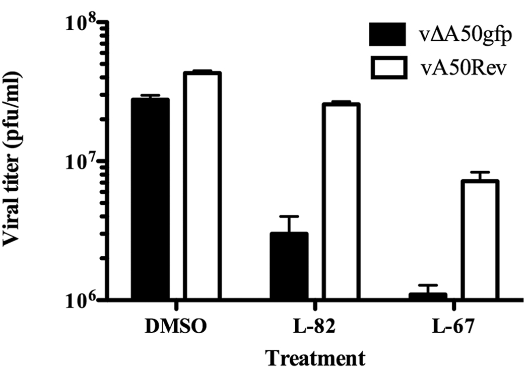
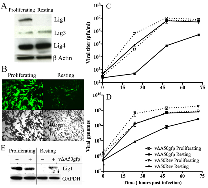
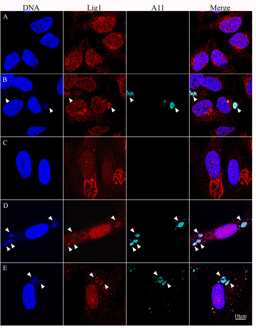
Vaccinia virus (VACV) encodes DNA polymerase and additional proteins that enable cytoplasmic replication. We confirmed the ability of VACV DNA ligase mutants to replicate and tested the hypothesis that cellular ligases compensate for loss of viral gene expression. RNA silencing of human DNA ligase I expression and a small molecule inhibitor of human DNA ligase I [corrected] severely reduced replication of viral DNA in cells infected with VACV ligase-deficient mutants, indicating that the cellular enzyme plays a complementary role. Replication of ligase-deficient VACV was greatly reduced and delayed in resting primary cells, correlating with initial low levels of ligase I and subsequent viral induction and localization of ligase I in virus factories. These studies indicate that DNA ligation is essential for poxvirus replication and explain the ability of ligase deletion mutants to replicate in dividing cells but exhibit decreased pathogenicity in mice. Encoding its own ligase might allow VACV to "jump-start" DNA synthesis.
Figures
Similar articles
-
Vaccinia virus DNA ligase recruits cellular topoisomerase II to sites of viral replication and assembly.J Virol. 2008 Jun;82(12):5922-32. doi: 10.1128/JVI.02723-07. Epub 2008 Apr 16. J Virol. 2008. PMID: 18417590 Free PMC article.
-
A DNA ligase gene in the Copenhagen strain of vaccinia virus is nonessential for viral replication and recombination.Virology. 1990 Nov;179(1):267-75. doi: 10.1016/0042-6822(90)90295-3. Virology. 1990. PMID: 2219723
-
Vaccinia Virus BBK E3 Ligase Adaptor A55 Targets Importin-Dependent NF-κB Activation and Inhibits CD8+ T-Cell Memory.J Virol. 2019 May 1;93(10):e00051-19. doi: 10.1128/JVI.00051-19. Print 2019 May 15. J Virol. 2019. PMID: 30814284 Free PMC article.
-
Viral exploitation of the MEK/ERK pathway - A tale of vaccinia virus and other viruses.Virology. 2017 Jul;507:267-275. doi: 10.1016/j.virol.2016.12.011. Virology. 2017. PMID: 28526201 Review.
-
The interferon system and vaccinia virus evasion mechanisms.J Interferon Cytokine Res. 2009 Sep;29(9):581-98. doi: 10.1089/jir.2009.0073. J Interferon Cytokine Res. 2009. PMID: 19708815 Review.
Cited by
-
Human genome-wide RNAi screen reveals a role for nuclear pore proteins in poxvirus morphogenesis.Proc Natl Acad Sci U S A. 2013 Feb 26;110(9):3519-24. doi: 10.1073/pnas.1300708110. Epub 2013 Feb 11. Proc Natl Acad Sci U S A. 2013. PMID: 23401514 Free PMC article.
-
Genetic Confirmation that the H5 Protein Is Required for Vaccinia Virus DNA Replication.J Virol. 2015 Jun;89(12):6312-27. doi: 10.1128/JVI.00445-15. Epub 2015 Apr 8. J Virol. 2015. PMID: 25855734 Free PMC article.
-
Identifying Host Factors Associated with DNA Replicated During Virus Infection.Mol Cell Proteomics. 2017 Dec;16(12):2079-2097. doi: 10.1074/mcp.M117.067116. Epub 2017 Oct 2. Mol Cell Proteomics. 2017. PMID: 28972080 Free PMC article.
-
Evaluation of the role of the vaccinia virus uracil DNA glycosylase and A20 proteins as intrinsic components of the DNA polymerase holoenzyme.J Biol Chem. 2011 Jul 15;286(28):24702-13. doi: 10.1074/jbc.M111.222216. Epub 2011 May 13. J Biol Chem. 2011. PMID: 21572084 Free PMC article.
-
Poxvirus DNA replication.Cold Spring Harb Perspect Biol. 2013 Sep 1;5(9):a010199. doi: 10.1101/cshperspect.a010199. Cold Spring Harb Perspect Biol. 2013. PMID: 23838441 Free PMC article. Review.
References
-
- Applegren N, Hickey RJ, Kleinschmidt AM, Zhou Q, Coll J, Wills P, Swaby R, Wei Y, Quan JY, Lee MY, et al. Further characterization of the human cell multiprotein DNA replication complex. J. Cell. Biochem. 1995;59:91–107. - PubMed
-
- Broyles SS. A role for ATP hydrolysis in vaccinia virus early gene transcription. J. Biol. Chem. 1991;266:15545–15548. - PubMed
-
- Colinas RJ, Goebel SJ, Davis SW, Johnson GP, Norton EK, Paoletti E. A DNA ligase gene in the Copenhagen strain of vaccinia virus is nonessential for viral replication and recombination. Virology. 1990;179:267–275. - PubMed
Publication types
MeSH terms
Substances
Grants and funding
LinkOut - more resources
Full Text Sources
Research Materials
Miscellaneous
