

Quick shear-flow alignment of biological filaments for X-ray fiber diffraction facilitated by methylcellulose
- PMID: 20006950
- PMCID: PMC2794338
- DOI: 10.1016/j.bpj.2009.09.041
Quick shear-flow alignment of biological filaments for X-ray fiber diffraction facilitated by methylcellulose
Abstract
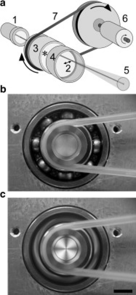
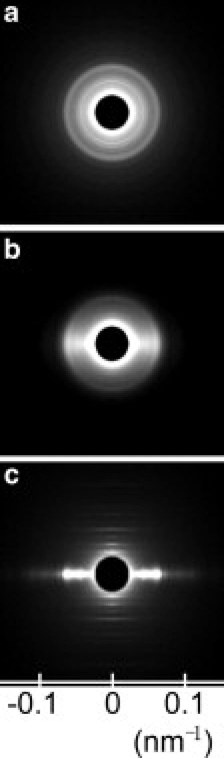
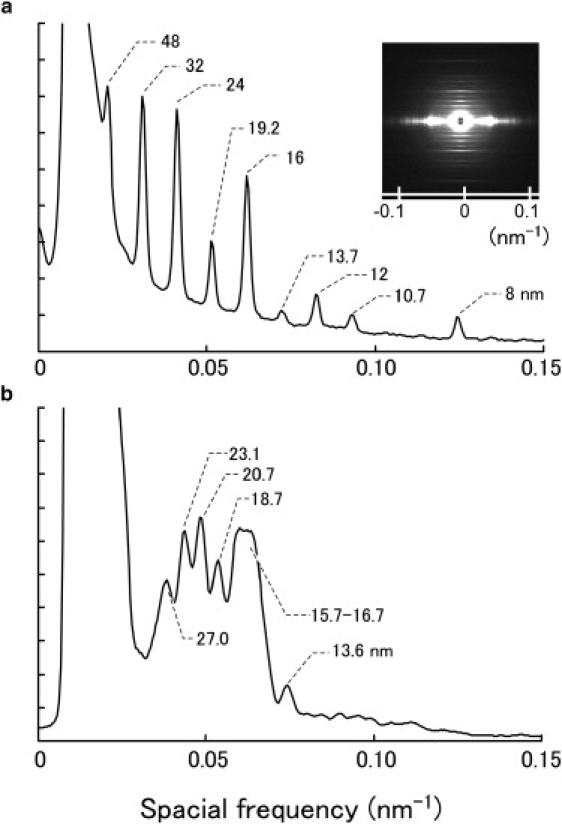
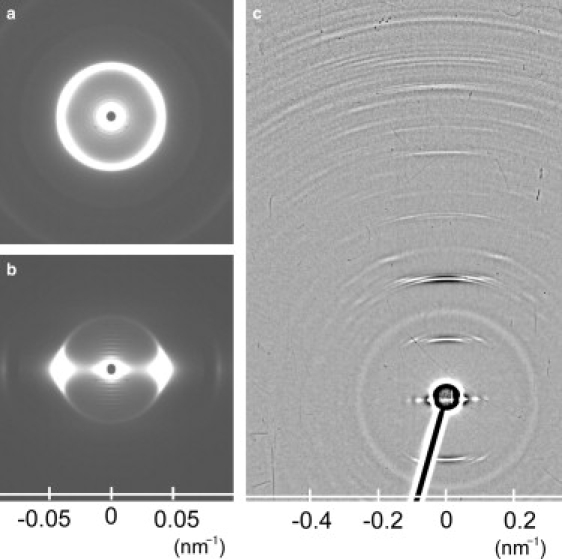
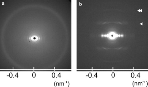
X-ray fiber diffraction is one of the most useful methods for examining the structural details of live biological filaments under physiological conditions. To investigate biologically active or labile materials, it is crucial to finish fiber alignment within seconds before diffraction analysis. However, the conventional methods, e.g., magnetic field alignment and low-speed centrifugations, are time-consuming and not very useful for such purposes. Here, we introduce a new alignment method using a rheometer with two parallel disks, which was applied to observe fiber diffractions of axonemes, tobacco mosaic tobamovirus, and microtubules. We found that fibers were aligned within 5 s by giving high shear flow (1000-5000 s(-1)) to the medium and that methylcellulose contained in the medium (approximately 1%) was essential to the accomplishment of uniform orientation with a small angular deviation (<5 degrees). The new alignment method enabled us to execute structure analyses of axonemes by small-angle x-ray diffraction. Since this method was also useful for the quick alignment of purified microtubules, as well as tobacco mosaic tobamovirus, we expect that we can apply it to the structural analysis of many other biological filaments.
Figures
Similar articles
-
X-Ray Fiber Diffraction Recordings from Oriented Demembranated Chlamydomonas Flagellar Axonemes.Biophys J. 2015 Jun 16;108(12):2843-53. doi: 10.1016/j.bpj.2015.04.039. Biophys J. 2015. PMID: 26083924 Free PMC article.
-
X-ray diffraction recording from single axonemes of eukaryotic flagella.J Struct Biol. 2012 Jun;178(3):329-37. doi: 10.1016/j.jsb.2012.03.011. Epub 2012 Apr 5. J Struct Biol. 2012. PMID: 22503702
-
X-ray fiber diffraction studies on flagellar axonemes.Methods Cell Biol. 2009;91:89-109. doi: 10.1016/S0091-679X(08)91005-0. Epub 2009 Dec 1. Methods Cell Biol. 2009. PMID: 20409782
-
Modern X-ray scattering studies of complex biological systems.Curr Opin Biotechnol. 2013 Aug;24(4):716-23. doi: 10.1016/j.copbio.2013.01.005. Epub 2013 Jan 29. Curr Opin Biotechnol. 2013. PMID: 23369441 Review.
-
Molecular dynamics applied to X-ray structure refinement.Acc Chem Res. 2002 Jun;35(6):404-12. doi: 10.1021/ar010034r. Acc Chem Res. 2002. PMID: 12069625 Review.
Cited by
-
Fiber Diffraction and Small-Angle Scattering for Structural Investigation of Bacterial Amyloids.Methods Mol Biol. 2022;2538:95-107. doi: 10.1007/978-1-0716-2529-3_7. Methods Mol Biol. 2022. PMID: 35951295
-
X-Ray Fiber Diffraction Recordings from Oriented Demembranated Chlamydomonas Flagellar Axonemes.Biophys J. 2015 Jun 16;108(12):2843-53. doi: 10.1016/j.bpj.2015.04.039. Biophys J. 2015. PMID: 26083924 Free PMC article.
-
Hard X-ray Fourier transform holography from an array of oriented referenced objects.J Synchrotron Radiat. 2011 Jul;18(Pt 4):564-8. doi: 10.1107/S0909049511009836. Epub 2011 May 11. J Synchrotron Radiat. 2011. PMID: 21685672 Free PMC article.
-
X-ray diffraction recording from a small amount of fibrous protein materials oriented by a micro shear-flow cell.Biophys Physicobiol. 2024 Apr 20;21(2):e210014. doi: 10.2142/biophysico.bppb-v21.0014. eCollection 2024. Biophys Physicobiol. 2024. PMID: 39206128 Free PMC article.
-
Alternative Approaches to Understand Microtubule Cap Morphology and Function.ACS Omega. 2023 Jan 13;8(4):3540-3550. doi: 10.1021/acsomega.2c06926. eCollection 2023 Jan 31. ACS Omega. 2023. PMID: 36743020 Free PMC article. Review.
References
-
- Stubbs G. Developments in fiber diffraction. Curr. Opin. Struct. Biol. 1999;9:615–619. - PubMed
-
- Torbet J. Using magnetic orientation to study structure and assembly. Trends Biochem. Sci. 1987;12:327–330.
-
- Yamashita I., Suzuki H., Namba K. Multiple-step method for making exceptionally well-oriented liquid-crystalline sols of macromolecular assemblies. J. Mol. Biol. 1998;278:609–615. - PubMed
-
- Popp D., Lednev V.V., Jahn W. Methods of preparing well oriented sols of f-actin containing filaments suitable for fiber diffraction. J. Mol. Biol. 1987;197:679–684. - PubMed
Publication types
MeSH terms
Substances
LinkOut - more resources
Full Text Sources
