. 2010 Mar 1;398(1):87-97.
doi: 10.1016/j.virol.2009.11.033. Epub 2009 Dec 16.

Identification of two auto-cleavage products of nonstructural protein 1 (nsp1) in porcine reproductive and respiratory syndrome virus infected cells: nsp1 function as interferon antagonist

Affiliations

Identification of two auto-cleavage products of nonstructural protein 1 (nsp1) in porcine reproductive and respiratory syndrome virus infected cells: nsp1 function as interferon antagonist

Z Chen et al. Virology. .

Abstract

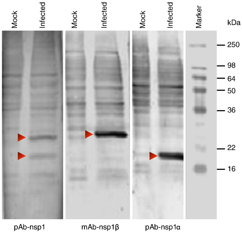
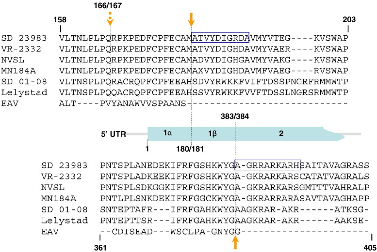
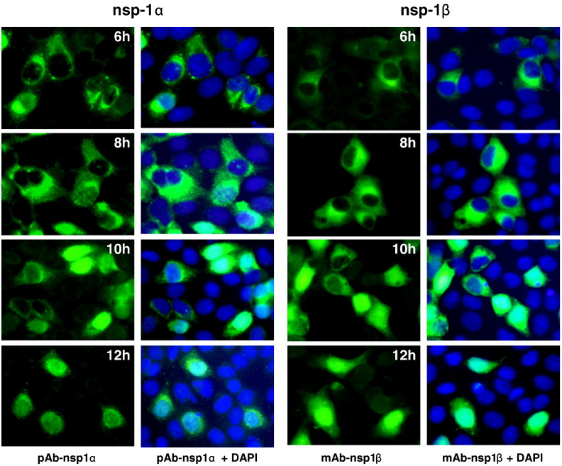
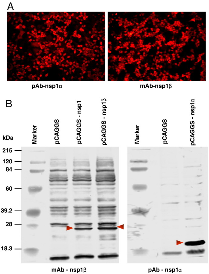
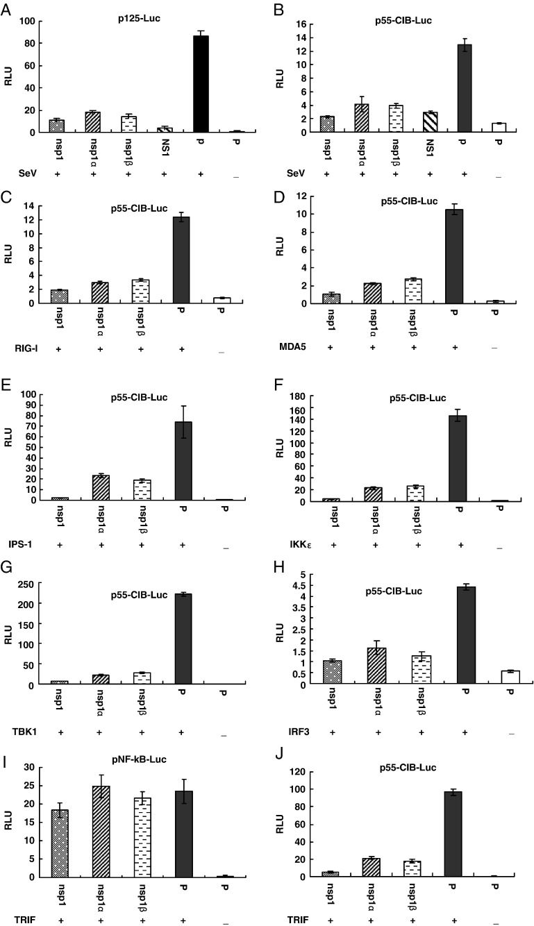
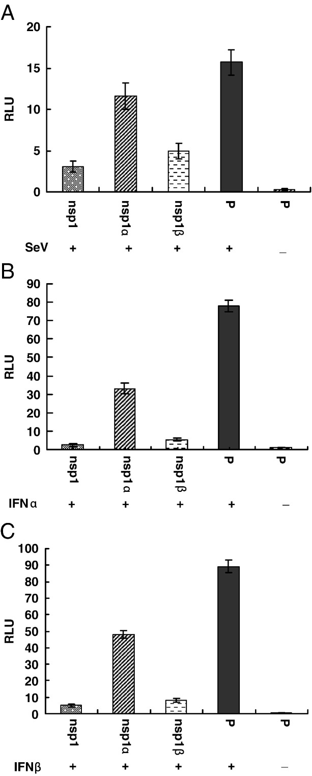
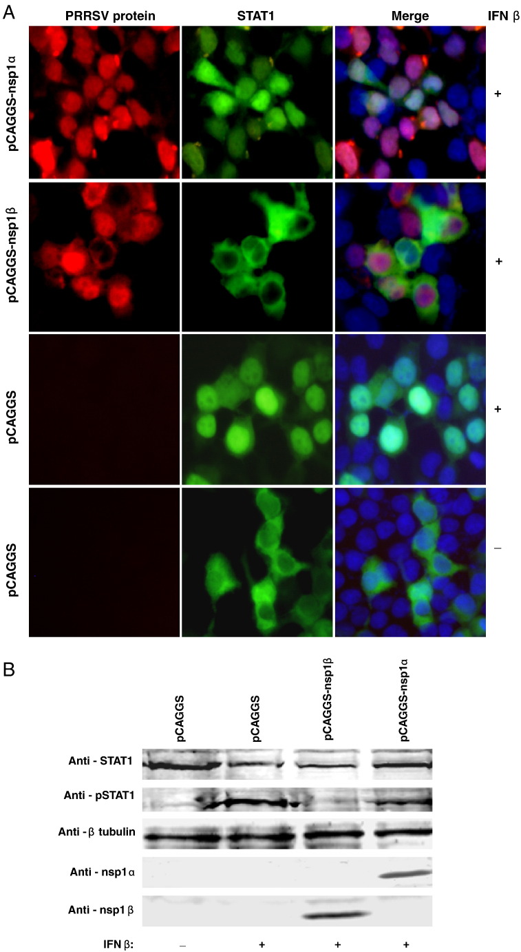
The porcine reproductive and respiratory syndrome virus nsp1 is predicted to be auto-cleaved from the replicase polyprotein into nsp1alpha and nsp1beta subunits. In infected cells, we detected the actual existence of nsp1alpha and nsp1beta. Cleavage sites between nsp1alpha/nsp1beta and nsp1beta/nsp2 were identified by protein microsequencing analysis. Time course study showed that nsp1alpha and nsp1beta mainly localize into the cell nucleus after 10 h post infection. Further analysis revealed that both proteins dramatically inhibited IFN-beta expression. The nsp1beta was observed to significantly inhibit expression from an interferon-stimulated response element promoter after Sendai virus infection or interferon treatment. It was further determined to inhibit nuclear translocation of STAT1 in the JAK-STAT signaling pathway. These results demonstrated that nsp1beta has ability to inhibit both interferon synthesis and signaling, while nsp1alpha alone strongly inhibits interferon synthesis. These findings provide important insights into mechanisms of nsp1 in PRRSV pathogenesis and its impact in vaccine development.


Figures

Fig. 1
Fig. 1
Identification of PRRSV nsp1 auto-cleavage products nsp1α and nsp1β in infected cells by Western blot. MARC-145 cells were infected with SD23983 virus or mock-infected. Viral proteins from cell lysate were separated by 15% SDS-PAGE gel and transferred to a nitrocellulose membrane. The membrane was probed with rabbit polyclonal anti-sera against nsp1 (pAb-nsp1) or nsp1α (pAb-nsp1α), or monoclonal antibody against nsp1β (mAb-nsp1β). Arrows point to specific PRRSV nsp1 proteins.
Fig. 2
Fig. 2
Comparative sequence analysis of the nsp1α/nsp1β and nsp1β/nsp2 cleavage sites. Partial ORF1a amino acid sequence of six PRRSV strains corresponding to SD23983 ORF1a amino acid position 160–204 and 361–409 was aligned with EAV nsp1. The alignment was generated by ALIGNX program of Vector NTI Suite software (InforMax, Inc.). Two boxes depicted in the map of amino acid sequence comparison represent the result of nsp1β and nsp2 protein N-terminal sequencing, which was determined by sequential Edman degradation of 10 cycles for each viral protein. The upward and downward solid arrows point to the identified cleavage site of nsp1β/nsp2 and nsp1α/nsp1β, respectively. The downward dashed arrow points to the predicted nsp1α/nsp1β cleavage site.
Fig. 3
Fig. 3
Detection of nsp1α and nsp1β expression in virus-infected cells by indirect immunofluorescence assay. MARC-145 cells were infected with PRRSV SD23983 and fixed at 6, 8, 10 and 12 hpi. Cells were stained with a PRRSV protein specific primary antibody, pAb-nsp1α or mAb-nsp1β. FITC-conjugated goat anti-rabbit or anti-mouse antibody was used as secondary antibody. Cell nucleus was stained with DAPI. Images were obtained by fluorescence and phase-contrast microscopy using a 40× objective.
Fig. 4
Fig. 4
Identification of nsp1α and nsp1β expression in transfected cells. HEK293T cells were transfected with either control plasmid, pCAGGS or plasmids expressing PRRSV nsp1 proteins, including pCAGGS, pCAGGS-nsp1α or pCAGGS-nsp1β. At 24 h post-transfection, cells were fixed for immunofluorescence assay or harvested for Western blot analysis. (A) Immunofluorescent detection of nsp1α and nsp1β expression in HEK293T cells. Cells were stained with a specific antibody as indicated at the bottom of the picture, and DyLight 549-conjugated goat anti-rabbit or anti-mouse antibody was used as the secondary antibody. Images were taken by fluorescence and phase-contrast microscopy using a 20× objective. (B) Western blotting analysis nsp1, nsp1α and nsp1β expression in 293T cells. Membranes were probed with an nsp1 protein specific antibody as indicated at the bottom of each membrane. Arrows point to the specific PRRSV proteins.
Fig. 5
Fig. 5
PRRSV nsp1 proteins inhibit IFN-β production. (A, B) HEK293T cells cultured in 24-well plates were cotransfected with a plasmid expressing nsp1 proteins, a plasmid expressing influenza virus NS1, or pCAGGS empty vector (P), pRL-SV40, and a luciferase reporter plasmid p125-Luc (A) or pCIB-55-Luc (B). At 20 h post transfection, cells were infected with Sendai virus (SeV) for 16 h to stimulate the production of interferon. (C–H, J) HEK293T cells in 24-well plates were cotransfected with the plasmid pEFneo-RIG-I (C), pEFneo-MDA5 (D), pEGFP-IPS-1 (E), pEFneo-TBK1 (F), pEFneo-IKKɛ (G), or pCAGGS-IRF3 (H), or pcDNA3-TRIF (J), along with pRL-SV40, pCAGGS expressing nsp1 proteins, and pCIB-55 plasmid for 20–24 h. (I) HEK293T cells were cotransfected with pNF-kB-luc, pcDNA3-TRIF, pRL-SV40 and pCAGGS expressing nsp1 proteins or pCAGGS empty vector (P) for 20 h. Cells were harvested and measured for firefly and Renilla luciferase activities. Relative luciferase activity is defined as a ratio of firefly luciferase reporter activity to Renilla luciferase activity. Each data point shown represents a mean value from three experiments. Error bars show standard deviations of the normalized data.
Fig. 6
Fig. 6
PRRSV nsp1 proteins inhibit expression from an ISRE promoter. HEK293T cells were cotransfected with pISRE-luc, pRL-SV40 and pCAGGS expressing nsp1 proteins or pCAGGS empty vector (P) for 20 h. Cells were then infected with Sendai virus (A) or treated with IFN-α (B) and IFN-β (C) for 20 h. The cells were harvested and measured for firefly and Renilla luciferase activities. Relative luciferase activity is defined as a ratio of firefly luciferase reporter activity to Renilla luciferase activity. Each data point shown represents a mean value from three experiments. Error bars show standard deviations of the normalized data.
Fig. 7
Fig. 7
Analysis of PRRSV nsp1 protein's effect on STAT1 translocation and phosphorylation. (A) HEK293T Cells were transfected with the indicated plasmid and STAT1-GFP for 20 h and then treated with IFN-β for 2 h. Cells were fixed and stained with pAb-nsp1α or mAb-nsp1β. DyLight 549-conjugated goat anti-rabbit or anti-mouse antibody (red fluorescence) was used as the secondary antibody. Cell nucleus was stained with DAPI (blue fluorescence). The protein localization was analyzed by fluorescence phase-contrast microscopy using a 40× objective. (B) HEK293T cells were transfected with the indicated plasmid for 24 h and then treated with IFN-β for 2 h. Cells were harvested, and lysates were analyzed by Western blot using antibodies recognizing total and phosphorylated forms of STAT1, pAb-nsp1α, mAb-nsp1β and anti-β-tubulin as a loading control.

Similar articles

Cited by

References

    1. Allende R., Lewis T.L., Lu Z., Rock D.L., Kutish G.F., Ali A., Doster A.R., Osorio F.A. North American and European porcine reproductive and respiratory syndrome viruses differ in non-structural protein coding regions. J. Gen. Virol. 1999;80:307–315. - PubMed
    1. Bauhofer O., Summerfield A., Sakoda Y., Tratschin J.D., Hofmann M.A., Ruggli N. Classical swine fever virus Npro interacts with interferon regulatory factor 3 and induces its proteasomal degradation. J. Virol. 2007;81:3087–3096. - PMC - PubMed
    1. Benfield D.A., Nelson E., Collins J.E., Harris L., Goyal S.M., Robison D., Christianson W.T., Morrison R.B., Gorcyca D., Chladek D. Characterization of swine infertility and respiratory syndrome (SIRS) virus (isolate ATCC VR-2332) J. Vet. Diagn. Invest. 1992;4:127–133. - PubMed
    1. Brown E., Lawson S., Welbon C., Murtaugh M.P., Nelson E.A., Zimmerman J.J., Rowland R.R.R., Fang Y. Antibody response of nonstructural proteins: implication for diagnostic detection and differentiation of Type I and Type II porcine reproductive and respiratory syndrome virus. Clin. Vaccine Immunol. 2009;16:628–635. - PMC - PubMed
    1. Buddaert W., Van Reeth K., Pensaert M. In vivo and in vitro interferon (IFN) studies with the porcine reproductive and respiratory syndrome virus (PRRSV) Adv. Exp. Med. Biol. 1998;440:461–467. - PubMed

Publication types

MeSH terms