. 2010 Mar;95(3):415-23.
doi: 10.3324/haematol.2009.010785. Epub 2009 Dec 8.

Autocrine IGF-1/IGF-1R signaling is responsible for constitutive PI3K/Akt activation in acute myeloid leukemia: therapeutic value of neutralizing anti-IGF-1R antibody

Affiliations

Autocrine IGF-1/IGF-1R signaling is responsible for constitutive PI3K/Akt activation in acute myeloid leukemia: therapeutic value of neutralizing anti-IGF-1R antibody

Nicolas Chapuis et al. Haematologica. 2010 Mar.

Abstract

Background: Alterations in the PI3K/Akt pathway are found in a wide range of cancers and the development of PI3K inhibitors represents a promising approach to cancer therapy. Constitutive PI3K activation, reflecting an intrinsic oncogenic deregulation of primary blast cells, is detected in 50% of patients with acute myeloid leukemia. However, the mechanisms leading to this activation are currently unknown. As we previously reported IGF-1 autocriny in acute myeloid leukemia cells, we investigated whether IGF-1 signaling was involved in the constitutive activation of PI3K.

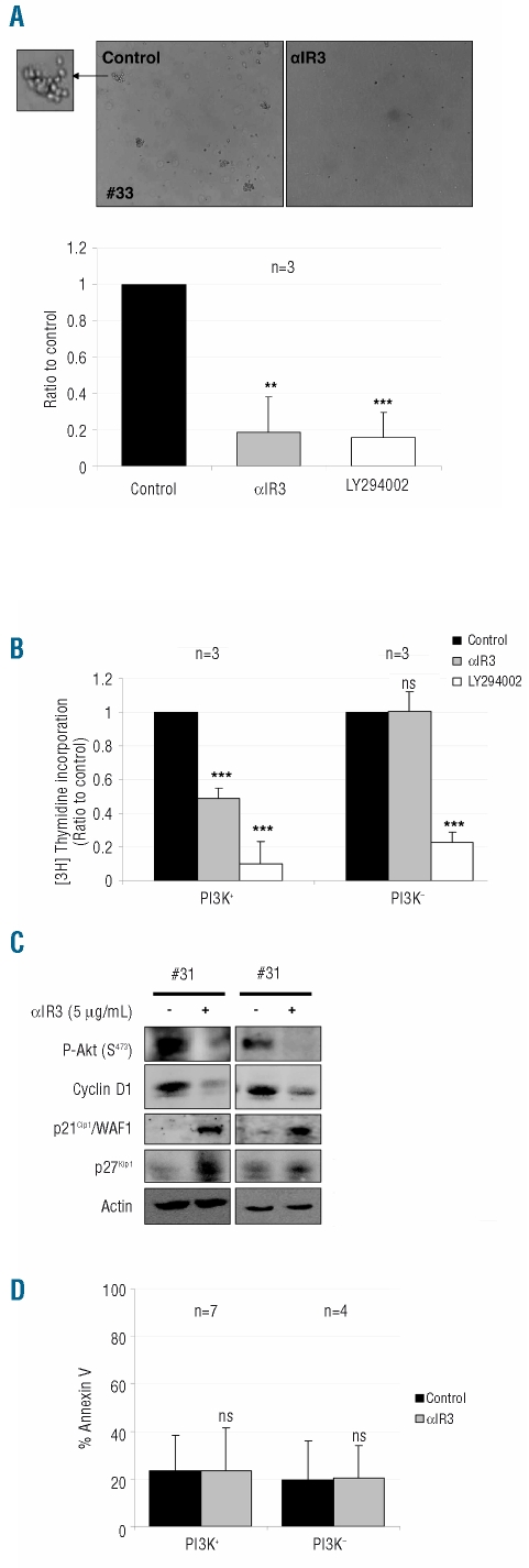
Design and methods: We analyzed the IGF-1/IGF-1R signaling pathway and PI3K activity in 40 acute myeloid leukemia bone marrow samples. Specific inhibition of IGF-1/IGF-1R signaling was investigated using neutralizing anti-IGF-1R, anti-IGF-1 antibodies or IGF-1 short interfering RNA. The anti-leukemic activity of the neutralizing anti-IGF-1R was tested by analyzing its effects on leukemic progenitor clonogenicity, blast cell proliferation and survival.

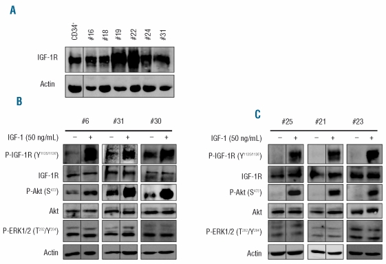
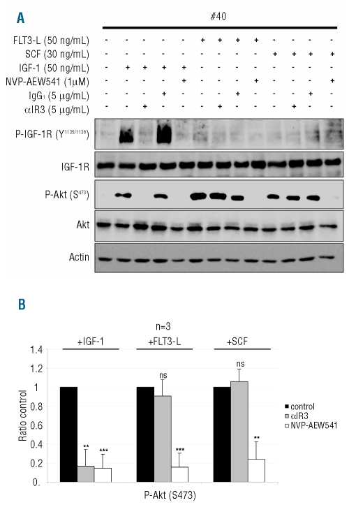
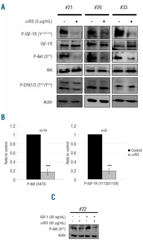
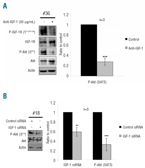
Results: In all samples tested, we found that functional IGF-1R was constantly expressed in leukemic cells. In the acute myeloid leukemia samples with PI3K activation, we found that the IGF-1R was constitutively phosphorylated, although no IGF-1R activating mutation was detected. Specific inhibition of IGF-1R signaling with neutralizing anti-IGF-1R strongly inhibited the constitutive phosphorylation of both IGF-1R and Akt in 70% of the PI3K activated samples. Moreover, both incubation with anti-IGF-1 antibody and IGF-1 short interfering RNA inhibited Akt phosphorylation in leukemic cells. Finally, neutralizing anti-IGF-1R treatment decreased the clonogenicity of leukemic progenitors and the proliferation of PI3K activated acute myeloid leukemia cells.

Conclusions: Our current data indicate a critical role for IGF-1 autocriny in constitutive PI3K/Akt activation in primary acute myeloid leukemia cells and provide a strong rationale for targeting IGF-1R as a potential new therapy for this disease.


Figures

Figure 1.
Figure 1.
The IGF-1/IGF-1R pathway is functional and constitutively activated in PI3K+ AML blast cells. (A) Expression of IGF-1R in total cell lysates from six AML samples was compared with that in normal CD34+ hematopoietic progenitors. Protein extracts from 106 cells were analyzed by western blotting. (B) After purification, PI3K+ or (C) PI3K AML cells were starved for 4 h in cytokine and serum-free medium and then stimulated or not with 50 ng/mL IGF-1 for 10 min. Protein extracts from 106 cells were analyzed by western blotting.
Figure 2.
Figure 2.
Specific inhibition of IGF-1R signaling with αIR3. (A) PI3K-AML blast cells were cultured for 4 h in cytokine- and serum-free medium with or without αIR3 (5 μg/mL), mouse IgG1 (5 μg/mL) for isotype negative control or NVP-AEW541 (1 μM) during the last hour. AML blast cells were then stimulated for 10 min with either IGF-1 (50 ng/mL), FLT3-L (50 ng/mL) or SCF (30 ng/mL). Protein extracts from 106 cells were then analyzed by western blotting. (B) Phospho-Akt S473 signal intensity was quantified and normalized to Akt signal intensity in three PI3K AML samples. Results are expressed as a ratio to the control incubation without αIR3 or NVP-AEW541. Vertical bars indicate standard deviations. The statistical significance was calculated by Student’s t test. **P<0.01, ***P<0.001, ns: not significant.
Figure 3.
Figure 3.
PI3K constitutive activation is due to IGF-1/IGF-1R interaction in 70% of AML. After purification, AML blast cells were cultured for 4 h in cytokine- and serum-free medium. (A) During the last hour of starvation, cells were treated with or without αIR3 (5μg/mL) and (C) stimulated or not with IGF-1 (50 ng/mL). Protein extracts from 106 cells were analyzed by western blotting. (B) Phospho-Akt S473 and phospho-IGF-1R (Y1135/1136) were quantified and normalized to Akt and IGF-1R signal intensity in 14 and 6 PI3K+ samples, respectively. Results are expressed as a ratio to the control incubation without αIR3. Vertical bars indicate standard deviations. ***P<0.001.
Figure 4.
Figure 4.
Autocrine IGF-1 production is responsible for PI3K constitutive activation in AML samples. (A) During the last hour of starvation, blast cells from three AML samples were treated with or without an anti-human IGF-1 antibody (50 μg/mL). Protein extracts from 106 cells were analyzed by western blotting and phospho-Akt S473 signal intensity was quantified as described in Figure 3B. ***P<0.001. (B) AML blast cells from five patients were transfected with IGF-1 siRNA or control siRNA, then incubated for 24 h in 10% FCS MEM. IGF-1 mRNA expression was assessed by quantitative RT-PCR and results are expressed as a ratio to the incubation with control siRNA. Protein extracts from 106 cells were analyzed by western blotting and phospho-Akt S473 signal intensity was quantified as described in Figure 3B. **P<0.01; ***P<0.001.
Figure 5.
Figure 5.
Inhibition of IGF-1 signaling with αIR3 impairs the clonogenic growth of leukemic progenitors and the cell cycle progression of PI3K+ AML blast cells. (A) Blast cells from three PI3K+ AML samples were cultured at 105 cells/mL in H4230 methyl cellulose medium, supplemented with 10% 5637 conditioned medium with or without the different inhibitors: αIR3 (5 μg/mL), LY2942002 (25 μM). Cells were then plated in 35-mm Petri dishes in duplicate, and incubated for 7 days. At day 7, CFU-L were scored under an inverted microscope. Upper panel: the picture illustrates the results obtained for sample #33. Lower panel: results are expressed as a percentage of the control for each condition. Vertical bars indicate standard deviations. **P<0.01; ***P<0.001. (B) For proliferation assays, AML blast cells from three PI3K+ and three PI3K samples were cultured for 48 h at 105 cells/mL, in triplicate, in 5% FCS, without or with αIR3 (5 μg/mL) or LY2942002 (25 μM), and then pulsed for 6 h with 1 μCi (37 kBq) [3H]thymidine. The amounts of radioactivity were determined after trichloracetic acid precipitation. Results are expressed as a ratio to the control, for each condition. Vertical bars indicate standard deviations. ***P<0.001; ns: not significant (C) AML cells from two PI3K+ AML samples were incubated at 106/mL in FCS 10%, without or with αIR3 for 24 h. Protein extracts from 106 cells were analyzed by western blotting. (D) AML blast cells from seven PI3K+ and four PI3K samples were incubated at 5×10⁵ cells/mL for 48 h in 10% FCS, without or with αIR3 (5 μg/mL) and then stained with annexin V-phycoerythrin. Results are expressed as a percentage of annexin-V stained cells. Vertical bars indicate standard deviations and “ns” means that the difference is not significant.

Similar articles

Cited by

References

    1. Appelbaum FR, Rowe JM, Radich J, Dick JE. Acute myeloid leukemia. Hematology Am Soc Hematol Educ Program. 2001:62–86. - PubMed
    1. Ravandi F, Burnett AK, Agura ED, Kantarjian HM. Progress in the treatment of acute myeloid leukemia. Cancer. 2007;110(9):1900–10. - PubMed
    1. Heinrich MC, Corless CL, Duensing A, McGreevey L, Chen CJ, Joseph N, et al. PDGFRA activating mutations in gastrointestinal stromal tumors. Science. 2003;299(5607):708–10. - PubMed
    1. Lee JW, Soung YH, Kim SY, Lee HW, Park WS, Nam SW, et al. PIK3CA gene is frequently mutated in breast carcinomas and hepatocellular carcinomas. Oncogene. 2005;24(8):1477–80. - PubMed
    1. Cairns P, Okami K, Halachmi S, Halachmi N, Esteller M, Herman JG, et al. Frequent inactivation of PTEN/MMAC1 in primary prostate cancer. Cancer Res. 1997;57(22):4997–5000. - PubMed

Publication types

MeSH terms