Review
. 2010 Mar;38(5):1415-30.
doi: 10.1093/nar/gkp1117. Epub 2009 Dec 8.

5-methylcytosine in RNA: detection, enzymatic formation and biological functions

Affiliations
Review

5-methylcytosine in RNA: detection, enzymatic formation and biological functions

Yuri Motorin et al. Nucleic Acids Res. 2010 Mar.

Abstract

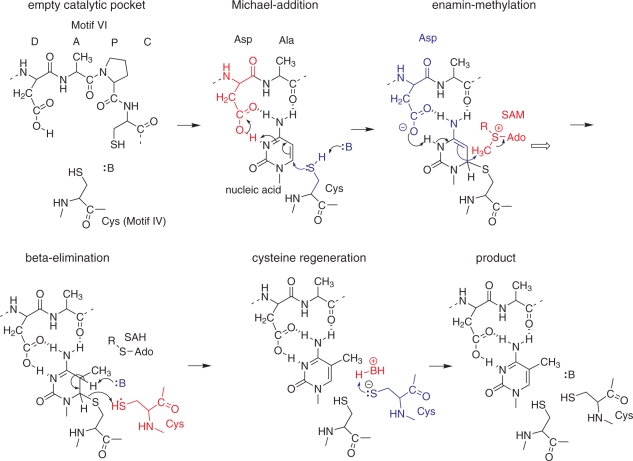
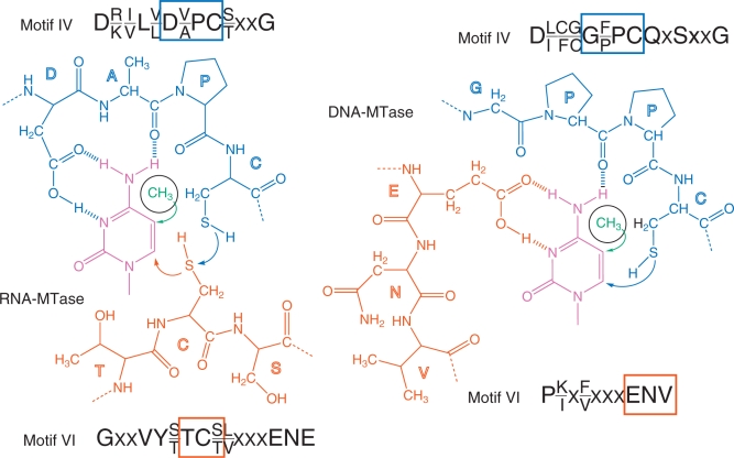
The nucleobase modification 5-methylcytosine (m(5)C) is widespread both in DNA and different cellular RNAs. The functions and enzymatic mechanisms of DNA m(5)C-methylation were extensively studied during the last decades. However, the location, the mechanism of formation and the cellular function(s) of the same modified nucleobase in RNA still remain to be elucidated. The recent development of a bisulfite sequencing approach for efficient m(5)C localization in various RNA molecules puts ribo-m(5)C in a highly privileged position as one of the few RNA modifications whose detection is amenable to PCR-based amplification and sequencing methods. Additional progress in the field also includes the characterization of several specific RNA methyltransferase enzymes in various organisms, and the discovery of a new and unexpected link between DNA and RNA m(5)C-methylation. Numerous putative RNA:m(5)C-MTases have now been identified and are awaiting characterization, including the identification of their RNA substrates and their related cellular functions. In order to bring these recent exciting developments into perspective, this review provides an ordered overview of the detection methods for RNA methylation, of the biochemistry, enzymology and molecular biology of the corresponding modification enzymes, and discusses perspectives for the emerging biological functions of these enzymes.


Figures

Figure 1.
Figure 1.
(A) Chemical reactivity of cytidine and m5C. The methyl group of m5C is shown in purple. Attack sites on the nucleobase are indicated by arrows, where black indicates oxidizing agents, red indicates alkylating electrophiles and blue arrows indicate nucleophiles. Numbering of the nucleobase atoms 1–6 is indicated inside the ring. Ribose carbon atom numbering is indicated by primed (′) numbers (B) Bisulfite mediated conversion of cytosine to uracil. Nucleophiles, bases and nucleophilic attacks are indicated in blue, acids are indicated in red.
Figure 2.
Figure 2.
Principle of m5C detection by RNA bisulfite sequencing and combination with next-generation sequencing. The DNA primer sequence for reverse transcription is adapted for C to U conversions in the analyzed RNA.
Figure 3.
Figure 3.
Occurrence of m5C residues in tRNAs and rRNAs. The frequency of m5C occurrence is color-coded in tRNA: yellow, <10%; orange, between 10 and 30%; green, between 30 and 60%; blue >60%. Numbers in square brackets show the number of m5C and number of unmodified or other modified C at a given position [m5C/C] The same color code is used for global distribution in all sequenced tRNAs, numbers are not shown for clarity. Abbreviations: CY, cytoplasm; MI, mitochondria; ANI, animals; SIN, single cell eukaryota; PLA, plants; ARCHAE, Archaea. The lower panel shows rRNA fragments from different species with m5C residues highlighted in blue.
Figure 4.
Figure 4.
Subcellular localization of m5C residues in RNAs and m5C-MTases in bacteria (upper left), lower eukaryota (lower left) and higher eukaryota (right). Known m5C residues in RNAs are shown in white circles, together with the corresponding enzyme (if known). DNA-MTases and Dnmt2 are colored in purple, the RsmB/Nol1 family in blue, the YebU (RsmF) family in orange, the Ynl022 family in green and NSUN6 in grey.
Figure 5.
Figure 5.
Catalytic mechanism of m5C-MTases. Nucleophiles, bases and nucleophilic attacks are indicated in blue; acids and electrophiles are indicated in red.
Figure 6.
Figure 6.
Catalytic pocket of RNA- (left) and DNA-MTases (right). The target cytidine residue is indicated in purple, the sequences of conserved motifs IV and VI are given for both classes of enzymes. Motif IV is in blue and motif VI is in orange. Boxes highlight residues belonging to the respective sequence motifs.

References

    1. Hotchkiss RD. The quantitative separation of purines, pyrimidines, and nucleosides by paper chromatography. J. Biol. Chem. 1948;175:315–332. - PubMed
    1. Luciano DJ, Mirsky H, Vendetti NJ, Maas S. RNA editing of a miRNA precursor. RNA. 2004;10:1174–1177. - PMC - PubMed
    1. Blow MJ, Grocock RJ, van Dongen S, Enright AJ, Dicks E, Futreal PA, Wooster R, Stratton MR. RNA editing of human microRNAs. Genome Biol. 2006;7:R27. - PMC - PubMed
    1. Yang Z, Ebright YW, Yu B, Chen X. HEN1 recognizes 21-24 nt small RNA duplexes and deposits a methyl group onto the 2′ OH of the 3′ terminal nucleotide. Nucleic Acids Res. 2006;34:667–675. - PMC - PubMed
    1. Yu B, Yang Z, Li J, Minakhina S, Yang M, Padgett RW, Steward R, Chen X. Methylation as a crucial step in plant microRNA biogenesis. Science. 2005;307:932–935. - PMC - PubMed

Publication types

MeSH terms