. 2010 Apr 22;277(1685):1289-98.
doi: 10.1098/rspb.2009.1884. Epub 2009 Dec 9.

Atractiellomycetes belonging to the 'rust' lineage (Pucciniomycotina) form mycorrhizae with terrestrial and epiphytic neotropical orchids

Affiliations

Atractiellomycetes belonging to the 'rust' lineage (Pucciniomycotina) form mycorrhizae with terrestrial and epiphytic neotropical orchids

Ingrid Kottke et al. Proc Biol Sci. .

Abstract

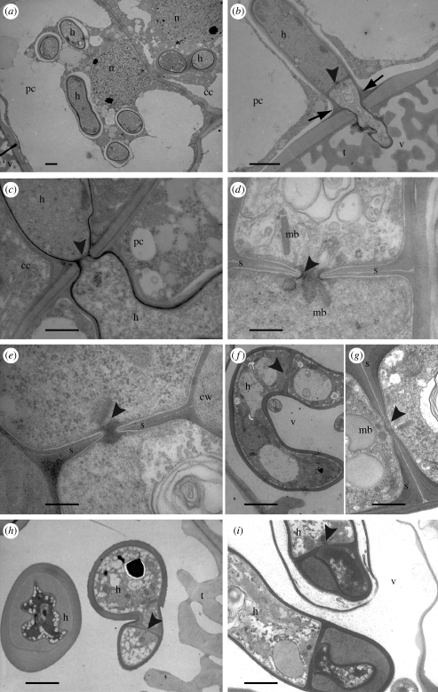
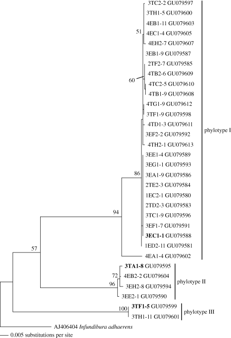
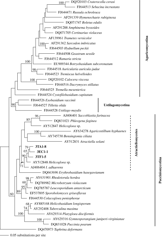
Distinctive groups of fungi are involved in the diverse mycorrhizal associations of land plants. All previously known mycorrhiza-forming Basidiomycota associated with trees, ericads, liverworts or orchids are hosted in Agaricomycetes, Agaricomycotina. Here we demonstrate for the first time that Atractiellomycetes, members of the 'rust' lineage (Pucciniomycotina), are mycobionts of orchids. The mycobionts of 103 terrestrial and epiphytic orchid individuals, sampled in the tropical mountain rainforest of Southern Ecuador, were identified by sequencing the whole ITS1-5.8S-ITS2 region and part of 28S rDNA. Mycorrhizae of 13 orchid individuals were investigated by transmission electron microscopy. Simple septal pores and symplechosomes in the hyphal coils of mycorrhizae from four orchid individuals indicated members of Atractiellomycetes. Molecular phylogeny of sequences from mycobionts of 32 orchid individuals out of 103 samples confirmed Atractiellomycetes and the placement in Pucciniomycotina, previously known to comprise only parasitic and saprophytic fungi. Thus, our finding reveals these fungi, frequently associated to neotropical orchids, as the most basal living basidiomycetes involved in mycorrhizal associations of land plants.


Figures

Figure 1.
Figure 1.
(ac) Light micrographs of transverse section through root of Pleurothallis sp. 3TC1 displaying fungal colonization of velamen and cortical cells. (a) Overview (scale bar, 30 µm). (b) Cortical cell with coils (pelotons) of living hyphae (scale bar, 10 µm). (c) Cortical cell with collapsed, lysed hyphae and pycnoid nucleus (scale bar, 10 µm). (di) TEM of orchid-mycorrhiza structures and details of fungal features in unidentified orchids 3EB1 (d,e) and 3EE2 (f), Maxillaria sp. 4EC1 (g) and Pleurothallis sp. 3TC1 (h,i); (d) living hyphae embedded in dense root cytosol separated by plasma membrane and interfacial matrix (scale bar, 3 µm); (e) enlargement of hypha with well-preserved interfacial matrix (arrows; scale bar, 1 µm). (f) Hypha in root cortical cell displaying simple-pored septum without membrane caps, the pore plugged by a microbody (arrow head; scale bar, 1 µm); (g) enlargement of simple septal pore (arrow head) surrounded by several membrane-coated microbodies in dense cytosol (scale bar, 0.5 µm); (h) symplechosome in hypha of simple-septate basidiomycete (scale bar, 0.2 µm); (i) hypha branching without clamp formation displaying simple-pored septum (scale bar, 1 µm). cc, Cortical cell; cv, cortical cell vacuole; cy, cortical cell cytosol; dp, degenerating hyphal peloton; ex, exodermis; h, hypha; m, mitochondrion; mb, microbody; n, nucleus; p, living hyphal peloton; pc, passage cell; s, septum; st, stele; t, tilosomes; v, velamen.
Figure 2.
Figure 2.
TEM showing series of root colonization in Pleurothallis sp. 3TC1 by the simple-septate basidiomycetes. (a) Overview of colonization of passage cell and adjacent cortical cell, note velamen with tilosomes at lower left corner (scale bar, 3 µm); (b) simple-septate basidiomycete (arrow head) passing from velamen among tilosomes into the passage cell, note narrowing of hyphal diameter, formation of septum and host cell wall apposition at the entry point (arrows; scale bar, 3 µm); (c) fungus passing from passage cell into cortical cell, note narrowing of hyphal diameter and formation of septum (scale bar, 2 µm); (d) enlargement of simple-pored septum with microbodies of micrograph (b) (scale bar, 0.5 µm); (e) enlargement of simple-pored septum of micrograph (c), pore plugged by electron dark material (scale bar, 0.5 µm); (fi) simple-septate basidiomycete in the velamen: (f) large hypha in velamen (scale bar, 5 µm); (g) enlargement of the simple-pored septum with microbodies of micrograph (f) (scale bar, 1 µm); (h) square section through thick-walled hypha and hypha branching with septum formation (scale bar, 2 µm); (i) hypha with partial cell wall thickening and simple-pored septum, indicating that the thick-walled hyphae belong to the respective fungus (scale bar, 2 µm). For abbreviations see figure 1.
Figure 3.
Figure 3.
ML analysis of Atractiellomycetes sequences (ITS + nucLSU rDNA, 1550 bp) obtained from orchid mycorrhizae with I. adhaerens as outgroup. Bootstrap values are given for 1000 replicates, values below 50 per cent are omitted. The names of the sequences correspond to the orchid IDs giving site (1–4), habitat (Epiphytic or Terrestrial), plot (A–H), plant individual (1–5) and clone (corresponding to electronic supplementary material, table S1e). Sequences in bold were selected as representatives of the phylotypes.
Figure 4.
Figure 4.
Phylogenetic placement of the fungal representatives of phylotypes I–III associated with orchids (figure 3) within Basidiomycota based on ML analysis from an alignment of partial nuclear large subunit rDNA sequences. Bootstrap values are given for 1000 replicates; values below 50 per cent are omitted.

References

    1. Aime M. C., et al. 2007An overview of the higher-level classification of Pucciniomycotina based on combined analysis of nuclear large and small subunit rDNA sequences. Mycologia 98, 896–905 (doi:10.3852/mycologia.98.6.896) - DOI - PubMed
    1. Altschul S. F., Madden T. L., Schäffer A. A., Zhang J., Zhang Z., Miller W., Lipman D. J.1997Gapped BLAST and PSI-Blast: a new generation of protein database search programs. Nucleic Acids Res. 25, 3389–3402 (doi:10.1093/nar/25.17.3389) - DOI - PMC - PubMed
    1. Barroso J., Pais M. S.1985Cytochimie—caractérisation cytochimique de l'interface hôte/endophyte des endomycorrhizes d’Ophrys lutea. Rôle de l'hôte dans la synthèse des polysaccharides. Ann. Sci. Nat., Bot. 13, 237–244
    1. Bauer R., Oberwinkler F.1991The symplechosome: a unique cell organelle of some basidiomycetes. Botanica Acta 104, 93–97
    1. Bauer R., Begerow D., Sampaio J. P., Weiß M., Oberwinkler F.2006The simple-septate basidiomycetes: a synopsis. Mycol. Progr. 5, 41–66 (doi:10.1007/s11557-006-0502-0) - DOI

Publication types

LinkOut - more resources