The NS segment of an H5N1 highly pathogenic avian influenza virus (HPAIV) is sufficient to alter replication efficiency, cell tropism, and host range of an H7N1 HPAIV
- PMID: 20007264
- PMCID: PMC2812369
- DOI: 10.1128/JVI.01668-09
The NS segment of an H5N1 highly pathogenic avian influenza virus (HPAIV) is sufficient to alter replication efficiency, cell tropism, and host range of an H7N1 HPAIV
Abstract
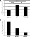
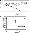
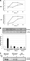
A reassortant avian influenza virus (designated FPV NS GD), carrying the NS-segment of the highly pathogenic avian influenza virus (HPAIV) strain A/Goose/Guangdong/1/96 (GD; H5N1) in the genetic background of the HPAIV strain A/FPV/Rostock/34 (FPV; H7N1), was rescued by reverse genetics. Remarkably, in contrast to the recombinant wild-type FPV (rFPV), the reassortant virus was able to replicate more efficiently in different human cell lines and primary mouse epithelia cells without prior adaptation. Moreover, FPV NS GD caused disease and death in experimentally infected mice and was detected in mouse lungs; in contrast, rFPV was not able to replicate in mice effectively. These results indicated an altered host range and increased virulence. Furthermore FPV NS GD showed pronounced pathogenicity in chicken embryos. In an attempt to define the molecular basis for the apparent differences, we determined that NS1 proteins of the H5N1 and H7N1 strains bound the antiviral kinase PKR and the F2F3 domain of cleavage and polyadenylation specificity factor 30 (CPSF30) with comparable efficiencies in vitro. However, FPV NS GD infection resulted in (i) increased expression of NS1, (ii) faster and stronger PKR inhibition, and (iii) stronger beta interferon promoter inhibition than rFPV. Taken together, the results shed further light on the importance of the NS segment of an H5N1 strain for viral replication, molecular pathogenicity, and host range of HPAIVs and the possible consequences of a reassortment between naturally occurring H7 and H5 type HPAIVs.
Figures


Similar articles
-
The NS segment of H5N1 avian influenza viruses (AIV) enhances the virulence of an H7N1 AIV in chickens.Vet Res. 2014 Jan 25;45(1):7. doi: 10.1186/1297-9716-45-7. Vet Res. 2014. PMID: 24460592 Free PMC article.
-
Reassortment of NS segments modifies highly pathogenic avian influenza virus interaction with avian hosts and host cells.J Virol. 2013 May;87(10):5362-71. doi: 10.1128/JVI.02969-12. Epub 2013 Mar 6. J Virol. 2013. PMID: 23468508 Free PMC article.
-
NS reassortment of an H7-type highly pathogenic avian influenza virus affects its propagation by altering the regulation of viral RNA production and antiviral host response.J Virol. 2010 Nov;84(21):11323-35. doi: 10.1128/JVI.01034-10. Epub 2010 Aug 25. J Virol. 2010. PMID: 20739516 Free PMC article.
-
The genetics of highly pathogenic avian influenza viruses of subtype H5 in Germany, 2006-2020.Transbound Emerg Dis. 2021 May;68(3):1136-1150. doi: 10.1111/tbed.13843. Epub 2020 Sep 29. Transbound Emerg Dis. 2021. PMID: 32964686 Review.
-
Accessory Gene Products of Influenza A Virus.Cold Spring Harb Perspect Med. 2021 Dec 1;11(12):a038380. doi: 10.1101/cshperspect.a038380. Cold Spring Harb Perspect Med. 2021. PMID: 32988983 Free PMC article. Review.
Cited by
-
Zoonotic Potential of Influenza A Viruses: A Comprehensive Overview.Viruses. 2018 Sep 13;10(9):497. doi: 10.3390/v10090497. Viruses. 2018. PMID: 30217093 Free PMC article. Review.
-
The NS segment of H5N1 avian influenza viruses (AIV) enhances the virulence of an H7N1 AIV in chickens.Vet Res. 2014 Jan 25;45(1):7. doi: 10.1186/1297-9716-45-7. Vet Res. 2014. PMID: 24460592 Free PMC article.
-
Exposure to a low pathogenic A/H7N2 virus in chickens protects against highly pathogenic A/H7N1 virus but not against subsequent infection with A/H5N1.PLoS One. 2013;8(3):e58692. doi: 10.1371/journal.pone.0058692. Epub 2013 Mar 4. PLoS One. 2013. PMID: 23469288 Free PMC article.
-
Development of an Enhanced High-Yield Influenza Vaccine Backbone in Embryonated Chicken Eggs.Vaccines (Basel). 2023 Aug 15;11(8):1364. doi: 10.3390/vaccines11081364. Vaccines (Basel). 2023. PMID: 37631932 Free PMC article.
-
H5N1 influenza virulence, pathogenicity and transmissibility: what do we know?Future Virol. 2015;10(8):971-980. doi: 10.2217/fvl.15.62. Future Virol. 2015. PMID: 26617665 Free PMC article.
References
-
- Alexander, D. J. 2000. A review of avian influenza in different bird species. Vet. Microbiol. 74:3-13. - PubMed
-
- Alexander, D. J. 2007. Summary of avian influenza activity in Europe, Asia, Africa, and Australasia, 2002-2006. Avian Dis. 51:161-166. - PubMed
-
- Bonin, J., and C. Scholtissek. 1983. Mouse neurotropic recombinants of influenza A viruses. Arch. Virol. 75:255-268. - PubMed
-
- Bonnet, M. C., C. Daurat, C. Ottone, and E. F. Meurs. 2006. The N-terminus of PKR is responsible for the activation of the NF-kappaB signaling pathway by interacting with the IKK complex. Cell Signal. 18:1865-1875. - PubMed
Publication types
MeSH terms
Substances
Grants and funding
LinkOut - more resources
Full Text Sources
Other Literature Sources
Medical
Miscellaneous

